Abstract
Chronic obstructive pulmonary disease (COPD) is associated to cardiovascular morbidity and mortality, and abnormalities of the autonomic nervous system have been described in subjects with severe disease. We studied heart rate variability (HRV) in COPD patients at rest and during the 6–minute Walk Test (6mWT) and the association with lung function impairment taking into account systemic inflammation. Thirty outpatients with stable COPD underwent lung function measurements, blood gas analysis, ECG Holter and transcutaneous pulse oximetry during 6mWT and then they were classified by BODE index. Also C-reactive protein (CRP) was measured. At rest, we observed a significant reduction of HRV for increasing BODE index. During the 6mWT, HRV tended to decrease in BODE 1 subjects whereas an increase was observed in BODE 2 and BODE 3-4 subjects. Subjects with elevated CRP values had a significant reduction in Standard Deviation of all normal RR intervals at rest (SDNN: p = 0.013), Total Power (TFA: p = 0.04) and Very Low Frequency band (VLF: p = 0.041). At rest, subjects with Inspiratory Capacity-to-Total Lung Capacity ratio (IC/TLC) < 36% had a significant reduced SDNN (p = 0.004), TFA (p = 0.001), VLF (p = 0.001), Low Frequency band (p = 0.007). During 6mWT, changes of HRV parameters were significantly related to airflow obstruction and static hyperinflation indices. At rest and during submaximal exercise, COPD patients with moderate and severe disease had an abnormal cardiac autonomic modulation which was related to both systemic inflammation and lung function impairment.
Introduction
Chronic obstructive pulmonary disease (COPD) is the only major cause of death for which morbidity and mortality are increasing, partially because of associated cardiovascular involvement (Citation1). During two last decades, several studies clearly evidenced that patients with COPD are two to three times more at risk for cardiovascular mortality (Citation2, 3). In the Lung Health Study (Citation4) 2.5% of the original cohort died, and 25% of them died of a cardiovascular event; furthermore cardiovascular events accounted for 42% of the first hospitalizations and 48% of the second hospitalizations. All-cause mortality increased by 14%, for every 10% decrease in FEV1, whereas cardiovascular mortality increased by 28%, and nonfatal coronary events increased by almost 20%. Data coming from the Torch study clearly showed that sudden death accounts for the vast majority of cardiovascular deaths (Citation5–7). Moreover, Inspiratory Capacity–to–Total Lung Capacity ratio (IC/TLC), a measure of static lung hyperinflation, has been found a predictor of mortality and a risk factor for acute exacerbation (Citation8,9).
Natural history of COPD is also influenced by the evidence of subclinical systemic inflammation. Indeed, increased serum levels of C-reactive protein (CRP) have been found to be linked to increased risk of hospitalization or death as well as to the risk of cardiovascular events in COPD patients (Citation10, 11). Heart rate variability (HRV) expresses cyclic fluctuations of heart rate (HR) and reflects autonomic modulation by sympathetic and parasympathetic efferent nervous impulses of heart rhythm. Numerous clinical studies have showed that depressed HRV is an expression of sympatho-vagal balance towards a sympathetic predominance and is a predictor of mortality in post–myocardial infarction (Citation12–14). HRV has been found to be reduced in subjects with COPD (Citation15) and related to the severity of the disease in patients with PiZ α1-antitrypsin deficiency (Citation16). Moreover, depressed HRV in power spectral analysis was demonstrated in cachexia due to severe COPD (Citation17). Lastly, cardiac autonomic modulation during peak exercise has been found different in COPD patients compared to normal subjects (Citation18).
In this pilot study we aimed at investigating two main issues in COPD patients: (Citation1) whether cardiac autonomic function, as assessed by HRV at rest and during physical activity, was influenced by the severity of the disease, defined by the BODE Index (Body-Mass Index, Airflow Obstruction, Dyspnea, and Exercise Capacity Index) and lung mechanical impairment, and (Citation2) whether this relation could be influenced by systemic inflammation.
Methods
The study was approved by the local ethical committee of Università Cattolica del Sacro Cuore, Roma (Approval number: 57-A.1853). Thirty outpatients with stable COPD were recruited, after written informed consent was obtained. We adopted very selective exclusion criteria to investigate HRV in COPD patients with minimal confounding effects deriving from other medical conditions or drugs. This heavily influenced the number of enrolled patients. Patients with a history of cardiovascular disease, diabetes, use of systemic drugs able to influence the nervous autonomic system and/or systemic inflammation (i.e., beta-blockers, aspirin, antidepressants) were excluded. Patients stopped respiratory drugs 24 h before the 6–minute Walk Test (day 2 of the study, see later).
The study lasted 2 days for every patient. On day 1 spirometry, lung volumes and single-breath diffusion capacity for carbon monoxide were measured according to recent ATS/ERS guidelines (Citation19–22). Lung function values were expressed as absolute and as percent of the predicted. A blood gas analysis at rest was also performed. Body Mass Index (BMI, kg/m2) and dyspnoea grade (modified Medical Research Council scale) were evaluated in order to define the BODE index.
On day 2 we obtained a venous blood sample for CRP (lowest detection limit = 0.05 mg/L) and N-terminal B-type natriuretic peptide (NT_proBNP) measurement. Then, all patients underwent, simultaneously: 1) transcutaneous oximetry, with wearable digital Pulse Oximeter; 2) electrocardiographic (ECG) Holter monitoring, using 3-channel digital recorders and monitoring 3 bipolar chest leads; 3) a six–minute Walk Test (6mWT).
HRV was assessed both in the time domain and in the frequency domain. Time domain HRV parameters included the standard deviation of all normal to normal RR intervals (SDNN) and the square root of the mean of the sum of the squares of the differences between adjacent RR intervals (r-MSSD). Frequency domain HRV variables included the amplitude of RR changes in the total spectrum (TFA, 0-0.5 Hz) and in the very low frequency (VLF) band (0.003-0.04 Hz), in the low frequency (LF) band (0.04-0.15 Hz), and in the high frequency (HF) band (0.15-0.4 Hz). The LF/HF ratio was also determined. Additional details about the exclusion criteria and methods of measurement of lung function, 6mWT, CRP and HRV are provided in the online data supplement.
Statistical analysis
Patients were divided into 3 groups according to BODE Index. Subjects with score 0-2 and those with score 3-4 were included in the BODE 1 and BODE 2 group, respectively; whereas subjects with a score greater than 4 were grouped together in BODE 3-4 group.
The HRV measures showed a skewed distribution and were log-transformed for the analysis. Baseline values were analyzed using analysis of variance (ANOVA). Association between HRV and both lung function measures and systemic inflammation marker was then tested by linear regression. In a preliminary analysis, we performed linear regression to identify the lung function parameter which showed the most significant relation with HRV at rest. Measures of static lung hyperinflation (RV/TLC, FRC/TLC and IC/TLC ratios) were found to show better association than airflow obstruction indices (FEV1, FEV1/FVC ratio). Thus, IC/TLC ratio was chosen as index of lung function impairment.
Subjects were then categorized as regards both IC/TLC ratio and CRP value. Distribution of IC/TLC ratio was divided in tertiles: greater than 44%, between 43 and 36% and less than 36%, being the first category the reference group. Patients were also categorized in 3 groups according to CRP levels: up to 5 mg/L (reference group), between 5 and 10 mg/L and greater than 10 mg/L. A multiple regression analysis was then applied to assess the association of IC/TLC and CRP categories with HRV parameters adjusting for oxygen haemoglobin saturation (SatO2), as derived from arterial blood analysis, and for NT proBNP, as marker of cardiac overload. Results were given as adjusted means of each CRP and IC/TLC category. Repeated measures ANOVA was applied to test the changes of HRV variables during the 6mWT test and in each BODE group.
Finally, to assess the influence of lung function indices on HRV changes during the 6mWT, a univariate linear regression analysis was performed taking the difference between the value of each HRV parameter during the test and the baseline value as dependent variable and lung function indices as independent variables.
All analyses were performed using STATA software (Stata Corporation, College Station, Texas).
Results
The main characteristics of patients, according to the BODE index, are shown in . As expected, there was a progressive decrement of lung function with the increase of BODE index: the reduction of airways caliber was found to be parallel to the change of resting hyperinflation indices (RV/TLC, FRC/TLC and IC/TLC ratios). No significant differences were found as regards to arterial pressure of O2, whereas arterial pressure of CO2 was significantly higher in subjects of BODE group 3-4 compared to patients of BODE 1 and BODE 2 groups (F = 5.59, p = 0.009).
Table 1: Characteristics of the sample according to BODE Index.
HRV parameters at rest
shows baseline measurements of HRV parameters. At rest, RR interval became shorter as BODE grading increased. Time-domain HRV measures decreased with the increase of BODE index, although statistical significance was only achieved for SDNN (p = 0.024). Similarly, frequency-domain HRV measures also showed lower values with higher BODE indices with statistical significance found for TFA (p = 0.018) and VLF band (p = 0.002).
Table 2. Baseline values (geometric mean and 95% Confidence Intervals) of HRV parameters according to BODE group.
Correlation analysis showed that SDNN (r = 0.47; p = 0.007), TFA (r = 0.52; p = 0.003), VLF band (r = 0.53; p = 0.002) and LF band (r = 0.43; p = 0.017) at rest were directly related to IC/TLC ratio. Moreover, SDNN (r = 0.40; p = 0.028), r-MSSD (r = 0.48; p = 0.008), and HF amplitude (r = 0.36; p = 0.047) were found to be inversely related to CRP serum levels.
shows the adjusted means of HRV parameters at baseline according to IC/TLC ratio and CRP categories. Subjects with a reduced IC/TLC ratio tended to have reduced HRV parameters at rest, with a significant reduction of SDNN, TFA, VLF, LF in those with IC/TLC ratio lower than 36% (). Furthermore, subjects with a CRP value greater than 10 mg/L had significant lower values of several HRV parameters than subjects with CRP < 5 mg/L.
Figure 1. Change of HRV parameters according to IC/TLC ratio.
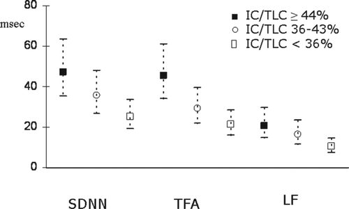
Table 3. Adjusted means of HRV parameters at baseline according to the categories of IC/TLC ratio and CRP.
HRV during 6mWT and recovery time
provides a description of the changes of HRV parameters during the 6mWT. RR interval showed a significant decrease in all 3 BODE groups. However, a different pattern was observed with regard to HRV parameters. Overall, a decrease in HRV indices during the 6mWT and a return to basal or higher values were observed in BODE 1 group. On the other hand, in BODE 3-4 group, an increase during the 6mWT was observed for the HRV parameters that assess global or long-term HRV (i.e., SDNN, TFA, VLF) and there was also a tendency to a lower reduction during the test for the short-term HRV parameters (i.e. r-MSSD, LF and HF). Mixed results were observed in the intermediate BODE 2 group.
Table 4. Changes of HRV parameters during 6mWT within each BODE group.
The relation of lung function indices with the changes of HRV parameters during the 6mWT is shown in . Indices of airflow obstruction were significantly inversely related to the change of SDNN, TFA and VLF (i.e., the greater was the reduction of the airways caliber the greater was the increase of HRV parameters). Moreover, indices of pulmonary hyperinflation (RV/TLC, FRC/TLC and IC/TLC ratios) were significantly related to LF and HF changes and IC/TLC showed an inverse association to the change of r-MSSD, TFA and VLF although the statistical significance was not reached (p values < 0.10).
Table 5: Univariate linear regression analysis between lung function parameters and CRP and change of HRV parameters.
Discussion
Our data demonstrate that COPD is associated with an abnormal cardiac autonomic modulation at rest. Indeed, HRV parameters were progressively reduced with the increase of COPD severity, as assessed both by BODE index and IC/TLC ratio. Of note, a significant correlation was found between the impaired cardiac autonomic function and the evidence of increased subclinical systemic inflammation, suggesting a possible pathophysiologic link.
Importantly, our data also provide evidence of a different response of cardiac autonomic function to submaximal exercise according to COPD severity. HRV parameters, indeed, further decreased during 6mWT in patients with mild COPD (BODE 1), whereas were found to increase or show a less consistent reduction in those with higher degrees of the disease (BODE 2 and 3-4). This behaviour seemed to mainly depend on airways caliber as the HRV increase was related to the degree of bronchial obstruction.
A lot of studies have been performed on HRV in COPD (Citation16–18, Citation23, 24), but most have included subjects with severe disease. In this study we enrolled patients belonging to various categories of COPD severity. In agreement with previous studies we show that COPD patients have reduced HRV (Citation15). However, our study also shows that HRV is already impaired in patients with milder forms of COPD (BODE 2) and that the entity of HRV impairment reflects the severity of the disease (Citation16).
It should be observed that the differences among BODE groups mainly concerned HRV indices that assess longer-term variability of RR interval, such as SDNN in the time-domain and VLF amplitude in the frequency-domain, whereas no differences have been noticed for HRV indices assessing shorter-term variability of RR interval (i.e., r-MSSD, and LF and HF amplitude) suggesting an effect by the disease on selective aspects of the variability of heart rate.
The progressive impairment of cardiac autonomic function related to COPD severity may have several explanations. First, a reduction in lung function, as suggested by the IC/TLC ratio, may lead to increased adrenergic activation, which notoriously results in depressed HRV (Citation13). Abnormal oxygen supply to cardiac sinus node in severe disease might also facilitate abnormal response of sinus node to autonomic stimuli. Finally, the increased inflammatory state shown in our study might also contribute to the reduction of HRV.
The relation between inflammation and autonomic nervous function is, in fact, complex. Experimental findings have shown that vagal stimulation inhibits inflammatory reactions (Citation25), whereas the effects of sympathetic function are more complex, but can in several case favour inflammation (Citation26). However, inflammatory cytokines have also been found to influence autonomic centers causing inhibition of vagal activity (Citation27). Thus, although the increased CRP levels in our patients might, at least in part, be determined by the depressed vagal function, it may also possible that they are a stimulus for the depressed HRV.
HRV during 6–minute walk test
In this study, for the first time we assessed the response of cardiac autonomic tone to exercise in COPD patients according to the severity of the disease. Interestingly, we found different behaviour of HRV variables depending on BODE category. Indeed, in mildly diseased patients (BODE 1) a significant reduction in HRV during moderate exercise (6mWT) was observed, in agreement with the exercise induced vagal inhibition/adrenergic activation. In contrast, severe COPD (BODE category 3-4) was paradoxically associated with an increase in global/long-term HRV variables, which suggests vagal activation during the exercise, with BODE 2 category showing intermediate behaviour.
An increase in HRV variables during exercise in patients with severe COPD was previously reported by Bartels et al. (Citation18), who performed a cardiopulmonary exercise test in patients with average FEV1% predicted of 35% showing an increase of LF and HF bands. In this study, during 6mWT, we found a significant increase in TFA and VLF, but not in LF and HF, that actually decreased. The reasons for these discrepancies are not clear but differences in COPD severity as well as in presence of co-morbidities and in the kind of exercise may have influenced the results.
A reason for the vagal activation associated with exercise in severe COPD might be, at least in part, related to the reduction of airways caliber shown in our patients. Mechanisms by which reduced airways caliber can alter the sympathetic–parasympathetic balance could be the stimulation of vagal stretch–sensitive mechanosensors of the lung (i.e., slowly adapting receptors [SAR], and rapidly adapting receptors [RAR]), which are dependent on the rate and depth of breathing (Citation28). During exercise in severe COPD patients a dynamic hyperinflation is likely to occur because of both a reduction of expiratory time and expiratory airflows, due to airway narrowing, and the increase of respiratory rate, thus resulting in an increased activity of SAR and RAR fibers.
This hypothesis could support our results as during exercise indices of airways caliber were inversely related to HRV, suggesting a vagal activation.Moreover, the different behaviour of HRV variables during 6mWT could reflect different exercise tolerance. Porszasz et al. (Citation35) showed that exercise training in patients with severe COPD decreased dynamic hyperinflation and, recently, Camillo et al. demonstrated an improvement of HRV after intensive exercise training due to improved biceps and triceps brachialis muscle force (Citation36). Thus, exercise training could have a further therapeutic role in pulmonary rehabilitation program.
Association of IC/TLC ratio with HRV
We found that the IC/TLC ratio was strongly associated to HRV at rest and was related to the change of HRV induced by submaximal exercise. FRC/TLC, RV/TLC and IC/TLC ratios are considered markers of static lung hyperinflation in COPD being the last two indices proposed as predictors of mortality (Citation8, Citation33). Recently, Watz et al. (Citation34) studied the relationship between lung function with heart size and heart dysfunction in a sample of COPD patients and they found that static hyperinflation indices showed stronger association with cardiac chamber sizes then airway obstruction indices being IC/TLC an independent predictor of cardiac chamber sizes. In our sample subjects with reduced respiratory reserve had a significant lower HRV than subjects with normal value. Our results and data of Watz et al. suggested that lung hyperinflation could play a key role in determining heart involvement in the natural history of COPD patients.
Association of CRP with HRV
We found that COPD patients with high level of CRP had a significantly reduced HRV at rest. CRP has been found inversely related to HRV in several clinical contexts, including patients with unstable angina pectoris (Citation29), heart failure (Citation30), or even apparently healthy subjects (Citation31, 32), suggesting a pathophysiologic link between autonomic dysfunction and inflammation.
In this study we show that this relation also exists in COPD patients, although we cannot establish whether there is any causal association between the two kinds of abnormalities or they merely depend on a common independent cause. However, the association seemed largely independent of lung function, oxygen saturation and cardiac overload.
Limitations of the study
Our study had some limitations which should be discussed. First of all, the sample size could be underpowered. We adopted very selective exclusion criteria and this heavily influenced the number of enrolled patients. This is a pilot study and further studies with much larger sample could be necessary to fully investigate this clinical aspect of the management of COPD patients with cardiovascular comorbidity. As the study was focused on 6–minutes Walk Test, HRV was evaluated at rest only for 6 minutes, which was a very short period of time and, thus, results could not reflect the HRV measured over the 24 hours period. We did not include a group of healthy controls as the aim was to evaluate the HRV during the 6mWT and the association to the degree of severity of COPD. We found that COPD seems to affect HRV at rest and during the test even in subjects with moderate disease (i.e. BODE 2 subjects), but the lack of control group did not allow to exclude that the autonomic control could be altered even in subjects with mild disease (i.e. BODE 1 subjects).
Conclusions
In conclusion, our results show that COPD patients have an abnormal cardiac autonomic modulation at rest and during submaximal exercise, and that the degree of the abnormality is related to the degree of the disease. Moreover, both systemic inflammation (i.e., CRP) and mechanical impairment of lung function have been found to be associated with a reduced HRV at rest. On the other hand, a vagal activation has been found to occur during mild exercise in COPD patients with a severe impairment of airways caliber. The exact pathophysiologic links among the abnormal findings detected in our study and their clinical implications in COPD patients deserve assessment in further studies.
Declaration of Interest
The authors declare that they have no conflict of interest. The authors are responsible for the content and the writing of this paper.
References
- Sin DD, Man SFP. Chronic obstructive pulmonary disease as a risk factor for cardiovascular morbidity and mortality. Proc Am Thorac Soc 2005; 2:8–11.
- Jousilahti P, Vartiainen E, Tuomilehto J, Puska P. Symptoms of chronic bronchitis and the risk of coronary disease. Lancet 1996; 348:567–72.
- Engström G, Wollmer P, Hedblad B, Juul-Möller S, Valind S, Janzon L. Occurrence and prognostic significance of ventricular arrhythmia is related to pulmonary function: a study from “men born in 1914”, Malmö, Sweden. Circulation 2001; 103: 3086–91.
- Anthonisen NR, Connett JE, Enright PL, Manfreda J. Lung Health Study Research Group. Hospitalizations and mortality in the Lung Health Study. Am J Respir Crit Care Med 2002; 166: 333–9.
- Ferguson GT, Calverley PMA, Anderson JA, Incidence of cardiac events in COPD patients in the TORCH study. Am J Respir Crit Care Med 2008; 177:A962.
- Vestbo J. Torch Study Group. The TORCH (towards a revolution in COPD health) survival study protocol. Eur Respir J 2004; 24:206e10.
- McGarvey LP, John M, Anderson JA, Zvarich M, Wise RA; TORCH Clinical Endpoint Committee. Ascertainment of cause-specific mortality in COPD: operations of the TORCH Clinical Endpoint Committee. Thorax 2007; 62(5):411–5.
- Casanova C, Cote C, de Torres JP Inspiratory-to-total lung capacity ratio predicts mortality in patients with chronic obstructive pulmonary disease. Am J Respir Crit Care Med 2005; 171(6):591–7.
- Zaman M, Mahmood S, Altayeh A. Low inspiratory capacity to total lung capacity ratio is a risk factor for chronic obstructive pulmonary disease exacerbation. Am J Med Sci 2010; 339(5):411–4.
- Dahl M, Vestbo J, Lange P, Bojesen SE, Tybjaerg-Hansen A, Nordestgaard BG. C-reactive protein as a predictor of prognosis in chronic obstructive pulmonary disease. Am J Respir Crit Care Med 2007; 175(3):250–5.
- Sin DD, Man SF. Systemic inflammation and mortality in chronic obstructive pulmonary disease. Can J Physiol Pharmacol 2007; 85(1):141–7.
- Casolo G, Balli E, Taddei T, Amuhasi J, Gori C. Decreased spontaneous heart rate variability in congestive heart failure. Am J Cardiol 1989; 64:1162–7.
- Kleiger RE, Stein PK, Bigger JT. Heart rate variability: measurement and clinical utility. Ann Noninvasive Electrocardiol 2005; 10(1):88–101.
- Task Force of the European Society of Cardiology and the North American Society of Pacing and Electrophysiology. Heart rate variability. Standards of measurement, physiological interpretation, and clinical use. Eur Heart J 1996; 17(3):354–81.
- Volterrani M, Scalvini S, Mazzuero G, Decreased heart rate variability in patients with chronic obstructive pulmonary disease. Chest 1994; 106(5):1432–7.
- Stein PK, Nelson P, Rottman JN, Heart rate variability reflects severity of COPD in PiZ alpha1-antitrypsin deficiency. Chest 1998; 113(2):327–33.
- Takabatake N, Nakamura H, Minamihaba O, A novel pathophysiologic phenomenon in cachexic patients with chronic obstructive pulmonary disease: the relationship between the circadian rhythm of circulating leptin and the very low-frequency component of heart rate variability. Am J Respir Crit Care Med 2001; 163(6):1314–9.
- Bartels MN, Jelic S, Ngai P, Basner RC, DeMeersman RE. High-frequency modulation of heart rate variability during exercise in patients with COPD. Chest 2003; 124(3):863–9.
- Miller MR, Crapo R, Hankinson J, ATS/ERS Task Force. General considerations for lung function testing. Eur Respir J 2005; 26(1):153–61.
- Miller MR, Hankinson J, Brusasco V, ATS/ERS Task Force. Standardisation of spirometry. Eur Respir J 2005; 526(2):319–38.
- Wanger J, Clausen JL, Coates A, Standardisation of the measurement of lung volumes. Eur Respir J 2005; 26(3):511–22.
- Macintyre N, Crapo RO, Viegi G, Standardisation of the single-breath determination of carbon monoxide uptake in the lung. Eur Respir J 2005; 26(4):720–35.
- Camillo CA, Pitta F, Possani HV, Heart rate variability and disease characteristics in patients with COPD. Lung 2008; 186(6):393–401.
- Antonelli Incalzi R, Corsonello A, Trojano L, Heart rate variability and drawing impairment in hypoxemic COPD. Brain Cogn 2009; 70(1):163–70.
- Borovikova LV, Ivanova S, Zhang M, Vagus nerve stimulation attenuates the systemic inflammatory response to endotoxin. Nature 2000; 405:458–62.
- März P, Cheng JG, Gadient RA, Sympathetic neurons can produce and respond to interleukin 6. Proc Natl Acad Sci USA 1998; 95:3251–6.
- Banks WA, Kastin AJ. Blood to brain transport of interleukin links the immune and central nervous systems. Life Sci 1991; 48:PL117–21.
- Undem BJ, Kollarik M. The role of vagal afferent nerves in chronic obstructive pulmonary disease. Proc Am Thorac Soc 2005; 2(4):355–360; discussion371–2.
- Lanza GA, Sgueglia GA, Cianflone D, , SPAI (Stratificazione Prognostica dell'Angina Instabile) Investigators. Relation of heart rate variability to serum levels of C-reactive protein in patients with unstable angina pectoris. Am J Cardiol Jun 2006; 15;97(12):1702–6.
- Aronson D, Mittleman MA, Burger AJ. Interleukin-6 levels are inversely correlated with heart rate variability in patients with decompensated heart failure. J Cardiovasc Electrophysiol 2001; 12:294–300.
- Lampert R, Bremner JD, Su S, Decreased heart rate variability is associated with higher levels of inflammation in middle-aged men. Am Heart J 2008; 156(4):759. e1–759.e7.
- Sajadieh A, Nielsen OW, Rasmussen V, Hein HO, Hansen JF. C-reactive protein, heart rate variability and prognosis in community subjects with no apparent heart disease. J Intern Med 2006; 260(4):377–87.
- Martinez FJ, Foster G, Curtis JL, , NETT Research Group. Predictors of mortality in patients with emphysema and severe airflow obstruction. Am J Respir Crit Care Med 2006; 173(12):1326–34.
- Watz H, Waschki B, Meyer T, Decreasing cardiac chamber sizes and associated heart dysfunction in COPD: role of hyperinflation. Chest 2010; 138(1):32–8.
- Porszasz J, Emtner M, Goto S, Exercise training decreases ventilatory requirements and exercise-induced hyperinflation at submaximal intensities in patients with COPD. Chest 2005; 128(4):2025–34.
- Camillo CA, Laburu Vde M, Gonçalves NS, Improvement of heart rate variability after exercise training and its predictors in COPD. Respir Med 2011; 105(7):1054–62.