
Abstract
A new single-beat three-dimensional (3D) real time echocardiographic semi-automatic images processing (4D Auto LVQ) allows accurate assessment of left ventricular function, but whether it is suitable for the evaluation of right ventricular function remains unknown. To evaluate the feasibility of this procedure for assessing right ventricular volumes and function, right ventricular end-diastolic volumes (RVEDV), end-systolic volumes (RVESV) and ejection fraction (RVEF), stroke volumes (SV) and cardiac output (CO) were computed in 49 patients with chronic obstructive pulmonary disease (COPD) using 4D Auto LVQ. The myocardial performance index (MPI) was obtained by Doppler tissue imaging. The RV function parameters were compared with MPI by linear correlation analysis. A comparison of the performance of these RV function parameters in discrimination between MPI at a value of >0.45 or not was done. Compared with normal subjects, patients with COPD had significantly greater RVEDV, RVESV, MPI and significantly lower RVEF. Significant correlations were found between RVEF and MPI (r = –0.67, p < 0.001). The areas under the receiver operating characteristic curve for RVEF in discrimination between MPI at a value of >0.45 or not were 0.72, while they were 0.55 for SV and 0.57 for CO, respectively. The overall sensitivity, specificity and accuracy for RVEF analysis in predicting a >0.45 MPI in patients with COPD was 78.57%, 66.67% and 73.46%, respectively. These data suggest that 4D Auto LVQ is a feasible method for right ventricular volumes and function quantification in patients with COPD. Further studies are needed to improve the accuracy of the measurements.
| Abbreviations | ||
| CO Cardiac output test | = | |
| COPD Chronic obstructive pulmonary disease | = | |
| ET Ejection time | = | |
| HR Heart rate | = | |
| IVCT Isovolumic contraction time | = | |
| IVRT Isovolumic relaxation time | = | |
| MPI Myocardial performance index | = | |
| RAD Right atrium end-systolic diameter | = | |
| RVD Right ventricular end-diastolic diameter | = | |
| RVEDV Right ventricular end-diastolic volume | = | |
| RVEF Right ventricular ejection fraction | = | |
| RVESV Right ventricular end-systolic volume | = | |
| SV Stroke volume | = | |
| 3DE Three dimensional echocardiography | = | |
| WT The middle myocardial thickness of right ventricular free wall | = | |
Introduction
Chronic obstructive pulmonary disease (COPD) is an important cause of morbidity and mortality worldwide. The reduction of right ventricular (RV) function characterizes patients with COPD. Up to date, recognizing this situation, particularly in acute settings, remains a clinical challenge (Citation1,2). Echocardiography is the first available imaging modality for assessing the RV function (Citation3), and three-dimensional echocardiography (3DE) has become a feasible and reproducible method for RV dimension and function quantification in recent years (Citation4–7).
The most commonly used 3DE method for volumes and function measurement is to use real-time ECG-gated volume stitching from four consecutive 4B with the purpose to maintain an acceptable spatial and temporal resolution (Citation8). However, the multibeat modality in comparison to single-beat requires breath-hold technique and regular heart rhythm that could limit the use of this technique in patients with COPD due to the low cooperation rate of the patients and the inherent stitching artifact of multibeat modality, especially in those with arrhythmia, such as atrial fibrillation. Single-beat real-time 3DE allows accurate assessment of heart volumes and function from the same cardiac cycle, which will further advance this assessment by improving the speed of acquisition and reducing stitching artifacts (Citation9).
Myocardial performance index (MPI) is a parameter unaffected by RV geometry, which can be measured non-invasively by both pulsed-wave Doppler and tissue Doppler imaging (TDI). It has been proved to correlate well with invasive procedures such as catheterization (Citation10,11), and it has high sensitivity and negative predictive value for detecting abnormal RV systolic function (Citation12). MPI measured by TDI has the advantage of simultaneously recording the time intervals from the same cardiac cycle (Citation13), which has been more frequently used for the assessment of RV function. In patients with COPD, it is not affected by the abnormal location of the heart.
The study aimed to evaluate the feasibility of a new single-beat real time 3D echocardiographic semi automatic images processing (4D Auto LVQ) for assessing RV volumes and function, and to compare it to MPI obtained by TDI in patients with COPD.
Patients and methods
Study population
Forty-nine inpatients with clinically diagnosed stable COPD of different severity (24 men, 25 women; 61.7 ± 16.43 years, range: 46–78 years), and 49 age and sex-matched healthy volunteers (23 men, 26 women; 64.67 ± 17.24 years, range: 47–82 years) were investigated between February 2011 and December 2012 at Department of Respiratory Medicine in our institution. The COPD criteria is based on spirometry test (forced expiratory volumes in 1 second [FEV1]/forced vital capacity [FVC] < 70%) according to the Global Initiative for Chronic Obstructive Lung Disease (Citation14,15). Patients with coronary heart disease, a valvular dysfunction of the left heart exceeding a mild degree, atrial fibrillation, complete right or left bundle branch block or pacemaker, and a left ventricular systolic ejection fraction of <50% were excluded. The study was approved by the local ethics committee of Yancheng, and free informed consent was obtained from all the subjects.
Echocardiographic measurements
A complete 2DE, 3DE and Doppler study was performed in all subjects, using a commercially available Vivid E9 ultrasound machine (GE Healthcare, Horten, Norway) equipped with M5S single-crystal matrix-array transducer and 3V matrix-array transducer. All acquisitions were performed by two experienced operator who were blinded to all clinical data and previous reading. Data sets were stored digitally for off-line analysis using commercially available software (EchoPAC PC version 108.1.4, GE Healthcare). All values for each parameter were obtained by averaging measurements from three successive cardiac cycles.
The subjects were lying in the left lateral position with the electrocardiography recorded simultaneously. First, right ventricular (RV) end-diastolic diameters (RVD), right atrium (RA) end-systolic diameters (RVD) and the middle myocardial thickness of right ventricular free wall (WT) were measured by 2 DE.
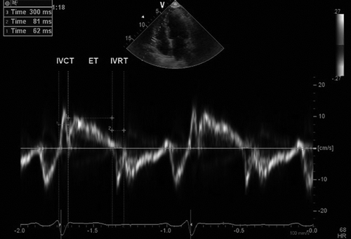
Second, the TDI function was activated, gains were adjusted at the minimal optimal level to minimize noise, and the filter settings were kept low (50 Hz). In the apical four-chamber view, a 3.5-mm sample volume was placed at the lateral tricuspid annulus. A Doppler velocity range of −20 cm/s to 20 cm/s was used. The systolic velocity duration was measured as ejection time (ET), whereas the time between the end of the late diastolic velocity and the beginning of systolic velocity was measured as isovolumic contraction time (IVCT), and the time between the end of the systolic velocity and the beginning of early diastolic velocity was measured as isovolumic relaxation time (IVRT). MPI was calculated by the following formula: (IVCT + IVRT)/ET ().
Figure 1. Intervals measured from pulsed tissue Doppler obtained from the lateral tricuspid annulus. ET, ejection time; IVCT, isovolumic contraction time; IVRT, isovolumic relaxation time; MPI, myocardial performance index = (IVCT+IVRT)/ET.
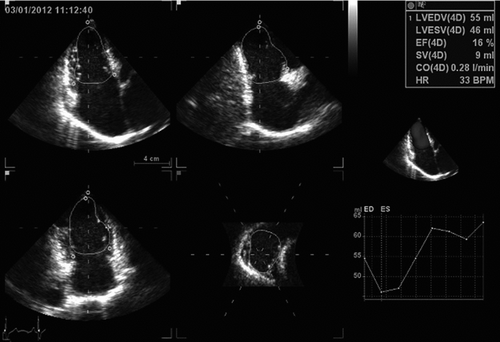
Finally, a full volume scan was acquired from one cardiac cycle, single-beat () during end expiration breath-hold as soon as gain, time gain compensation and compression control settings were optimized to obtain a satisfactory 3D image. Volume analysis was made using a commercially available semi-automated analysis tool, 4D auto LV volume quantification (4DLVQ, EchoPAC PC version 108.1.4, GE Healthcare). The ED frames for contour detection were automatically displayed in quad-view: apical four-, two-chamber, three-chamber and LV short axis plane (). Manual positioning by translating the four-chamber plane was first performed in order that the corresponding intersection line of all planes was placed in the middle of RV cavity, crossing the RV apex and the center of tricuspid valve opening in each view. The software required manual input of three points for each of the three apical planes (two points at tricuspid annulus borders, and one at the apex) first in ED frames, and then continuing for ES frames. The software automatically delineates the RV endocardial border in a 3D-model from ED and ES phases. In cases where the automatic delineation of the endocardial border was considered suboptimal the borders could be adjusted manually. RVEDV, RVESV, RVEF, SV and CO was finally displayed.
Figure 2. The measurement of right ventricular volumes and function using single-beat real time three-dimensional echocardiographic semi automatic images processing software (4D Auto LVQ). Volume time-plot and quantitative analysis and three-dimensional model are presented in the right panel.
Reproducibility
Intraobserver variability was assessed in 30 selected randomly participants by repeating the measurements on two occasions (3 days apart) under the same basal conditions. To test the interobserver variability, the measurements were performed on the same subject by a second blinded observer. Variability was calculated as the mean percentage error, derived as the difference between the two sets of measurements, divided by the mean observations.
Statistical analysis
Data were expressed as the mean ± SD. The differences between the two groups were tested using an unpaired two tailed t test. A receiver operating characteristic curve (ROC) analysis was used to evaluate and compare the performance of the right ventricular function parameters obtained by 4D Auto LVQ in discrimination between MPI of RV at a value of >0.45 or not in patients with COPD. The sensitivity, specificity, positive predictive value, negative predictive value and accuracy of the RV function parameters as a predictor of MPI of RV at a value of >0.45 were measured in the traditional manner. A value of p < 0.05 was considered statistically significant. All statistical analysis was performed with SPSS version 13 software for Windows (SPSS Inc., Chicago, IL).
Results
Echocardiographic parameters
MPI analyses were successfully completed in all subjects, and 3DE analyses were successfully completed in 48 normal subjects and 45 patients with COPD. One normal subjects and four patients with COPD were excluded from the study due to poor acoustic window or invisible endocardial border (two segments or more).
As shown in , RAD, RVD, WT, RVEDV, RVESV, ET and MPI in patients with COPD were significantly greater than those in normal subjects (p < 0.01), whereas IVCT, IVRT and RVEF in this cohort were significantly lower than those in normal subjects (p < 0.05). In addition, SV and CO did not differ between the normal subjects and patients with COPD (p > 0.05).
Table 1. Echocardiographic parameters of normal subjects and patients with chronic obstructive pulmonary disease (COPD)
The Pearson correlation analysis in patients with COPD
As shown in , RVEF correlated significantly with MPI (r = −0.67, p < 0.001), IVCT (r = −0.408, p = 0.012) and ET(r = 0.411, p = 0.01). Significant correlations were also found between CO and ET (r = −0.372, p = 0.023) and MPI (r = 0.336, p = 0.042). However, SV was lacking in significant correlation with IVCT, IVRT, ET and MPI.
Table 2. The Pearson correlation analysis for right ventricular function based on single-beat, three-dimensional echocardiography and isovolumic contraction time, isovolumic relaxation time, ejection time and myocardial performance index of right ventricle obtained by tissue Doppler imaging in patients with chronic obstructive pulmonary disease
Receiver operating characteristic curve (ROC) analysis
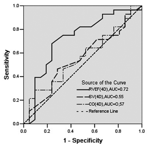
With the use of MPI > 0.45 as the criteria to distinguish right ventricular dysfunction or not, the areas under the ROC (AUC) for RVEF in discrimination between MPI of RV at a value of >0.45 or not were 0.72, while they were 0.55 for SV and 0.57 for CO, respectively. The AUC for RVEF was maximal, which was far higher than that of SV and CO (p < 0.05, ).
Figure 3. Receiver operating characteristic curve showing the performance of right ventricular ejection fraction (RVEF), stroke volumes (SV) and cardiac output (CO) obtained by 4D auto LVQ software in discrimination between myocardial performance index of right ventricle at a value of >0.45 or not in patients with chronic obstructive pulmonary disease. AUC, the area under receiver operating characteristic curve.
Discriminant analysis of RVEF obtained by 4D Auto LVQ for predicting MPI of RV at a value of >0.45
Discriminant analysis of RVEF obtained by 4D Auto LVQ for predicting MPI of RV at a value of >0.45 in patients with COPD was conducted. The best cutoff value for predicting MPI of RV at a value of >0.45 was RVEF of less than 39% (sensitivity, 78.57%; specificity, 66.67%; positive predictive value, 75.86%; negative predictive value, 70% and accuracy, 73.46%; p < 0.001).
Reproducibility
Intraobserver and interobserver variability for RVEDV and RVESV ranged from 6.7% to 8.3%. Intraobserver and interobserver variability for RVEF were 6.5% ± 3.2%, 7.4% ± 2.9%, respectively. Intraobserver and interobserver variability for MPI were 3.2% ± 1.9% and 3.5% ± 2.3%, respectively.
Discussion
The results presented here indicate that 4D Auto LVQ is a feasible method for RV volumes and function quantification in patients with COPD.
Magnetic resonance imaging (MRI) is a gold standard for the evaluation of right ventricular volumes and function (Citation6,Citation16). But it is not always readily available. For patients with COPD, particularly in acute settings, they often can not be moved or lie in the supine position, and the oxygen equipment also limit MRI examination. The echocardiography examination is convenient and flexible, which can be carried out at the bedside.
Our results showed that MPI analyses were successfully completed in all subjects, but 3DE analyses not. This result can be explained by a fact that the heart locations of patients with COPD often move down and the acoustic windows are poor or endocardial border visualization are not clear due to emphysema. Volumetric methods of image acquisition by 3DE is often difficult to perform in this condition, yet MPI obtained by TDI is less subject to the factors mentioned here, because TDI is used to measure the motion (velocity) of the myocardial tissue and is not affected by RV geometry, which is less subject to background noise. Even so, 4D Auto LVQ analyses showed that patients with COPD had significantly greater RVEDV, RVESV and significantly lower RVEF than normal subjects, which objectively reflected the volumes and function changes of right ventricular in patients with COPD.
The Pearson correlation analysis showed that RVEF correlated significantly with MPI, IVCT and ET (especially MPI), and CO correlated significantly with MPI and ET. These results further indicate that 4D Auto LVQ can effectively quantify the RV function in patients with COPD. To rank RVEF, CO and SV obtained by 4D Auto LVQ against the reference of MPI of RV as obtained in a broad clinical population by TDI, a ROC analysis for their performance to distinguish between MPI at a value of > 0.45 or not was done. The AUC for RVEF was maximal, which was far higher than that of SV and CO. Therefore, REVF (4D) is a reliable index when using 4D Auto LVQ software for the evaluation of right ventricular function.
In our study, we did not find a better correlation(r ≥ −0.7, p < 0.001) between RVEF and MPI as the previous report (Citation2,Citation11,Citation13). This results from the inherent defects of 4D Auto LVQ, such as dependence on semi-automated delineation of the endocardial border, and the poor acoustic windows in patients with COPD. Optimization of the endocardial borders in right ventricular is maybe inadequate, whether it is done automatically or manually. Further studies are needed to improve the accuracy of the measurements.
Conclusion
In this study we evaluated the feasibility of a new 3D real time echocardiographic semi automatic images processing (4D Auto LVQ) for assessing right ventricular volumes and function in patients with COPD. We have shown that 4D Auto LVQ can objectively reflect the RV volumes and function changes of patients with COPD, and REVF(4D) is a reliable index when using this software for the evaluation of RV function. Although some limitations were mentioned here, this method still holds considerable clinical promise for the assessment of RV volumes and function in patients with COPD.
Declaration of Interest Statement
The authors have no conflict of interest to declare. The authors alone are responsible for the content and writing of the paper.
Acknowledgments
The authors gratefully acknowledge the technical assistance and helpful discussion of J. Li, X.Q. Huang, and G.X. Fan at the Department of Ultrasound, The First People's Hospital of Yancheng, Jiangsu Province, P.R. China.
References
- Gariani K, Delabays A, Perneger TV, Agoritsas T. Use of brain natriuretic peptide to detect previously unknown left ventricular dysfunction inpatients with acute exacerbation of chronic obstructive pulmonary disease. Swiss Med Wkly 2011; 141: w 13298. doi: 10.4414/ smw.2011.13298.
- Seyfarth HJ, Pankau H, Winkler J, Wirtz H.Correlation of TEI Index and invasive parameters of right heart function in PAH. Pneumologie 2004; 58:217–221.
- Bleeker GB, Steendijk P, Holman ER, Assessing right ventricular function: the role of echocardiography and complementary technologies. Heart 2006; 92:Suppl 1:i19–26.
- Tamborini G, Brusoni D, Torres Molina JE, Feasibility of a new generation three-dimensional echocardiography for right ventricular volumestric and functional measurements. Am J Cardiol 2008; 102:499–505.
- Dragulescu A, Mertens LL. Developments in echocardiographic techniques for the evaluation of ventricular function in children. Arch Cardiovasc Dis 2010; 103:603–614.
- Grewal J, Majdalany D, Syed I, Pellikka P, Warnes CA. Three-dimensional echocardiographic assessment of right ventricular volume and function in adult patients with congenital heart disease: comparison with magnetic resonance imaging. J Am Soc Echocardiogr 2010; 23:127–133.
- van der Hulst AE, Roest AA, Holman ER, Real-time three-dimensional echocardiography: segmental analysis of the right ventricle in patients with repaired tetralogy of fallot. J Am Soc Echocardiogr 2011; 24:1183–1190.
- Kühl HP, Schreckenberg M, Rulands D, High-resolution transthoracic real-time three-dimensional echocardiography: quantitation of cardiac volumes and function using semi-automatic border detection and comparison with cardiac manetic resonance imaging. J Am Coll Cardiol 2004; 43:2083–2090.
- Shahgaldi K, Manouras A, Abrahamsson A, Gudmundsson P, Brodin LA, Winter R. Three-dimensional echocardiography using single-heartbeat modality decreases variability inmeasuring left ventricular volumes and function in comparison to four-beat technique in atrialfibrillation. Cardiovasc Ultrasound 2010; 8:45.
- Seyfarth HJ, Pankau H, Winkler J, Wirtz H. Correlation of TEI Index and invasive parameters of right heart function in PAH. Pneumologie 2004; 58:217–221.
- Su HM, Lin TH, Voon WC, Correlation of Tei index obtained from tissue Doppler echocardiography with invasive measurements of left ventricular performance. Echocardiography 2007; 24:252–257.
- Miller D, Farah MG, Liner A, Fox K, Schluchter M, Hoit BD. The relation between quantitative right ventricular ejection fraction and indices of tricuspid annular motion and myocardial performance. J Am Soc Echocardiogr 2004; 17:443–447.
- Zimbarra CI, Ruisanchez C, Dawson D, Right ventricular function in patients with pulmonary hypertension; the value of myocardial performance index measured by tissue Doppler imaging. Eur J Echocardiogr 2010; 11:719–724.
- Pistelli R, Ferrara L, Misuraca C, Bustacchini S. Practical management problems of stable chronic obstructive pulmonary disease in the elderly. Curr Opin Pulm Med 2011; 17 Suppl 1:S43–48.
- Obase Y, Mouri K, Shimizu H, Nutritional deficits in elderly smokers with respiratory symptoms that do not fulfill the criteria for COPD. Int J Chron Obstruct Pulmon Dis 2011; 6:679–683.
- Pavlicek M, Wahl A, Rutz T, Right ventricular systolic function assessment: rank of echocardiographic methods vs. cardiac magnetic resonance imaging. Eur J Echocardiogr 2011; 12:871–880.