
Abstract
Right ventricular (RV) systolic failure is rare in patients with COPD, but they often develop RV diastolic dysfunction. Left ventricular (LV) diastolic dysfunction is also common in this population. Nevertheless, data are scarce regarding the effect of diastolic dysfunction on the functional capacity in patients with COPD. We investigated the correlation between echocardiographic parameters of RV and LV diastolic function and the exercise capacity in COPD, by using conventional echocardiographic methods and tissue Doppler imaging. 65 patients with COPD (61 ± 9 years) in stages GOLD II-IV were investigated. Functional capacity was measured with 6-minute walk test (6MWT). Right (RA) and left atrial (LA) area index were measured; collapsibility index inferior vena cava was calculated. Parameters of the mitral and tricuspid inflow (E, A) as well as annular systolic (S), early- (e’) and late- (a’) diastolic myocardial longitudinal velocities were measured. E/A, E/e’ and e’/a’ ratios were calculated. 6MWT distance was 330 ± 76 m. LV diastolic dysfunction was found in 48 (74%) patients. LV and RV filling pressures were elevated in 28 (43%) and in 29 (45%) patients, respectively. In the left heart, LA area index showed significant correlation with the functional capacity (r = -0.319; p = 0.011). In stepwise multiple linear regression analysis tricuspid e’/a’ (r = 0.611; p = 0.000), collapsibility index (r = 0.505; p = 0.000), RA area index (r = -0.445; p = 0.000) and body surface area (r = 0.314; p = 0.011) were independent predictors of 6MWT distance. Right ventricular diastolic function and filling pressure have strong influence on the functional capacity in patients with COPD.
Introduction
Chronic obstructive pulmonary disease (COPD) is a growing global epidemic, which is forecasted to be the third leading cause of death by 2020 (Citation1,Citation2). Beside its impact on mortality, COPD represents large health care burden and it is important cause of disability Citation(3). Dyspnoea and reduced functional capacity are common consequences of COPD. The aetiology of reduced exercise tolerance is multifactorial thus parameters of the resting pulmonary function such as forced expiratory volume (FEV1) are poor predictors of the functional capacity (Citation4,Citation5). Dynamic lung hyperinflation is an important contributory factor that can be targeted for treatment (Citation5,Citation6). Depression, skeletal muscle weakness or pulmonary hypertension as well as alterations of the cardiac function may also serve as limiting factor to reduced functional capacity (Citation7,Citation8).
Right ventricular systolic failure with low cardiac output is rare in patients with COPD, but they often develop right ventricular diastolic dysfunction with stress induced or resting elevation of filling pressures (Citation9,Citation10). Left ventricular diastolic dysfunction is also reported to be common in the COPD population (Citation11–14). Nevertheless, data are scarce regarding the effect of the right and left ventricular diastolic dysfunction on the functional capacity in COPD.
Thus we aimed to investigate the relation between the echocardiographic parameters of the right and left ventricular diastolic function and the exercise capacity in patients with COPD. In addition to the conventional echocardiographic methods tissue Doppler imaging technique (TDI) was used offering a more sensitive assessment both of systolic and diastolic function.
Methods
Study population
Eighty outpatients with stable COPD of varying severity were consecutively screened for this study. Diagnosis and pulmonological management of the COPD were based on GOLD strategy document Citation(15). In patients with borderline FEV1/FVC (below 0.70 but above the lower limit of normal) the diagnosis of COPD was based on the clinical symptoms (dyspnea, chronic cough and/or sputum production) and a history of exposure to risk factors for the disease. All patients underwent spirometry within one month before the inclusion. They had to be free of exacerbations for the two months before inclusion. Patients with moderate-to-severe left ventricular (LV) systolic dysfunction (ejection fraction <45%), atrial fibrillation, neuromuscular disorders affecting exercise capacity, significant left sided valvular abnormalities or prosthetic valves were excluded. Detailed medical history was obtained. Significant ischemic heart disease was defined as coronary artery stenosis >50% proved by invasive measurements or as history of previous myocardial infarction. Heart failure was diagnosed when the patient was regularly treated with loop diuretics and/or at any symptoms of heart failure.
Data of 34 healthy volunteers without any cardiac disease were used as control. The study complied with the Declaration of Helsinki. The institutional ethics committee approved the study. All subjects had given written informed consent prior to inclusion.
Echocardiography
Echocardiography was performed using Philips CX50 ultrasound system (Philips Healthcare, Best, The Netherlands). Studies were performed by a single cardiologist blinded to all other data. 2D and M-mode echocardiographic data collected for analysis included: LV ejection fraction; LV mass corrected for body surface area (LVM index); maximal left and right atrial areas, corrected for body surface area (LA and RA area index); basal, mid-cavity, and longitudinal dimensions of the right ventricle (RV); tricuspid annular plane systolic excursion (TAPSE); RV fractional area change (RVFAC); maximal and minimal diameters of the inferior vena cava (IVC); collapsibility index (the percent decrease in the diameter during inspiration); RV wall thickness (Citation16, Citation17).
The following Doppler data were collected: spectral Doppler based myocardial performance (Tei) index, mitral and tricuspid E/A, calculated RV systolic pressure, myocardial systolic (S), early- (e′) and late- (a′) diastolic velocities at the lateral and septal mitral annulus and at the lateral tricuspid annulus, mitral and tricuspid E/e′ and e′/a′ ratios. Doppler measurements were obtained during end-expiratory apnoea Citation(16). LV diastolic dysfunction was identified if mitral lateral e′< 10 cm/s and septal e′< 8 cm/s. Elevated LV filling pressure was defined as mean E/e′≥ 9 Citation(18). Elevated RV filling pressure was diagnosed if tricuspid E/e′> 6 Citation(16).
Six-minute walk test
Functional capacity of the patients was measured with 6 minute walk test (6MWT), at the day of the echocardiography. Borg dyspnoea index (0–10) was used for subjective assessment of shortness of breath during the exercise Citation(19).
Statistical analysis
Categorical data were expressed as frequencies and percentages; continuous data were expressed as the mean ± SD. Intraclass correlation coefficient was calculated to assess intraobserver reliability. Comparisons of data between two groups were performed using independent-sample t-tests for continuous variables and chi square tests for categorical variables. Comparisons of data between more groups were performed using ANOVA with LSD post hoc test. Univariate predictors of 6MWT distance were assessed using linear regression analysis. A p-value of < 0.05 was considered significant.
Multiple stepwise linear regression analysis was performed by entering those variables that were considered significant (p < 0.05) on univariate analysis. In this method the variable with the smallest probability of its F-statistic (p ≤ 0.05) is entered into the model first. This process continues to add variables to the model until there are no variables left that have F statistics that meet the criteria. As this process progresses, the F statistics for variables already in the model may change. If the significance level of these F statistics exceeds the criterion (if p ≥ 0.1), then these variables are removed from the model. Variance Inflation Factor (VIF) values above 2.5 were considered to have potential multicollinearity. IBM SPSS 22 statistical software was used.
Results
Of a total of 80 patients with COPD, 65 were eligible for the study. Fifteen patients were excluded due to atrial fibrillation (4 patients), poor acoustic window Citation(2), severe sinus tachycardia Citation(1), moderate-to-severe left-sided valve disease (1 aortic stenosis, 1 mitral regurgitation), severely reduced LV systolic function (1 patient with dilated cardiomyopathy, 1 patient with previous myocardial infarction), and severe pericardial constriction Citation(1). Three patients (all in GOLD stage IV) did not attempt to perform 6MWT because of their weak physical condition.
Systemic hypertension, heart failure and diabetes were common in our COPD population. In 1 case percutaneous coronary intervention was performed while 4 patients underwent coronary artery bypass surgery. In 3 cases prior myocardial infarction was reported, but coronary intervention was not feasible. Clinical and echocardiographic data of the 65 patients are reported in
Table 1. Baseline characteristics of the COPD population and comparison with healthy subjects.
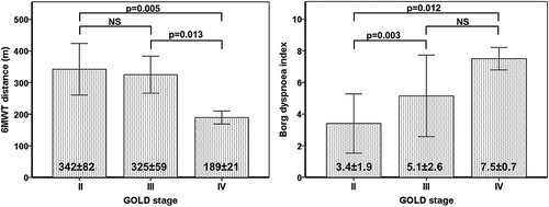
Thirty-five patients were in GOLD stage II, 27 patients in GOLD stage III, and 3 patients in GOLD stage IV. Borg dyspnoea index was significantly higher in GOLD III patients compared with GOLD II. On the other hand, no significant difference was found between the 6MWT distances covered by the patients in GOLD stage II and III ().
Figure 1. 6MWT and Borg dyspnoea index results in different GOLD stages (Mean ± SD; ANOVA with LSD post hoc test).
Spectral Doppler and TDI measurements were feasible in all patients. Intraclass correlation coefficients were 0.988 and 0.981 for mitral and tricuspid E, respectively. Intraclass correlation coefficients for e′, a′ and S were 0.984, 0.984 and 0.917 on mitral lateral annulus; 0.983, 0.978 and 0.977 on mitral septal annulus as well as 0.970, 0.982 and 0.986 on tricuspid annulus.
Comparison of COPD population with healthy controls
Our patients and healthy controls were matched in age and gender distribution (). Body surface area was significantly larger in patients with COPD, but the difference was clinically not remarkable. LV ejection fraction was preserved (≥ 55%) in 59 (91%), while mildly reduced (45–54%) in 6 (9%) patients. LVM index was significantly higher in patients with COPD. Both lateral and septal myocardial early diastolic velocities (e′) were significantly lower, while mean E/e′ was significantly higher in the COPD population. LV diastolic dysfunction was found in 48 (74%), while LV filling pressure was elevated in 28 (43%) patients. (Grade I and grade II diastolic dysfunction in 20 (31%) and in 28 (43%) patients, respectively.)
Assessment of the tricuspid regurgitation's velocity was feasible in 41 (63%) patients. RV systolic pressure was significantly higher in patients with COPD. Pulmonary hypertension (RV systolic pressure ≥ 35 mmHg) was found in 9 (14%) patients. RV wall thickness, RA area and RV basal diameter were significantly increased in patients with COPD. IVC was dilated, while collapsibility index was significantly lower in the COPD group. TAPSE and myocardial longitudinal systolic velocity (tricuspid S) were significantly lower in our patients as compared with controls. RV systolic dysfunction was rare in the COPD population: RVFAC < 35%, TAPSE < 16 mm and tricuspid S < 10 cm/s were found in 1 (1.5%), 3 (5%) and 7 (11%) patients, respectively. Myocardial performance index was prolonged (> 0.4) in 31 (47%) patients. Tricuspid e′ and tricuspid e′/a′ were significantly decreased in the COPD population. RV filling pressure was elevated in 29 (45%) patients.
Determinants of functional capacity in patients with COPD
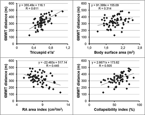
Among all clinical parameters, 6MWT distance showed significant negative correlation with age and significant positive correlation with body surface area. FEV1% and FEV1/FVC did not show significant correlation with 6MWT distance, but were significant predictors of Borg dyspnoea index (FEV1%: r = -0.474; p = 0.000 and FEV1/FVC: r = -0.374; p = 0.002). Among all echocardiographic parameters, significant negative correlation was found between LA area index and 6MWT distance. Other parameters of LV systolic and diastolic function did not show correlation with the functional capacity. A number of parameters representing RV size, RV diastolic function and filling pressure were proven to be significant predictors of 6MWT. No correlation was found between 6MWT results and the echocardiographic parameters of RV systolic function.
Univariate and multivariate predictors of the 6MWT distance are reported in . In stepwise multiple linear regression analysis tricuspid e′/a′, body surface area, RA area index and collapsibility index were independent predictors of 6MWT distance (multiple r = 0.764; p = 0.000; F = 19.269) (). VIF values for all variables were below 2.5.
Table 2. Predictors of the 6MWT distance (m) in patients with COPD: univariate and multivariate regression analyses (Unstandardized (B) and standardized (β) regression coefficients).
Figure 2. Main predictors of the 6MWT distance in patients with COPD.
Discussion
Multiple mechanisms lead to the restriction of functional capacity in COPD. Our hypothesis was that left and right ventricular diastolic dysfunction is a contributing factor to exercise intolerance in this disease.
LV systolic and diastolic function: correlation with 6MWT distance
LV ejection fraction was preserved or mildly reduced in our COPD population. Mitral annular S values, however, were significantly decreased as compared to healthy subjects. This subclinical impairment of the LV systolic function commonly occurs in patients with impaired LV diastolic function as the evidence of the interdependence between contraction and relaxation Citation(20).
More mechanisms may explain the presence of LV diastolic dysfunction in patients with COPD. Significant reduction of the LV diastolic diameter and consequential impairment of LV filling was reported in pulmonary hyperinflation Citation(21). Abnormal patterns of LV diastolic filling have been also described in patients with COPD and elevated pulmonary pressure, due to ventricular interdependence Citation(22). In addition, hypoxemia and systemic inflammation may directly impair LV myocardial function Citation(23). Cardiovascular comorbidities, such as systemic hypertension or ischaemic heart disease, are common in COPD (Citation24, Citation25), which may be also responsible for the LV diastolic dysfunction.
COPD on CT scan was associated with reduced pulmonary vein cross-sectional area. These findings suggest that impaired LV filling in COPD may be predominantly due to reduced LV preload from upstream pulmonary causes rather than intrinsic diastolic dysfunction Citation(26). The mitral inflow pattern (E/A) is in fact dependent on loading conditions. Mitral annular TDI parameters, however, are able to verify the presence of relaxation abnormalities by reflecting the intrinsic features of the LV myocardium Citation(27). In patients with COPD this new technique was used for the assessment of the diastolic function only in few recent studies (Citation12–14).
These results are not completely comparable, since different approaches were used to estimate the frequency of the diastolic dysfunction. Nevertheless, these works have concordantly proved that LV diastolic dysfunction is common in COPD. We applied the recent ASE/EACVI recommendation for the evaluation of the left ventricular diastolic function Citation(18). Mild or moderate form of left ventricular diastolic dysfunction was found in 74% of our COPD patients, what is in accordance with the previous findings (Citation12–14). In addition to the manifest diastolic and subclinical systolic RV dysfunction, LV diastolic dysfunction may be also responsible for the large number of heart failure cases in our population.
Lopez-Sanchez et al. Citation(13) reported significant correlation between E/A ratio and the functional capacity of the patients while in the work of Cuttica et al. Citation(14) E/e′ and the degree of diastolic dysfunction were predictors of the 6MWT distance in univariate analysis. In our population, LA area index was the only parameter in the left heart, which showed significant correlation with 6MWT distance. Although E/e′ reflects the momentary value of the LV filling pressure, LA size is considered as a reliable indicator of the cumulative effects of the elevated LV filling pressure over time Citation(28) and also predicts abnormal elevation of LV filling pressure during exercise in patients with normal resting LV filling pressure Citation(29). These facts may serve as explanation for the superiority of LA size over E/e′ in the prediction of the functional capacity of the patients.
RV systolic and diastolic function: correlations with 6MWT distance
RVFAC was preserved in our COPD population, while TAPSE and tricuspid S, the parameters of RV longitudinal systolic function, were significantly decreased as compared to healthy subjects, suggesting subclinical impairment of RV systolic function. These results are in line with the data of Hilde at al., who reported the decrease of the RV longitudinal strain in a COPD population without pulmonary hypertension Citation(30).
Prolonged myocardial performance index as well as significantly decreased tricuspid e′ and tricuspid e′/a′ were found as the signs of RV diastolic dysfunction. Enlarged RA suggested chronic or intermittent elevation of the RV filling pressure Citation(31), while the dilated IVC and lower collapsibility index were the signs of the elevated RA pressure in our patients.
Cuttica et al. have already demonstrated that structural changes in the right heart are associated with reduced functional capacity in patients with COPD. In their study, RA size and RV wall thickness were independent predictors of 6MWT distance. Tricuspid annular TDI parameters, however, were not included in their work Citation(14). In our study an even more comprehensive analysis of this topic was performed investigating all the available echocardiographic (2D, spectral Doppler and TDI) parameters of the left and right heart. With this approach we provided further evidence suggesting that right ventricular diastolic function and filling pressure have a major impact on the functional capacity in patients with COPD.
Aetiology of RV dysfunction in patients with COPD
Heart failure with preserved ejection fraction may be accompanied by RV dysfunction. In these cases impaired RV function is related to both primary myocardial impairment and elevated RV afterload Citation(32). LV diastolic dysfunction and elevated LV filling pressure were common in our COPD population. This fact suggests that RV dysfunction of our patients is not necessarily related to COPD, but may be the consequence of the LV disease. On the other hand, all independent echocardiographic predictors of the 6MWT distance represented the right heart. It does not support the primary role of the left heart disease in the reduced functional capacity of the patients with COPD.
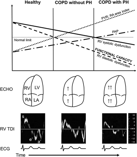
Pulmonary hypertension is a well-known comorbidity in advanced COPD. In patients with severe COPD waiting for lung volume reduction surgery or lung transplantation, pulmonary hypertension and consequential RV dysfunction was present in the half of the patients Citation(33). In unselected COPD patients with moderate-to-severe disease the incidence of the resting pulmonary hypertension is much lower. Subclinical systolic and manifest diastolic dysfunction of the RV, however, is already present in patients without pulmonary hypertension (Citation30,Citation34). The possible explanation for this phenomenon is the elevated pulmonary vascular resistance, which may be unmasked by exercise, when the pulmonary circulation no longer has the capacity to adapt and the pulmonary pressure increases parallel with the increasing cardiac output Citation(35). Kovacs et al. reported that borderline resting pulmonary arterial pressure is associated with decreased exercise capacity in patients with systemic sclerosis. In their work, 6MWT distance showed significant negative correlation with pulmonary vascular resistance measured at peak exercise Citation(36). demonstrates the hypothesized changes in the typical parameters of the RV systolic and diastolic function parallel with the progression of the pulmonary vascular disease in patients with COPD.
Figure 3. Schematic diagram demonstrating the hypothesized changes in the typical echocardiographic and hemodynamic parameters parallel with the course of the disease in patients with COPD.
Limitations of the study
Some limitations of our study are to be acknowledged. The extent of the dynamic lung hyperinflation was not investigated in our patients. This may be the cause that not any measures of respiratory function show significant correlation with the 6MWT distance in our study.
The number of patients with resting pulmonary hypertension was low in our COPD population and the elevation of the pulmonary pressure was mild in the majority of these cases. In addition, few patients with GOLD stage IV disease participated in the study. A higher proportion of patients with considerable pulmonary hypertension or more advanced lung disease may also alter our results.
Our patients did not undergo right heart catheterization, therefore the pulmonary arterial pressure was estimated noninvasively, and no data were available about the pulmonary vascular resistance of the patients. Due to the lack of invasive measurements, left and right ventricular filling pressures were also estimated by Doppler methods. Unfortunately, these parameters are less reliable in the evaluation of filling dynamics than the invasive measurements.
Conclusion
The mechanism of the exercise intolerance is complex in COPD, thus it is difficult to identify the true contributors. Nevertheless, our study suggests that RV diastolic function and filling pressure have strong influence on the functional capacity in patients with COPD.
Declaration of interest The authors report no conflicts of interest. The authors alone are responsible for the content and writing of the paper.
Funding
This research was supported by the European Union and the State of Hungary, co-financed by the European Social Fund in the framework of TÁMOP 4.2.4. A/2-11-1-2012-0001 “National Excellence Program” to R.F.
References
- Mathers CD, Boerma T, Ma Fat D. Global and regional causes of death. Br Med Bull 2009; 92:7–32.
- Murray CJ, Lopez AD. Alternative projections of mortality and disability by cause 1990–2020: Global Burden of Disease Study. Lancet 1997; 349(9064):1498–1504.
- Chapman KR, Mannino DM, Soriano JB, Vermeire PA, Buist AS, Thun MJ, et al. Epidemiology and costs of chronic obstructive pulmonary disease. Eur Respir J 2006; 27(1):188–207.
- Bauerle O, Chrusch CA, Younes M. Mechanisms by which COPD affects exercise tolerance. Am J Respir Crit Care Med 1998; 157(1):57–68.
- Marin JM, Carrizo SJ, Gascon M, Sanchez A, Gallego B, Celli BR. Inspiratory capacity, dynamic hyperinflation, breathlessness, and exercise performance during the 6-minute-walk test in chronic obstructive pulmonary disease. Am J Respir Crit Care Med 2001; 163(6):1395–1399.
- Laveneziana P, Guenette JA, Webb KA, O'Donnell DE. New physiological insights into dyspnea and exercise intolerance in chronic obstructive pulmonary disease patients. Expert Rev Respir Med 2012; 6(6):651–662.
- Nici L. Mechanisms and measures of exercise intolerance in chronic obstructive pulmonary disease. Clin Chest Med 2000; 21(4):693–704.
- Barnes PJ. Chronic obstructive pulmonary disease: effects beyond the lungs. PLoS Med 2010; 343(3):269–280.
- Hoeper MM, Barbera JA, Channick RN, Hassoun PM, Lang IM, Manes A, et al. Diagnosis, assessment, and treatment of non-pulmonary arterial hypertension pulmonary hypertension. J Am Coll Cardiol 2009; 54(1 Suppl):S85–96.
- Scharf SM, Iqbal M, Keller C, Criner G, Lee S, Fessler HE, et al. Hemodynamic characterization of patients with severe emphysema. Am J Respir Crit Care Med 2002; 166(3):314–322.
- Freixa X, Portillo K, Pare C, Garcia-Aymerich J, Gomez FP, Benet M, et al. Echocardiographic abnormalities in patients with COPD at their first hospital admission. The Eur Respir J 2013; 41(4):784–791.
- Schoos MM, Dalsgaard M, Kjaergaard J, Moesby D, Jensen SG, Steffensen I, et al. Echocardiographic predictors of exercise capacity and mortality in chronic obstructive pulmonary disease. BMC Cardiovasc Disord 2013; 13:84.
- Lopez-Sanchez M, Munoz-Esquerre M, Huertas D, Gonzalez-Costello J, Ribas J, Manresa F, et al. High prevalence of left ventricle diastolic dysfunction in severe COPD associated with a low exercise capacity: A cross-sectional study. PLoS One 2013; 8(6):e68034.
- Cuttica MJ, Shah SJ, Rosenberg SR, Orr R, Beussink L, Dematte JE, et al. Right heart structural changes are independently associated with exercise capacity in non-severe COPD. PLoS One 2011; 6(12):e29069.
- Vestbo J, Hurd SS, Agusti AG, Jones PW, Vogelmeier C, Anzueto A, et al. Global strategy for the diagnosis, management, and prevention of chronic obstructive pulmonary disease: GOLD executive summary. Am J Respir Crit Care Med 2013; 187(4):347–365.
- Rudski LG, Lai WW, Afilalo J, Hua L, Handschumacher MD, Chandrasekaran K, et al. Guidelines for the echocardiographic assessment of the right heart in adults. J Am Soc Echocardiogr 2010; 23(7):685–713.
- Lang RM, Bierig M, Devereux RB, Flachskampf FA, Foster E, Pellikka PA, et al. Recommendations for chamber quantification. J Am Soc Echocardiogr 2005; 18(12):1440–1463.
- Nagueh SF, Appleton CP, Gillebert TC, Marino PN, Oh JK, Smiseth OA, et al. Recommendations for the evaluation of left ventricular diastolic function by echocardiography. J Am Soc Echocardiogr 2009; 22(2):107–133.
- ATS statement: guidelines for the six-minute walk test. Am J Respir Crit Care Med 2002; 166(1):111–117.
- Vinereanu D, Nicolaides E, Tweddel AC, Fraser AG. “Pure” diastolic dysfunction is associated with long-axis systolic dysfunction. Implications for the diagnosis and classification of heart failure. Eur J Heart Fail 2005; 7(5):820–828.
- Watz H, Waschki B, Meyer T, Kretschmar G, Kirsten A, Claussen M, et al. Decreasing cardiac chamber sizes and associated heart dysfunction in COPD: role of hyperinflation. Chest 2010; 138(1):32–38.
- Funk GC, Lang I, Schenk P, Valipour A, Hartl S, Burghuber OC. Left ventricular diastolic dysfunction in patients with COPD in the presence and absence of elevated pulmonary arterial pressure. Chest 2008; 133(6):1354–1359.
- Hannink JD, van Helvoort HA, Dekhuijzen PN, Heijdra YF. Heart failure and COPD: partners in crime? Respirology 2010; 15(6):895–901.
- Falk JA, Kadiev S, Criner GJ, Scharf SM, Minai OA, Diaz P. Cardiac disease in chronic obstructive pulmonary disease. Proc Am Thorac Soc 2008; 5(4):543–548.
- Sin DD, Anthonisen NR, Soriano JB, Agusti AG. Mortality in COPD: Role of comorbidities. Eur Respir J 2006; 28(6):1245–1257.
- Smith BM, Prince MR, Hoffman EA, Bluemke DA, Liu CY, Rabinowitz D, et al. Impaired left ventricular filling in COPD and emphysema: Is it the heart or the lungs? The Multi-Ethnic Study of Atherosclerosis COPD Study. Chest 2013; 144(4):1143–1151.
- Sohn DW, Chai IH, Lee DJ, Kim HC, Kim HS, Oh BH, et al. Assessment of mitral annulus velocity by Doppler tissue imaging in the evaluation of left ventricular diastolic function. J Am Coll Cardiol 1997; 30(2):474–480.
- Douglas PS. The left atrium: a biomarker of chronic diastolic dysfunction and cardiovascular disease risk. J Am Coll Cardiol 2003; 42(7):1206–1207.
- Hammoudi N, Achkar M, Laveau F, Boubrit L, Djebbar M, Allali Y, et al. Left atrial volume predicts abnormal exercise left ventricular filling pressure. Eur J Heart Fail 2014; 16(10):1089–1095.
- Hilde JM, Skjorten I, Grotta OJ, Hansteen V, Melsom MN, Hisdal J, et al. Right ventricular dysfunction and remodeling in chronic obstructive pulmonary disease without pulmonary hypertension. J Am Coll Cardiol 2013; 62(12):1103–1111.
- Do DH, Therrien J, Marelli A, Martucci G, Afilalo J, Sebag IA. Right atrial size relates to right ventricular end-diastolic pressure in an adult population with congenital heart disease. Echocardiography 2011; 28(1):109–116.
- Melenovsky V, Hwang SJ, Lin G, Redfield MM, Borlaug BA. Right heart dysfunction in heart failure with preserved ejection fraction. Eur Heart J 2014; 35(48):3452–3462.
- Thabut G, Dauriat G, Stern JB, Logeart D, Levy A, Marrash-Chahla R, et al. Pulmonary hemodynamics in advanced COPD candidates for lung volume reduction surgery or lung transplantation. Chest 2005; 127(5):1531–1536.
- Sabit R, Bolton CE, Fraser AG, Edwards JM, Edwards PH, Ionescu AA, et al. Sub-clinical left and right ventricular dysfunction in patients with COPD. Respir Med 2010; 104(8):1171–1178.
- Rubin LJ. Cor pulmonale revisited. J Am Coll Cardiol 2013; 62(12):1112–1113.
- Kovacs G, Maier R, Aberer E, Brodmann M, Scheidl S, Troster N, et al. Borderline pulmonary arterial pressure is associated with decreased exercise capacity in scleroderma. Am J Respir Crit Care Med 2009; 180(9):881–886.