Abstract
Prinzmetal (variant) angina may be associated with cardiac arrhythmias that can deteriorate to fatal ventricular arrhythmias. We present 2 patients with syncope where vasospastic angina and severe ventricular arrhythmias were found to be responsible for the syncopal episodes.
Keywords::
Introduction
Syncope is a common entity and its etiology is often difficult to determine, even after comprehensive investigation.
Prinzmetal (variant) angina is a well characterized entity with anginal symptoms at rest and electrocardiographic transient ST-segment elevation secondary to myocardial ischemia (Citation1). This syndrome may be associated with severe cardiac arrhythmias that can deteriorate to fatal ventricular arrhythmias in 3.5–6.5% of affected patients (Citation2). We present 2 patients with syncope where vasospastic angina and severe ventricular arrhythmias were found to be responsible for the syncopal episodes.
Case report
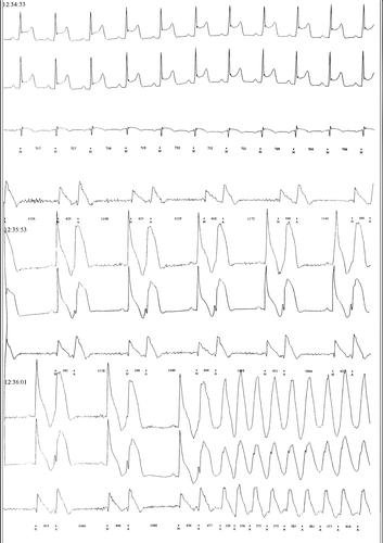
2 non-smoker male patients (aged 60 and 65 years) were hospitalized because of recurrent syncope, both patients complained of vague chest discomfort. Both patients fainted during hospitalization while being recorded with a Holter monitor. The Holter at the time of syncope showed transient and gradual ST elevation and at the time of maximal ST-segment elevation ventricular extra-systoles that deteriorated to severe ventricular arrhythmia were recor ded (, ).
Figure 1. Single channel Holter recording during syncope showing normal ST segment (1) and ST segment elevation (2); during ST segment elevation severe ventricular arrhythmias were detected (transient ventricular tachycardia and fibrillation). The long sinus pause after VF termination is most probably due to sinus node dysfunction secondary to transient global ischemia.
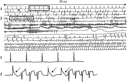
Figure 2. Three-channel Holter recording during syncope showing transient ST-segment elevation and ventricular extra-systoles, bigeminy and ventricular flutter at the time of maximal ST elevation.
Coronary angiography revealed non-significant coronary lesions with one of the patients developing severe spasm of the left anterior coronary artery during the procedure. Both patients received an implantable cardioverter defibrillator (ICD); and were treated concomitantly with nitrates and calcium channel blockers and have remained free of symptoms or ICD discharges during a follow up of three and two years respectively.
Discussion
Patients with variant angina are at greater risk of developing sudden death (Citation3). Calcium channel blockers and nitrates are an effective treatment to prevent coronary vasospasm with a good prognosis (89 to 97% survival) (Citation4), however, an ICD is indicated to prevent sudden death in selected patients and is usually reserved for patients who have experienced ischemia associated VF who continue to manifest ischemia despite maximal medical therapy (Citation1,Citation5). The difficulty to prevent new episodes of sudden death is also stressed by Meisel et al. (Citation5) in a follow-up of 8 patients with variant angina pectoris that developed ventricular fibrillation after chest pain with normal appearing coronary arteries despite treatment with calcium antagonists and recurrence of the symptoms in all patients.
Variant angina is usually characterized by resting angina, however, as shown in both our patients the main manifestation may be syncope related to ventricular arrhythmias. Both of our cases clearly demonstrate the relationship between transient ST elevation (due to coronary vasospasm) and ventricular arrhythmias. They also demonstrate the importance of Holter monitoring (of 24–48 h) or loop recorders (depending on the frequency of symptoms) illustrating both ischemic changes and the ventricular arrhythmias caused by this ischemic environment.
Conclusion
Variant angina should be considered in the differential diagnosis of syncope, especially when associated with chest discomfort. Holter monitoring or event recorders may be useful for the definite diagnosis.
Declaration of interest: The authors report no conflicts of interest. The authors alone are responsible for the content and writing of the paper.
References
- Canty JM. Prinzmetal variant angina. In: Bonow, Mann, Zipes, Libby, editors. Braunwald's Heart Disease 9th edition. Philadelphia: Elsevier Saunders, 2011. pp. 1195–8.
- Kerin NZ, Rubenfire M, Naimi M, Wajszezuk WJ, Pamatmat A, Cascade PN. Arrhythmias in variant angina pectoris, relationship of arrhythmias to ST segment elevation and R wave changes. Circulation 1979;60:1343–50.
- MacAlpin RN. Cardiac arrest and sudden unexpected death in variant angina: Complication of coronary spasm that can occur in the absence of severe organic stenosis. Am Heart J. 1993;125:1011–7.
- Bott-Silverman C, Heupler FA. Natural history of pure coronary spasm in patients treated medically. J Am Coll Cardiol. 1983; 2:200–5.
- Meisel SR, Mazur A, Chetboun I, Epstein M, Canetti M, Gallimidi J, . Usefulness of implantable cardioverter defibrillators in refractory variant angina pectoris complicated by ventricular fibrillation in patients with angiographically normal coronary arteries. Am J Cardiol. 2002;89:1114–6.