Abstract
Vessel perforation is an undesirable and life-threatening complication during vein graft angioplasty. We report on a case of vein graft rupture during angioplasty, which was successfully managed with deployment of a polytetrafluoroethylene-covered stent.
Keywords::
Case report
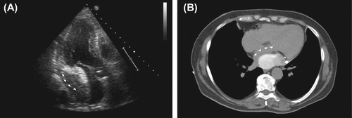
A 71-year-old female, with a history of coronary artery bypass grafting (CABG) 16 years ago and previous percutaneous interventions (PCI) for de novo and restenotic lesions of the saphenous vein graft (SVG) to obtuse marginal (OM) branch, presented with recurrence of exertional angina. Coronary angiography via the right femoral approach revealed a de novo lesion of the SVG between the previous stents (). A 6Fr AL1 guiding catheter was used to engage the ostium of the SVG and following administration of intragraft verapamil and deployment of a distal embolic protection device (FilterWire, Boston Scientific), the lesion was directly stented with a 4.0 × 16 mm Everolimus-eluting stent (Promus Element, Boston Scientific) to 16 atm (). Angiography following stent deployment showed a type III perforation of the vein graft (Figures 1C, D, Supplementary Videos 1, 2 to be found online at http://informahealthcare.com/doi/abs/10.3109/17482941.2013.835829.). Bleeding was temporarily controlled with inflation of the stent balloon at 10 atm until terminated due to ischaemia. However, the bleeding restarted and, therefore, was treated with a 4.0 × 26 polytetrafluoroethylene-covered stent (Jostent Graftmaster, Abbott) (). Final angiography showed TIMI III flow, with no residual extravasation (, Supplementary Video 3 to be found online at http://informahealthcare.com/doi/abs/10.3109/17482941.2013.835829.). Patient remained haemodynamically stable and asymptomatic throughout and following the procedure. Post-procedural echocardiography and CT chest demonstrated a loculated and localised haemopericardial effusion compressing the left atrium and left lower pulmonary vein which was treated conservatively (, Supplementary Video 4 to be found online at http://informahealthcare.com/doi/abs/10.3109/17482941. 2013.835829.). Serial transthoracic echocardiograms showed no increase in the size of the effusion and patient was discharged home 48 h later on lifelong dual antiplatelet therapy.
Figure 1. De novo SVG lesion directly stented with a drug eluting stent (A, B). Type III graft perforation revealed following stent deployment (C, D, arrowheads). Successfully treatment perforation with deployment of covered stent (E, F).
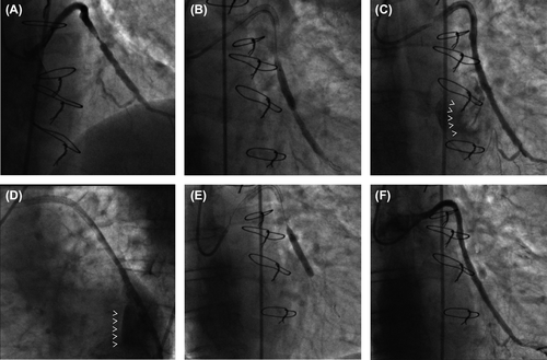
Figure 2. Transthoracic echocardiogram and chest computed tomography suggestive of a loculated and localized haemopericardial effusion compressing the left atrium and left lower pulmonary vein (arrowheads).
Discussion
Vessel perforation during coronary intervention is a rare, but well recognized complication with a reported incidence of 0.2% to 0.5% (Citation1). It can be associated with major adverse events such as cardiac tamponade, myocardial infarction, cardiogenic shock or death. Coronary perforation has been classified by Ellis et al. into three types: Type I, extraluminal crater without extravasation; Type II, pericardial or myocardial blushing; and Type III, perforation ≥ 1-mm diameter with contrast streaming and cavity spilling (Citation2). Degenerated vein grafts, highly calcified lesions, stent oversizing, balloon over-inflation and use of laser or rotational atherectomy are the most common risk factors (Citation3). Vessel perforation during stent deployment can be caused by rupture of the vessel wall by stent edges, which is most likely the cause of perforation in this case.
Heparin reversal with intravenous protamine and platelet transfusion, if a glycoprotein IIb/IIIa inhibitor has been used, may be necessary if extravasation is not adequately controlled or if percutaneous drainage of pericardial effusion is required. Initial balloon inflation upon recognition of vessel injury may correct the problem or allow time, as in this case, for a more definitive procedure. Polytetrafluoroethylene-covered stents are available as balloon expandable and self-expandable and their use is essential when prolonged balloon inflation fails to seal the perforation site (Citation4). Emergency CABG and pericardial drainage may be required if percutaneous management is unsuccessful.
Supplementary Videos 1–4
Download Microsoft Video (AVI) (1.8 MB)Supplementary Videos 1–4
Download Microsoft Video (AVI) (1.5 MB)Supplementary Videos 1–4
Download Microsoft Video (AVI) (1.2 MB)Supplementary Videos 1–4
Download Microsoft Video (AVI) (935 KB)Declaration of interest: The authors report no conflicts of interest. The authors alone are responsible for the content and writing of the paper.
References
- Subraya RG, Tannenbaum AK. Successful sealing of perforation of saphenous vein graft by coronary stent. Catheter Cardiovasc Interv. 2000;50:460–2.
- Ellis SG, Ajluni S, Arnold AZ, Popma JJ, Bittl JA, Eigler NL, et al. Increased coronary perforation in the new device era. Incidence, classification, management, and outcome. Circulation 1994;90:2725–30.
- Marmagkiolis K, Brilakis ES, Hakeem A, Cilingiroglu M, Bilodeau L. Saphenous vein graft perforation during percutaneous coronary intervention: a case series. J Invasive Cardiol. 2013;25:157–61.
- Baruah DK. Covered stent to treat saphenous venous graft perforation—a case report. Catheter Cardiovasc Interv. 2010; 76:844–46.