
Abstract
Objective: The purpose of this study was to evaluate long-term bond strengths of dentin adhesive systems, which include one-step self-etch adhesive systems (Optibond All-in-one, Kerr; Adper Prompt L-POP, 3 M ESPE), a three-step etch-and-rinse adhesive (Optibond FL, Kerr) and two-step self-etch adhesive (AdheSE Bond, Ivoclar), applied to pulp chamber dentin surfaces after 12-month water storage by using microtensile bond strength (µTBS) test.
Materials and methods: Dentin adhesive systems were applied to unprepared pulp chamber dentin surfaces according to manufacturer’s directions, respectively (n = 5). After applying adhesive systems, composite buildups were done incrementally. Bond strengths to pulp chamber dentin surfaces were determined using µTBS test after water storage for 24 h and 12 month. Kruskal–Wallis analysis and Mann–Whitney U-test for pairwise comparisons were used to determine statistical differences in µTBS between the groups at a significance level of 5%.
Results: There were no significant differences in µTBS between storage periods for tested adhesives regardless adhesive class.
Conclusion: Bond durability of tested adhesive systems, including one-bottle self-etch adhesives with pulp chamber dentin surfaces, may be considered stable after 12-month water storage. Therefore, one-step self-etch, also called “user-friendly” adhesives may perform and traditional three-step etch-and-rinse adhesives in the long-term when used for bonding to pulp chamber dentin surfaces.
Introduction
The teeth received root canal treatments can be restorated by using a resin composite restorative materials in couple of adhesive resin systems with high clinical success rate.[Citation1] The coronal restoration of endodontically treated teeth using resin composites permit a more conservative way to post and core restorations.[Citation2] Adhesive resin materials permit carrying of stresses through the resin–dentin/enamel interfaces to the supporting dental hard tissues, with restoring tooth structure after root canal treatment. However, the formation of dentinal hybrid layer across resin–dentin interfaces can seal dentin from oral fluids, thus reduce coronal microleakage.[Citation3,Citation4]
It was suggested that resin adhesive systems might be used to bonding to the pulp chamber dentin surfaces to seal the root canal to prevent microleakage of oral fluids and microorganism.[Citation3] However, dentin adhesive systems changed considerably on the way to being more user-friendly and less technique sensitive in the course of past decade. Current dental adhesive systems are divided two main categories either an etch-and-rinse adhesives or a self-etch adhesives. Self-etch adhesives do not require a separate etching step unlike to etch-and-rinse adhesives. They contain acidic monomers that simultaneously etch and prime dental hard tissues. The latest generations of self-etch adhesive are most simple-to-use one-step adhesives.[Citation5]
Extensive investigations are performed to assess bonding effectiveness of current dentin adhesive systems applied to prepared coronal dentin surfaces.[Citation6] The immediate bonding effectiveness of most current adhesive systems to coronal dentin is promising regardless of the adhesive used,[Citation7] although lower bond strengths were frequently reported for one-step self-etch adhesives.[Citation8] However, the limited knowledge is available in the literature regarding bonding effectiveness of current adhesive systems with pulp chamber dentin surfaces.
Bond strength tests are the most commonly used laboratory tests to rank dentin adhesive systems. Different bond strength tests are advanced. Currently, the shear bond strength and microtensile bond strength test methods are mostly used.[Citation6,Citation9] However, concerns regarding laboratory assessments involve only assessing initial bond strengths of dentin adhesives may not provide a valid in vitro data that in correlation with an actual clinical performances of dentin adhesives systems.[Citation10] Therefore, diverse laboratory aging techniques were used to obtain valid in vitro data.[Citation11] The most commonly used artificial aging technique is the water storage. In this technique, the bonded specimens are stored in watery medium for a certain period. Water storage period may vary from a few months [Citation12] up to 4 years [Citation13] or even longer up to 10 years.[Citation14] Most of these studies revealed significant reductions in bond strengths, even after quite short storage periods. The reduction in bonding after water storage was caused by the biodegradation of hybrid layer constituents by hydrolysis of poorly polymerized resin and an enzymatic degradation of collagen.[Citation15]
It seems that laboratory reports only provide information regarding to the bond durability of dentin adhesives applied to coronal dentin, currently. However, bond durability of dentin adhesive systems applied to pulp chamber dentin surfaces remains not well investigated. This is an important gap in literature, because pulp chamber dentin surfaces exhibit distinct surface morphology when compared to prepared coronal dentin surfaces.[Citation16–18] Prepared coronal dentin surfaces are covered with smear layers with varying thickness, which are supposed to exhibit a challenge against to bonding effectiveness of self-etch adhesive with mild pH.[Citation19,Citation20] Unlike to prepared dentin surfaces, pulp chamber dentin is generally not prepared during endodontic treatment and remains smear layer-free.[Citation16,Citation17] Therefore, the aim of this study was to evaluate potential degradations of bonds of different dentin adhesive systems applied to pulp chamber dentin surfaces by 12 month of water storage with the usage of microtensile bond strength test (µTBS). The null hypothesis tested found that there are no differences in the durability of the tested dentin adhesive systems bonded to pulp chamber dentin surfaces over 12-month in vitro.
Methods
In this study, four dental adhesive systems were tested as follows; a three-step etch-and-rinse adhesive system (Optibond FL, Kerr, Orange, CA), a two-step self-etch adhesives (AdheSE Bond, Ivoclar, Vivadent, Schaan, Liechtenstein) and two one-step self-etch adhesive systems (Optibond All-in-one, Kerr, Orange, CA; Adper Prompt L-POP, 3 M ESPE, Seefeld, Germany). Brand names, chemical compositions, manufacturer instructions of respective adhesive systems were presented in and , respectively.
Table 1. Chemical compositions and lot numbers of dental adhesive systems.
Table 2. Application protocols of dentin adhesive systems.
Specimen preparation
Ten bovine incisors, which were stored in the 0.02% sodium azide solution prior to usage at room temperature, were used to prepare pulp chamber dentin specimens to be bonded. Specimen preparation procedure used in the present study from bovine incisor teeth to obtain bonding substrate of pulp chamber dentin surfaces was shown in . Two subsequent sections, which were perpendicular to long axis of tooth, were performed using low-speed water-cooled diamond saw. The first section was performed at 5 mm above enamel–cement border; the other one was performed at 1 mm under enamel–cement border. Then, the crown segments with 6 mm heights were sectioned into halves using low-speed diamond saw. Pulp tissue remnants were removed from pulp chamber surfaces carefully with spoon-shaped excavator. Pulp chamber surfaces were examined using stereomicroscopy at 40 × magnification to avoid pulp calcifications and cracks. Exposed root dentin surfaces left after sectioning were covered with nail polish to demarcate pulp chamber dentin to be bonded.
Figure 1. Schematic presentation of specimen preparation for microtensile bond strength test for bonding to bovine pulp chamber dentin surfaces. (a) two subsequent sections, which were perpendicular to long axis of tooth, were performed using low speed diamond saw under water cooling. The first section was performed at 5 mm above enamel–cement border; the other one was performed at 1 mm under enamel–cement border. (b) The obtained crown segments with 6 mm heights were sectioned into halves using low speed diamond saw. Then, pulp chamber dentin surfaces in order to bond were exposed. (c) Following application of adhesives, composite buildups were done incrementally. (d) Bonded specimens were sectioned serially to obtain resin–dentin sticks to be used for microtensile bond strength test.
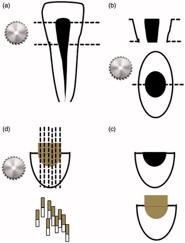
Twenty pulp chamber dentin specimens were divided into four groups randomly (n = 5). Each adhesive system was applied to pulp chamber dentin according to respective manufacturer instructions (). After adhesive procedures, resin composite (Valux Plus, 3 M ESPE, St Paul, MN) buildups in three layers up to a height of 4 mm were done on the surfaces. Each increment layer was cured for 20 s using the Elipar Freelight LED light (3 M ESPE, St Paul, MN) with a power of 1000 mW/cm2.
Microtensile bond strength test
All bonded teeth were stored in distilled water at 37°C for 24 h. Then, resin–dentin sticks with approximately 0.8 × 0.8 mm2 dimensions were obtained using diamond saw under copious water (Micracut 125, Metkon, Bursa, Turkey) running at 300 rpm. Obtained resin–dentin sticks were divided in two similar-sized subgroups according to μTBS test time (immediately and after 12-month water storage). Twenty sticks from each subgroup were selected randomly to perform μTBS test. Then, the sticks were fixed to jig with cyanoacrylate glue (Pattex, Henkel, Duesseldorf, Germany) and forced in tension at a crosshead speed of 1 mm/min using Bisco microtensile testing machine (Bisco Inc. Schaumburg, IL). Resting sticks were immersed in distilled water at 37°C for 12 month. No antibacterial agents were included to storage medium. The storage medium did not changed to maintain mechanical properties of dentin, as frequent changing of storage solution results significant mineral loss from dentin, thus decreasing mechanical properties of substrate.[Citation21] After 12-month storage, second μTBS test was performed. Then, the fractured specimens were removed, and the cross-sectional area at the site of fracture was measured with a digital caliper (Mitutoyo, Tokyo, Japan). Therefore, the μTBS was derived by dividing the enforced force at the time of fracture by the bond area (mm2). The mode of failure was determined by stereomicroscope under 40 × magnificence (Meade Bresser Biolux, Meade Bresser, Rhede, Germany) and recorded as “adhesive” or “cohesive” either dentin or resin and “mix” failures include more than one of the dentin and resin parts.
Statistical analyses
The distribution of radiopacity μTBS data was primarily analyzed for the normal distribution with the Kolmogorov–Smirnov test and equal variance with the Levene’s test. Although μTBS data were normally distributed, group variances were not equal. Thus, bond strength data (MPa) were analyzed by Kruskal–Wallis test, and post hoc comparison was done using the Mann–Whitney U-test. Statistical analyses were performed by using SPSS 18 software (SPSS Inc., Chicago, IL). The level of significance was set at p = 0.05.
Results
Mean microtensile bond strength (µTBS) values and failure modes of tested dental adhesive systems were presented in the . After 24 h of water storage, the mean µTBS values were ranged from 32.86 ± 13.38 MPa to 38.15 ± 10.38 MPa. Optibond FL, a three-step etch-and-rinse adhesive showed the numerically highest bond strength. After 12-month water storage, no significant reductions in bond strength were observed for any adhesive system tested (from 28.66 to 33.22 MPa). However, the shifts from mix failure to adhesive failure in dominant failure modes were observed after 12 month of water storage for AdheSE Bond and Optibond All-in-one.
Table 3. Mean µTBS values (n = 20) and failure modes.
Discussion
In the present study, the effect of 12 month of water storage on bond strengths of three-step etch-and-rinse adhesive, two-step self-etch adhesive and one-step self-etch adhesive systems applied to pulp chamber dentin surfaces. Coronal leakage is particularly significant in multirooted teeth in which accessory canals may be present in the furcation area.[Citation22] Therefore, the bond durability of adhesive systems applied to pulp chamber dentin is important factor for overall prognosis of tooth after root canal treatment, as the bond durability of adhesive systems may warrant an accomplished adhesive sealing at long term.
Bond durability of resin adhesive systems applied to prepared coronal dentin surfaces is extensively investigated in the literature. A current meta-analytical review shows the status of bond durability of dentin adhesives in detail.[Citation23] According to this report, Optibond FL that is a three-step etch-and-rinse adhesive exhibited the most favorable bond durability in the current literature. It is reported that the averaged bond strength mean of Optibond FL had reduced by only 10% after 12-month water storage. This adhesive exhibited better bond stability performances than its adhesive class since reduction rate for three-step etch-rinse adhesives was 17%. Two-step self-etch adhesives as an adhesive class exhibited similar bond stability performance with 13% reduction in bond strength after 12-month water storage. The worst performance in bond stability was owned to one-step self-etch adhesive systems with 33% reduction.
In the present study, with Optibond FL, bond strength of 38.15 ± 10.38 was determined at 24 h. Main bonding mechanism of etch-and-rinse adhesives was mechanical interlocking of diffused resin into demineralized collagen mesh and in situ polymerization. Thus, bond strength depends on hybridization of collagen mesh with infiltrated adhesive resin. After 12 month of water storage, 13% reduction was determined in bond strength, but this difference was not significant. The bond strength of AdheSE Bond that is a two-step self-etch adhesive also showed no significant deterioration by 12 month of water storage when applied to pulp chamber dentin surfaces, although 13% reduction was observed in bond strength. Therefore, it is concluded that differences in bonding substrate (prepared coronal dentin vs. pulp chamber dentin) might not affect bond stabilities of Optibond FL and AdheSE Bond adhesives when compared to status of this adhesive and adhesive class for prepared dentin surfaces.
Particular attention should be paid to one-step self-etch adhesive test in the present study. According to a review mentioned earlier, one-step self-etch adhesive test exhibited the worst bond durability performance. From the review,it was found that reduction rate was 33% for one-step self-etch adhesives.[Citation23] However, it was found that both Optibond All-in-one and Adper Prompt L-POP are one-step self-etch adhesive with different pH values, which shows a well-stabilized bond strength to pulp chamber dentin surfaces over 12 month of water storage in the present study. Reduction rates for Optibond All-in-one and Adper Prompt L-POP were 11% and 10%, respectively. Therefore, in case of one-step self-etch adhesives, dentin bond stability may depend on bonding substrate. Although huge numbers of parameters may influence assessment of dentin bond durability,[Citation24] this may arise from structural differences between pulp chamber dentin surfaces and prepared coronal dentin surfaces.
The structure of pulp chamber wall dentin differs from that of the other dentinal regions of teeth. Because it is not prepared during the endodontic procedures, this area does not have a smear layer. Therefore, the effectiveness and bond strength of the adhesive systems depend mostly on collagen-rich predentin of the wall, many enlarged tubules and a small amount of intertubular dentin.[Citation16,Citation18,Citation22] It is well known that the nature of smear layer significantly affected bond strength of mild self-etch adhesives to bur-cut dentin,[Citation19,Citation20] and its effect on long-term bond strength is still not well known. As pulp chamber dentin surfaces are free of smear layer, mild self-etch adhesives may able to etch profoundly enough sound dentin and encapsulate exposed collagen fibrils. Therefore, the favorable bond stability of Optibond All-in-one, which is an ultramild self-etch adhesive over 12 month of water storage (pH > 2.5), is attributed to smear layer-free property of pulp chamber dentin surface.
The increased hydrophilicity is another factor for reduced bond durability of these adhesives due to the absence of separate hydrophobic bonding layer.[Citation25] It is reported that one-step self-etching adhesives showed higher water sorption and solubility than two-step self-etching adhesives.[Citation26] However, it was revealed that the initial amount of water sorption and solubility might not affect the durability of the dentin bond of the one-step self-etch adhesives over one year of water storage.[Citation27] The aging effects of increased hydrophilicity might depend on adhesive brand and water storage time.[Citation14]
Numerous factors might influence reduction rate of resin–dentin bond strength after long-term water storage.[Citation24] The reason for the minimum reduction in bond strength values of adhesives after 12 months water storage was related to water volume and it was not changed regularly which caused ions supersaturation in the water at the beginning of the study, and thus, no leachable ions could migrate to the solution over the long-term storage. As the in vitro conditions are lack of remineralization effects of natural saliva, an equilibrium of calcium ion transfer between the dentin and unchanged storage solutions is established in the solution.[Citation21]
Several aspects for designing the present study that might reduce the reliability of the results are mentioned. First, the pulp chamber dentin surfaces were prepared by using bovine incisors as harvesting dentin samples with lesser heterogeneity is easy.[Citation28] However, bovine pulp chamber dentin surfaces may differ in terms of tubularity and mineral content from human pulp chamber dentin.[Citation29] Secondly, in clinical situations, before bonding to human, pulp chamber dentin surfaces are exposed to various chemicals. Therefore, further studies are needed to assess effects of various endodontic chemicals on bond durability of resin adhesives to pulp chamber dentin. Another important issue that might be considered as inherent problem with the assessment of bonding to pulp chamber dentin surfaces is that pulp chamber dentin surfaces are not flat and somewhat oblique. This might make tensile testing does not produce even forces across the interfaces. This may explain high variation within bond strength results.[Citation30]
Conclusion
The research regarding to dentin bond durability of current dental adhesive systems is highly complicated issue in which numerous factors that interact with each other might be involved. However, this is the first report on long-term bond strengths of resin–dentin adhesive systems applied to pulp chamber dentin surfaces over 12 month of water storage. It was found that bonds of all tested adhesive were quite durable with pulp chamber dentin surface regardless adhesive class and pH. Structural differences among prepared coronal dentin surfaces and pulp chamber dentin may contribute to these findings.
Declaration of interest
The author reports no conflicts of interest. The author alone is responsible for the content and writing of this article.
References
- Mannocci F, Cowie J. Restoration of endodontically treated teeth. Br Dent J. 2014;216:341–346
- Salvi GE, Siegrist Guldener BE, Amstad T, et al. Clinical evaluation of root filled teeth restored with or without post-and-core systems in a specialist practice setting. Int Endod J. 2007;40:209–215
- Belli S, Zhang Y, Pereira PN, et al. Adhesive sealing of the pulp chamber. J Endod. 2001;27:521–526
- Ebert J, Löffler C, Roggendorf MJ, et al. Clinical adhesive sealing of the pulp chamber following endodontic treatment: influence of thermomechanical loading on microleakage. J Adhes Dent. 2009;11:311–317
- Van Meerbeek B, Yoshihara K, Yoshida Y, et al. State of the art of self-etch adhesives. Dent Mater. 2011;27:17–28
- Scherrer SS, Cesar PF, Swain MV. Direct comparison of the bond strength results of the different test methods: a critical literature review. Dent Mater. 2010;26:e78–e93
- Inoue S, Vargas MA, Abe Y, et al. Microtensile bond strength of eleven contemporary adhesives to dentin. J Adhes Dent. 2001;3:237–245
- Van Meerbeek B, Peumans M, Poitevin A, et al. Relationship between bond-strength tests and clinical outcomes. Dent Mater. 2010;26:e100–e121
- Ayar MK. Reminder about long-term clinical trials as a gold standard for the bonding effectiveness of adhesive resins. J Restor Dent. 2015;3:54
- Hashimoto M, Nagano F, Endo K, et al. A review: biodegradation of resin–dentin bonds. Jpn Dent Sci Rev. 2011;47:5–12
- Deng D, Yang H, Guo J, et al. Effects of different artificial ageing methods on the degradation of adhesive-dentine interfaces. J Dent. 2014;42:1577–1585
- Sauro S, Watson TF, Thompson I, et al. One-bottle self-etching adhesives applied to dentine air-abraded using bioactive glasses containing polyacrylic acid: an in vitro microtensile bond strength and confocal microscopy study. J Dent. 2012;40:896–905
- De Munck J, Van Meerbeek B, Yoshida Y, et al. Four-year water degradation of total-etch adhesives bonded to dentin. J Dent Res. 2003;82:136–140
- Hashimoto M, Fujita S, Nagano F,et al. Ten-years degradation of resin-dentin bonds. Eur J Oral Sci. 2010;118:404–410
- Breschi L, Mazzoni A, Ruggeri A, et al. Dental adhesion review: aging and stability of the bonded interface. Dent Mater. 2008;24:90–101
- Ozturk B, Ozer F. Effect of NaOCl on bond strengths of bonding agents to pulp chamber lateral walls. J Endod. 2004;30:362–365
- Kijsamanmith K, Timpawat S, Harnirattisai C, et al. Micro-tensile bond strengths of bonding agents to pulpal floor dentine. Int Endod J. 2002;35:833–839
- Timpawat S, Nipattamanon C, Kijsamanmith K, et al. Effect of bleaching agents on bonding to pulp chamber dentine. Int Endod J. 2005;38:211–217
- Ermis RB, De Munck J, Cardoso MV, et al. Bond strength of self-etch adhesives to dentin prepared with three different diamond burs. Dent Mater. 2008;24:978–985
- Yiu CKY, Hiraishi N, King NM, et al. Effect of dentinal surface preparation on bond strength of self-etching adhesives. J Adhes Dent. 2008;10:173–182
- Kitasako Y, Burrow MF, Nikaido T, et al. The influence of storage solution on dentin bond durability of resin cement. Dent Mater. 2000;16:1–6
- Vertucci FJ, Anthony RL. A scanning electron microscopic investigation of accessory foramina in the furcation and pulp chamber floor of molar teeth. Oral Surg Oral Med Oral Pathol. 1986;62:319–326
- De Munck J, Mine A, Poitevin A, et al. Meta-analytical review of parameters involved in dentin bonding. J Dent Res. 2012;91:351–357
- Carvalho RM, Manso AP, Geraldeli S, et al. Durability of bonds and clinical success of adhesive restorations. Dent Mater. 2012;28:72–86
- Tay FR, Pashley DH. Have dentin adhesives become too hydrophilic? J Can Dent Assoc. 2003;69:726–731
- Ito S, Hoshino T, Iijima M, et al. Water sorption/solubility of self-etching dentin bonding agents. Dent Mater. 2010;26:617–626
- Itoh S, Nakajima M, Hosaka K, et al. Dentin bond durability and water sorption/solubility of one-step self-etch adhesives. Dent Mater J. 2010;29:623–630
- Wegehaupt, F, Gries D, Wiegand A, et al. Is bovine dentine an appropriate substitute for human dentine in erosion/abrasion tests? J Oral Rehabil. 2008;35:390–394
- Lopes MB, Sinhoreti MA, Gonini Júnior A, et al. Comparative study of tubular diameter and quantity for human and bovine dentin at different depths. Braz Dent J. 2009;20:279–283
- Ibarra G, Vargas MA, Armstrong SR, et al. Microtensile bond strength of self-etching adhesives to ground and unground enamel. J Adhes Dent. 2001;4:115–124