Abstract
Ecstasy (MDMA, 3,4-methylendioxymethamphetamine) and the stimulants methamphetamine (METH, speed) and amphetamine are popular drugs among young people, particularly in the dance scene. When given in high doses both MDMA and the stimulant amphetamines are clearly neurotoxic in laboratory animals. MDMA causes selective and persistent lesions of central serotonergic nerve terminals, whereas amphetamines damage both the serotonergic and dopaminergic systems. In recent years, the question of ecstasy-induced neurotoxicity and possible functional sequelae has been addressed in several studies in drug users. Despite large methodological problems, the bulk of evidence suggests residual alterations of serotonergic transmission in MDMA users, although at least partial recovery may occur after long-term abstinence. However, functional sequelae may persist even after longer periods of abstinence. To date, the most consistent findings associate subtle cognitive impairments with ecstasy use, particularly with memory. In contrast, studies on possible long-term neurotoxic effects of stimulant use have been relatively scarce. Preliminary evidence suggests that alterations of the dopaminergic system may persist even after years of abstinence from METH, and may be associated with deficits in motor and cognitive performance. In this paper, we will review the literature focusing on human studies.
El éxtasis (MDMA, 3,4 metilendioximetanfetamina), y los estimulantes metanfetaminkos (METH, “speed”) y anfetaminicos son drogas frecuentes entre los jóvenes, especialmenie en lugares de baile, Cuando el MDMA los estimulantes anfetamínicos son administradas en alias dosis a animales de laboratorio resultan claramente neurotóxicas. El MDMA produce lesiones selectivas y persistentes de los terminales nerviosos serotoninérgicos centrales, mientras que las anfetaminas dañan tanio los sisiemas serotoninérgicos como dopaminérgicos. En los últimos años la pregunia acerca de la neurotoxicidad inducida por el éxtasis y las posibles secuelas funcionales ha sido tema de algunos estudios con usuarios de drogas, A pesar de los grandes problemas metodológicos, la amplia evidencia sugiere que exisien alteraciones residuales de la transmisión serotoninérgica en usuarios de MDMA, aunque puede conseguirse cieria recuperatión parcial después de una abstinencia prolongada. Sin embargo, las secuelas funcionales pueden persistir aun después de largos périodes de abstinencia. A la fecha, los hallazgos más consisienies asocian los deierioros cognitivos levés, especialmente las alteraciones de memoria con el uso de éxtasis. En contraste, los estudios acerca de los posibles efectos neurotóxicos a largo plazo por el uso de estimulantes han sido relativamente escasos, La evidencia preliminar sugiere que las alteraciones del sistema dopaminérgico pueden persistir aun después de años de abstinencia de METH y pueden asociarse con déficit en el rendimiento motor y cognitivo. En este artículo se revisará la literatura dedicada a estudios en humanos.
L'ecstasy (MDMA, 3,4-méthylènedioxyméthamphétamine) et les stimulants méthamphêtaminiques (METH, speed) et amphétaminiques sont des drogues courantes chez les jeunes, surtout dans les milieux festifs. Administrées à dose élevée à des animaux de laboratoire, la MDMA et les amphétamines stimulantes sont clairement neurotoxiques, La MDMA provoque des lésions sélectives et persistantes des terminaisons nerveuses centrales sérotoninergiques et les amphétamines lèsent à la fois les systèmes sérotoninergique et dopaminergique. Ces dernières années, plusieurs études ont traité la question de la neurotoxicité de l'ecstasy et des séquelles éventuelles chez les consommateurs de cette drogue. Malgré d'importants problèmes méthodologiques, l'essentiel des arguments sont en faveur de modifications résiduelles de la transmission sérotoninergique chez les consommateurs de MDMA, récupérables en partie après une longue abstinence. Cependant, même après un arrêt prolongé, des séquelles fonctionnelles peuvent persister. Aujourd'hui les résultais les plus constants font état de déficits cognitifs subtils avec l'ecstasy, surtout mnésiques, À l'opposé, les études sur les éventuels effets neuroioxiques à long terme de la consommation de stimulant sont relativement rares. Des résultais préliminaires montrent que les modifications du système dopaminergique persistent même après des années d'abstinence de METH, persistance qui peut être associée à des déficits des performances motrices et cognitives. Dans cet article, nous passons en revue la littérature centrée sur les études chez l'homme.
Amphetamines and ring substituted methylenedioxyamphetamines are the most commonly used illicit drugs after cannabis. Amphetamines are psychostimulants, and methylenedioxyamphetamines are entactogens - psychoactive drugs with emotional and social effects. Both drug groups are derivatives of β-phenethylamine and they share chemical and pharmacological similarities 3, 4-methylenedioxymethamphetamine (MDMA, ecstasy) is the most popular entactogen, and methamphetamine (METH, speed) is the most popular stimulant. In Germany about 5% of young adults have used these drugs at least once.Citation1 However, this percentage is 5 to 10 times higher among people who regularly attend parties and raves, and seems to be generally higher in other countries such as the UK and USA.Citation2-Citation5
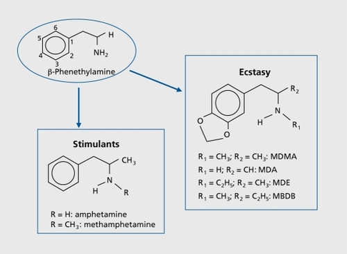
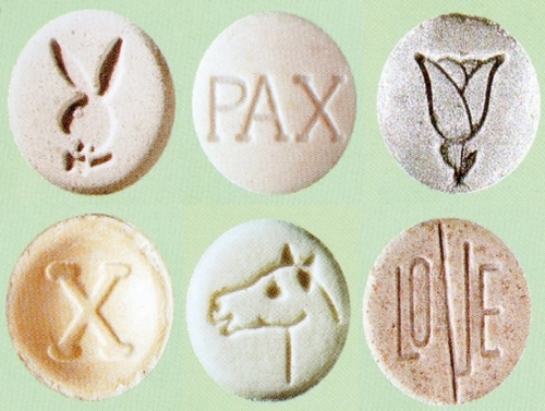
Both ecstasy and amphetamines are easy to manufacture in underground laboratories. Ecstasy is almost always sold as tablets or pills with various imprinted logos . The pills typically contain 70 to 120 mg of MDMA, although the concentration may sometimes be higher or lower. Occasionally ecstasy tablets will contain similarly acting analogues (3,4-methylenedioxy-Nethylamphetamine [MDE], 3,4-methylenedioxyamphetamine [MDA], or 3,4-methylenedioxy-alpha-ethylN-methylphenethylamine [MBDB], ) or amphetamines, and more rarely they may also contain substances from different classes. Amphetamines are mostly sold as powder which can be inhaled, smoked, ingested, or injected, although intranasal use (“snorting”) is now particularly common.
The acute pharmacology of MDMA and amphetamines has been widely studied.“ Among other actions, both drug groups bind to presynaptic monoamine transporters, and act as inhibitors on these sites and releasers of the endogenous monoamines from presynaptic terminals. The main mechanism of amphetamines is the enhanced release of dopamine (DA), particularly in the striatal system, and norepinephrine (NE). MDMA binds most strongly to the serotonin (5-HT) transporter (SERT) and induces rapid and powerful release of both 5-HT and DA.
Depending on the dose and route of administration, effects of stimulants may last from 3 to about 8 hours. They include increased drive, hypervigilance, pressure of ideas and speech, euphoria, and expansive behavior, but sometimes dysphoric mood, agitation, and aggression may occur. The psychological effects of MDMA last about 3 to 5 hours, and are more complex: they include relaxation, feelings of happiness, empathy, and closeness to other people, along with stimulant-like effects, alterations of perception, and other mild hallucinogenic effects.Citation7
The addictive potential of amphetamines is generally lower than that of cocaine or heroin, but it becomes high when the drugs are used intravenously. MDMA is considerably less addictive, and is mostly used as a recreational drug during weekends; however, a minority of about 15% to 20% of users develop a more frequent or compulsive use pattern, and they may ingest 10 or even more pills per occasion.Citation8 Beside the issue of addiction, there is a range of acute and subacute complications including drug-induced psychoses, seizures, myocardial infarction, or stroke resulting from hypertension and/or hemorrhage, hyperpyrexia with rhabdomyolysis, disseminated intravascular coagulation (DIG) and organ failure, toxic hepatitis, and many others. Moreover, amphetamines and MDMA have been shown to be neurotoxic in animal studies, particularly when given at high and repeated doses. This neurotoxic potential of the drugs may be relevant for humans. In the following sections we review the evidence for neurotoxicity in animal studies and in human populations.
Animal studies
Brain morphology and neurochemistry
Several studies in different laboratories and with different species demonstrate long-term alterations in brain 5-HT systems following high and repeated doses of MDMA. In studies with primates, even single doses of MDMA were found to elicit some degree of serotonergic depletion lasting over a few weeks;Citation4 However, the lowest MDMA dose which was shown to produce longterm neurotoxic effects that persisted over months and years has been 5 mg/kg given parenterally twice dailyover 4 days, ie, 40 mg/kg overall in 4 days.Citation9-Citation11 The alterations include depletion of 5-HT and its major metabolite 5-hydroxyindoleacetic acid (5-HIAA), reduced [3H]paroxetine binding, reflecting reduced density of SERT, and reduced serotonergic axonal density in several brain regions.Citation6-Citation12 All but one species tested so far, including nonhuman primates, have confirmed the pattern of selective neurotoxicity for serotonergic axons, with the sole exception of mice, which exhibit neurotoxic alterations of serotonergic and dopaminergic axons.Citation4 The rate of recovery was shown to be region-dependent. This probably corresponds to the very different distances that must be covered in the process of reinnervation. Axons need to be regrown from their origin in the serotonergic cell bodies in the raphe nuclei of the brain stem to the different terminal areas of the brain. In rats, full recovery was shown in most studies and most brain regions after 1 year, but some individual studies reported only partial recovery in the hippocampus and some cortical areas and hyperinnervation in the hypothalamus. In nonhuman primates, sensitivity to the neurotoxic effects of MDMA was shown to be more pronounced than in rodents, resulting in higher rates of 5-HT depletion with smaller doses of MDMA and persisting hypoinnervation patterns in most neocortical regions and the hippocampus in the range of 20% to 40% lower SERT binding depending on the brain region examined) for as long as 7 years post-treatment.Citation9-Citation11
Similarly to MDMA, stimulant amphetamines, particularly METH, have also been shown to be neurotoxic in rodent and nonhuman primate studies.Citation6,Citation13 Typical neurotoxic METH regimens are 5 to 10 mg/kg given parenterally 4 to 10 times within 1 to 4 days. Stimulant-related neurotoxicity is not restricted to the serotonergic system. High and/or repeated doses of METH induce widespread degeneration of presynaptic serotonergic axon terminals and degeneration of dopaminergic terminals, which is most prominent in the striatum. The consequences are 5-HT and dopamine (DA) depletion, and lower 5-HT and DA transporter densities (SERT and DAT) in brain tissue, with the effects being more pronounced on the striatal DA system.Citation6,Citation13-Citation15 A recent study in vervet monkeys used an escalating-dose METH exposure which models a common human abuse pattern, and demonstrated persistent changes in the presynaptic striatal DA system 3 weeks after abstinence (20% lower striatal DA content, 35% lower DAT binding).Citation16 However, METH toxicity to DA and 5-HT terminals had been previously shown to be considerably more long-lasting, and can persist for up to 4 years after drug administration in nonhuman primates.Citation17 The mechanism of neurotoxicity resulting from amphetamines and MDMA is not entirely understood. However, data from animal studies strongly suggest that the formation of free radicals is a key factor, that hyperthermia enhances the formation of free radicals, and that both hyperthermia and high ambient temperatures enhance the neurotoxic effects of the drugs.Citation4-Citation6
Functional consequences of neurotoxic drug regimens
Generally, the long-term functional abnormalities seen in laboratory animals after neurotoxic MDMA regimens have been only subtle.Citation18-Citation20 This may correspond to a complex role of the neuromodulator 5-HT in “fine tuning” and stabilizing neural transmission in cerebral networks.Citation21-Citation22 Broadly speaking, 5-HT appears to play important roles in several functional systems such as cognition, stimulus processing, psychological well-being, sleep control, and vegetative and neuroendocrine functions, without it being critical for the essential functioning of any of these domains. Nevertheless, some studies which used specialized behavioral test methods and pharmacological challenges reported subtle functional disturbances such as increased anxiety and poor memory performance in MDMA-treated rodents and monkeys.Citation19-Citation20,Citation23-Citation30 However, other studies reported normal or back-to-normal performance within 2 to 3 weeks following MDMA treatment,Citation31-Citation33 and studies which used behavioral tests for the assessment of anxiety and risktaking behavior yielded conflicting results.Citation27-Citation28,Citation34-Citation35 These data strongly suggest that if ecstasy users are indeed suffering neurotoxic damage to their serotonergic system, the functional consequences may be subtle.
Similarly, neurotoxic METH regimens which are sufficient to produce neurotoxicity were shown to induce only moderate, if any, alterations in behaviour of laboratory animals. These moderate effects may be best explained by the fact that METH-induced degeneration of DA and 5-HT axon terminals is incomplete and that long-term reductions in monoamine concentration levels and transporter densities are in the range of 20% to 45%.Citation16,Citation36 Indeed, higher reductions in the range of 80% to 95% may be required to produce gross abnormalities such as parkinsonian-like motor deficits. Accordingly, reduction of spontaneous locomotor activity was reported only 3 days after a neurotoxic METH regimen, but not after 1, 2, and 4 weeks in rodents.Citation33 However, using more subtle motor tests, persisting deficits in active avoidance performance (24% increase in response latency) and balance beam performance (2-to 3 -fold increase in footfalls) were demonstrated.Citation36 In mice, an impairment of consolidation of learned place preference was reported after neurotoxic METH doses.Citation37 Rats treated with a neurotoxic regimen of METH were impaired on a radial maze sequential learning task when tested after 3 weeks,Citation38 and on a novelty preference object recognition (OR) task when tested after 1 week and 4 weeks.Citation39-Citation40 Interestingly, a recent study reported than an escalating dose regimen which appears to mimic a common human pattern of escalating drug intake attenuates the neurotoxic effects and the OR deficits after METH treatment.Citation41 Similarly, in nonhuman primates progressive increases in METH doses in an escalating dose regimen induced abnormal behavior and decreases in social behavior on “injection” days with aggression decreasing throughout the study; however, after 3 weeks of abstinence no differences in baseline vs post-METH behaviors were observed.Citation16 These recent studies suggest that many METH users may not present with functional abnormalities despite residual dopaminergic toxicity; however, the extent of toxic damage and functional sequelae may well be more severe in heavy users with binge use behavior.
Are the animal data relevant for humans?
The key question is whether illicit drug users may suffer similar neurotoxic brain lesions as experimental animals. Over the last 10 to 15 years this question has received particular attention for MDMA,Citation42 while studies with amphetamine users very been relatively scarce. Two reasons may account for the relatively lower interest in amphetaminerelated neurotoxicity in humans: first, the neurotoxic doses in experimental animals are much higher than the typical human recreational doses of 20 to 40 mg of AMPPI or METH, and second, amphetamines have been used therapeutically for the treatment of attention deficit-hyperactivity disorder (ADHD) and narcolepsy for decades without clear evidence of long-term adverse effects.Citation43 Hence, the interest in possible long-term sequelae of neurotoxic drug use has focused highly on MDMA.
Compared with a neurotoxic MDMA regimen in primates (5 mg/kg twice daily over 4 days sc or ip the typical dose of a recreational MDMA weekend user (1 to 2 pills of 75 to 125 mg MDMA or analogue every 1 to 4 weeks) is still considerably lower.Citation44 However, according to some formulae for interspecies scaling, the recreational MDMA doses might well approach doses commonly given to animals in experimental studies.Citation4 Moreover, some heavy users take MDMA more frequently than just at weekends; they ingest up to 10 or even more pills in one night and they typically use MDMA over years, which may increase the risk for long-term cumulative neurotoxic effects. Although these heavy users are a minority, given the widespread use of MDMA, their absolute number is large. Interestingly, a study which looked at the effects of self-administration of MDMA in primates over a period as long as 18 months showed 5-HT depletions in the order of 25% to 50% lower (5-HT concentration depending on the region examined, in various cortical and subcortical regions.Citation45 These decrements in 5-HT content did not reach statistical significance, possibly due to the small sample in this study (n=3). Nevertheless, if the results are confirmed by further studies, they are clearly alarming.Citation45 Furthermore, the widespread parallel use of different neurotoxic substances such as MDMA, METH, and alcohol may act synergistically and enhance the neurotoxic effects of the single drugs. Finally, neurotoxicity may be enhanced by the typical conditions associated with MDMA and METH use such as hot, overcrowded surroundings and long periods of dancing, leading to further increases in body temperature.Citation46 In conclusion, it is possible that the animal data demonstrating MDMA and METH-induced neurotoxicity are indeed relevant for humans, and that club drug users may be exposing themselves to the risk of neurotoxic brain damage.
Studies in ecstasy users
Brain morphology and global brain function
In principle, it is rather unlikely that neurotoxic damage confined to the serotonergic system will be visible in routine brain imaging procedures in terms of loss of brain volume or atrophy, or that it will manifest itself as an alteration of global cerebral activity in positron emission tomography and single-photon emission tomography (PET and SPECT). However, serotonin is more than a neurotransmitter or neuromodulator in neuronal tissues; it also exerts powerful vasoconstrictive actions on small brain vessels,Citation47 has neurotrophic effects on brain tissue not confined to the period of brain maturation,Citation48 and has been shown to stimulate neurogenesis in the hippocampus throughout adulthood.Citation49
Routine structural magnetic resonance imaging (MRI), perfusion and diffusion MRI, SPECT with 133Xe, and 99mTc-hexamethylpropylene amine oxime (HMPAO) and H2150 PET were generally found to be normal in ecstasy users.Citation50-Citation53 However, one study reported an association between longer periods of MDMA use and decreased global brain volume,Citation50 and another studyCitation54 demonstrated reduced grey matter density in several cortical regions. In addition, studies with MR spectroscopy reported higher concentration of the glia marker myoinositole with heavier use of MDMA,Citation55 dose-dependent reductions of N-acetylaspartate (NAA) levels (NAAxreatine and NAAxholine ratios) in the frontal cortex of MDMA users,Citation56 and a tendency towards lower NAAxreatine ratios in the hippocampus of MDMA users compared with controls.Citation57 These findings could be related to neurotoxic damage and glial proliferation, indicating a repair mechanism. In addition, another small pilot studyreported a high diffusion coefficient (ADC) and high regional cerebral blood volume (rCBV) in the globus pallidus, a brain area that is particularly rich in 5-HT. This finding could be related to vasodilatation due to low serotonergic tone following degeneration of serotonergic axons.Citation52 Recently, a large study with 71 ecstasy polydrug users reported alterations in the thalamus associated specifically with MDMA use: decreased fractional anisotropy (FA) in diffusion tensor imaging (DTI) was suggestive of axonal loss; whereas increased regional cerebral blood volume (rCBV) in perfusion weighted imaging (PWI) may have been caused by 5-1 IT depletion.Citation58 In the same study no effects of ecstasy use on apparent diffusion coefficients and brain metabolites (MR spectroscopy) were detected.
Finally, an ambitious and methodologically sound prospective study examined a large number of young subjects who socialized in the drug scene, but had not yet used amphetamines or ecstasy (The Netherlands XTC Toxicity [NeXT] study).Citation59 After a mean period of 17 months' follow-up, neuroimaging was repeated in 59 incident ecstasy users and 56 matched persistent ecstasy-naives using multiple NMR techniques and SPECT for measurement of SERT availability. Although the novice MDMA users reported only very sporadic and low-dose use of MDMA in the follow-up period (mean 6.0, median 2.0 tablets), the MRI examinations showed decreases in rCBV in the globus pallidus and putamen (PWI), decreases in FA (indicator of axonal integrity) in the thalamus and frontoparietal white matter (DTI) and increases of FA in globus pallidus, and increase of apparent diffusion coefficient in the thalamus. Although relatively subtle, these findings are alarming, because they are in line with sustained effects of ecstasy on brain micro vasculature, white matter maturation, and possibly axonal damage, even after very low dosages of ecstasy.Citation59
Central serotonergic parameters
Reduced 5-HT concentration would be the expected outcome of widespread neurotoxic damage of serotonergic axon terminals in the brain tissue of MDMA users. As the 5-HT concentration cannot be measured in vivo in human brains, we may use the concentration of both 5-HT and its main metabolite, 5-HIAA, in cerebrospinal fluid (CSF) as a proxy for the concentration in the brain. An early study on a small number of ecstasy users reported normal levels of 5-HIAA in the CSF.Citation60 Since then several studies with larger samples showed reduced concentrations of 5-HIAA in cerebrospinal fluid of ecstasy users compared with control groups.Citation61-Citation64 However, only one study reported a correlation between the 5HIAA concentration and the extent of earlier ecstasy use.Citation62
PET and SPECT using suitable ligands make the in-vivo examination of brain tissue receptors and/ or binding sites feasible. An early PET study in 14 ecstasy users and the SERT ligand [11C] (+)McN5652 demonstrated a dose-dependent reduction in its binding, both globally and in most cortical and subcortical brain regions examined.Citation65 A further study in 10 ecstasy users also demonstrated reduced cortical SERT availability using SPECT and the SERT ligand β-CITCitation66 However, correlations between the SERT availability results, cumulative ecstasy consumption, and length of abstinence periods suggested a temporary occupation or downregulation of the binding site rather than structural neurotoxic damage.Citation66 Since then there has been some debate on the validity of SPECT and PET techniques with SERT ligands in measuring MDMA-related neurotoxicity and on additional subject-related methodological problems of these early studies. Nevertheless, all but one more recent studies with refined methodsCitation67 and larger samples (up to 61 current and former usersCitation68) confirmed reduced SERT availability at least in female current users with a relatively heavy use pattern (>50 pills),Citation58-Citation68-Citation72 and only one small study with 12 former MDMA users was negative.Citation73 All in all, alterations were less pronounced in male users, and were absent in former users following abstinence from MDMA use of at least 12 months. A small longitudinal study with two follow-up (+)McN5652-PET examinations confirmed the reversibility of alterations of SERT availability with a decrease in the intensity of MDMA consumption.Citation74 In summary, these studies indicate that women may be more susceptible to MDMAinduced alterations of the serotonergic system than men, and, in addition, they suggest at least some degree of recovery of the assumed serotonergic lesion following abstinence.
Interestingly, another SPECT study with the 5-HT2A receptor ligand [123I]-R91150 demonstrated reduced cortical binding in current ecstasy users with short-term abstinence and increased binding in former users who had not used ecstasy for an average of 5 months.Citation75 This pattern is in line with animal data showing temporary (up to 1 month) downregulation of postsynaptic 5-HT2 receptors resulting from high synaptic 5-HT concentration after administration of MDMA, and long-lasting upregulation of the same postsynaptic receptors following widespread presynaptic damage of serotonergic neurons leading to 5-HT depletion.Citation76-Citation77 Hence, unlike the SERT data, postsynaptic receptor data suggest that alterations of serotonergic systems may persist over long periods of time in abstinent MDMA users. Such subtle residual changes could be functionally important, and might contribute to clinical or subclinical alterations of psychological well-being and behavior of ecstasy users.
Serotonin-related functions
The neuromodulator 5-1 IT is involved in several functional systems of the CNS. Consequently, damage to the central serotonergic system could be theoretically followed by disturbances in different fields such as psychological well-being, neuroendocrine secretion, vegetative functions, processing of sensory stimuli, sleep architecture, and cognition. In the last 10 to 12 years there have been numerous studies demonstrating group differences between ecstasy users and controls in virtually all these fields, and differences favor the control groups in almost every study.Citation44 However, results have been inconsistent and several methodological problems (eg, pre-existing differences, polydrug use, differences in lifestyle) make it difficult and sometimes even impossible to draw firm conclusions from the data. The majority of studies report on psychopathology and cognition. Hence, in the following sections we will focus on these subjects.
Psychopathology
A low serotonergic tone has been widely associated with psychological disturbances, particularly with depression, suicidality, aggressiveness, and impulsiveness. There are several anecdotal reports of depressive syndromes, anxiety, and psychotic episodes associated with ecstasy use,Citation78 and high psychiatric comorbidity was established in studies with large samples of ecstasy-experienced polydrug users.Citation79-Citation80 A causal link between these disorders and ecstasy may exist at least in a predisposed subgroup of users. However, due to the widespread use of ecstasy and the parallel use of other substances no firm conclusion can be drawn from these reports. Moreover, results from a prospective-longitudinal investigation on a large representative sample of adolescents and young adults (n=2462) over 4 years confirmed a high psychiatric comorbidity in MDMA users, but demonstrated that the use of ecstasy started, in most cases, after the onset of the comorbid disorder.Citation81
Several studies used standardized psychometric instruments and demonstrated higher scores for impulsiveness, depressive mood, emotional instability, anxiety, noveltyseeking, hostility/ aggression, and an overall heightened level of psychological distress in mostly polydrug ecstasy users compared with control groups.Citation44 However, results have not been entirely consistent; for example, one studyreported reduced impulsiveness and aggression compared with the control group.Citation63 Two studies suggested a link between high scores and heavy parallel cannabis use.Citation82-Citation83 Moreover, in a recent study with a longitudinal design and a follow-up period of 18 months increases in self-rated psychopathology were associated with continued cannabis rather than continued ecstasy use.Citation84 Finally, in recent studies with relatively large samples of 234, 61, and 50 polydrug ecstasy users and controls using other drugs only, elevated psychopathology appeared to be associated with polydrug use in general and not specifically with ecstasy use.Citation68,Citation85-Citation86
All in all, it is still unclear whether the frequently reported emotional instability and impulsive features and/ or the overall high level of psychological distress result from ecstasy use or from the combined use of several substances, or whether alternatively these are factors predisposing to a general affinity to drugs. Interestingly, a recent combined SPECT and psychometric studyestablished decreased SERT availability only in current MDMA users, but elevated depression scores in current and former users:Citation87 In this study, higher depression scores were associated with higher lifetime MDMA dose, but there was no association of psychometric scores with SERT availability.Citation87 Finally, another study suggests an interaction between genetic factors and the effects of MDMA use on mood (high depression scores only in ecstasy users carrying the s allele of the SERT encoding gene but not in users with the 11 genotype).Citation88 These findings underline the complexity of the issue and are in line with animal data showing different long-term effects of MDMA on anxiety in rats depending on the level of their baseline anxiety, and only a loose association between the neurotoxic effects of MDMA and its long-term impact on anxiety-related behavior.Citation4,Citation27-Citation28,Citation89 In conclusion, the linkage between ecstasy-induced neurotoxicity and psychological problems does not seem to have been established at this stage.
Cognition
Although our understanding of the role of serotonin in cognitive processes is incomplete, there are indications that serotonergic neurotransmission may particularly interfere with an individual's cognitive style (impulsive vs systematic) as well as with memory and learning processes.Citation90-Citation91 Indeed, relative deficits of short-term or working memory, episodic memory and learning, as well as increased cognitive impulsivity and diminished executive control, were frequently reported in ecstasy users.Citation44,Citation92
To date, the most consistent finding is that of subtle deficits in episodic memory and learning abilities. Numerous cross-sectional studies demonstrated relative impairments of learning and memory performance and only a small minority of studies reported no differences between ecstasy users and controls or small and insignificant differences after adjusting for possible confounders.Citation44,Citation92-Citation93 In general, poor memory was associated with a heavier pattern of ecstasy use, although a minority of studies reported an association of memory deficits with the extent of the parallel use of cannabis or the combination of ecstasy and cannabis, rather than the use of ecstasy alone.Citation44 Elevated cognitive impulsivity and diminished executive control were also demonstrated in some studies; however, these results have been less consistent.Citation44,Citation94-Citation96 Although several studies and particularly the earlier studies suffered from significant methodological limitations such as polydrug use, short abstinence periods, poorly matched control groups, and lack of lexicological analyses for verification of the subjects' reports, a number of more recent investigations were carefully designed and conducted, and their results have been similar.Citation44-Citation92
The consistency of the data on memory functions and the association of performance with the extent of previous ecstasy use are highly suggestive of a residual neurotoxic effect of MDMA. It is possible that the hippocampus may be particularly vulnerable to the neurotoxic effects of MDMA, and this may explain why residual effects are most consistent in the memory domain.Citation97-Citation98 This interpretation is in line with the animal experimental data, which demonstrated particularly strong and long-lasting neurotoxic effects of MDMA in the hippocampus,Citation11 and a stimulatory role of 5-HT for neurogenesis in the hippocampus.Citation49
Interestingly, three studies in current and former MDMA users with an abstinence period of several months or even years reported similar or even poorer memory performance in the former MDMA users,Citation68,Citation70,Citation99 although SERT availability was only reduced in current users.Citation68-Citation70 Two longitudinal studies yielded conflicting results: a small study in 15 ecstasy users reported memory decline after continued use and improvement after abstinence over 36 months,Citation100-Citation101 but a larger study in 38 ecstasy users reported no further deterioration of memory performance after continued use and no improvement after abstinence over 18 months.Citation102 Although these results may be interpreted as evidence against neurotoxicity-related memory decline, it is still possible that memory deficits in ecstasy users persist even after 18 months of abstinence because, as shown in primate studies,Citation11 regeneration of serotonergic axons may take a long time and may remain incomplete. In addition, the functional consequences of neurotoxic lesions observed following a threshold use of ecstasy may manifest themselves in binary (yes/no) manner. Compensatory neural mechanisms that might develop could possibly explain the absence of functional deterioration despite subsequent “enlargement” of the neurotoxic lesions. This view would be in line both with findings of a dose-dependent memory deficit in cross-sectional studies comparing ecstasy users with control samples, and with the finding of stable performance in the larger within-subject longitudinal study.Citation102
Finally, findings from the only prospective study to date do support this view (part of the Netherlands XTC Toxicity [NeXT] study). A large number of young subjects who socialized in the drug scene, but had not yet used amphetamines or ecstasy, were followed up and reexamined after a mean period of 3 years' follow-up.Citation103 Although the 58 novice MDMA users reported only very sporadic and low-dose use of MDMA in the followup period (mean 3.2, median 1.5 tablets) they failed to demonstrate retest improvements in verbal memory shown by the persistent MDMA-naive group of 60 subjects.Citation103 This finding suggests that even very low MDMA doses may be associated with persisting alterations in memory and learning functions. Although the clinical relevance of this subtle finding is clearly limited, longterm negative consequences are conceivable. In conclusion, the linkage between ecstasy use and memorydecline is considered probable at this stage.
Studies in amphetamine users
Compared with MDMA, the literature on amphetamine related neurotoxicity in humans is limited, but the number of publications has been constantly increasing over the last few years. Initial small studies with PET (regional glucose metabolic rate (rMRGlu), DAT, and D2 receptor availability), SPECT (DAT availability) and MR spectroscopy techniques suggested that heavy use of stimulants may also be neurotoxic in humans and that alterations may persist over prolonged periods of time.Citation104-Citation109
Reduced levels of striatal DAT were found in former METH users even 3 years or more after last use,Citation104 and they were found to be associated with a longer duration of speed use.Citation106 In a preliminary longitudinal study in five former speed users, rMRGlu was assessed after 6 months and again after 12 to 17 months of abstinence. During this follow-up period the initially reduced MRGlu rose in the thalamus, but remained low in the striatum, caudate, and nucleus accumbens.Citation109
Two recent larger MR spectroscopy studies with 24Citation110 and 36Citation111 currently abstinent METH users reported lowlevels of the neuronal marker NAA (NAA/creatine ratio) in the anterior cingulate even after very long periods of abstinence of several years.Citation110 In contrast, the choline/NAA values were abnormally high in the users with relative short abstinence time, but they normalized after 1 year of abstinence. This finding suggests that following cessation of METH use, adaptive changes occur, which may contribute to some degree of normalization of neuronal structure and function.Citation110 A structural MRI study in 22 METH users and 21 controls revealed smaller hippocampal volumes and significant white-matter hypertrophy in the METII group.Citation112 Finally, the largest cross-sectional study so far demonstrated enlarged putamen and globus pallid us in 50 METH users compared with 50 controls.Citation113 Interestingly, within the METII group larger basal ganglia volumes were associated with better cognitive performance and less cumulative METH usage. Therefore, the authors argued that the enlarged putamen and globus pallidus might represent a compensatory response to maintain function.Citation113 A recent review of the literature reported enlarged striatal volumes, reduced concentrations of the neuronal marker NAA-acetylasparate and total creatine in the basal ganglia, reduced DAT density, and reduced dopamine D2 receptors in the striatum, lower levels of SERT density and vesicular monoamine transporters (VMAT2) across striatal subregions, and altered brain glucose metabolism in the limbic and orbitofrontal regions of METH users.Citation114
Theoretically, neurotoxic dopaminergic lesions could be associated with motor, cognitive, and psychopathological abnormalities. To date, gross motor disturbances have not been demonstrated in METH users.Citation115 However, more subtle motor deficits were reported in two studies.Citation106,Citation109 The literature on long-term psycho(patho)logical sequelae of stimulant use is inconclusive. Similarly, cross-sectional studies in chronic stimulant users demonstrated relatively low performance in short-term and episodic memory, frontal executive control, and planning abilities.Citation111,Citation116-Citation122 However, it is not clear whether these deficits are a consequence of the use of stimulants or whether they reflect pre-existing low cognitive abilities in people who become drug users later in life.
Nevertheless, reduced DAT densities and longer duration of METH use were associated with poorer performance in both fine motor and memory tasks in 15 currently abstinent METH users.Citation106 Also, the normalization of rMRGlu in the thalamus was associated with an improvement of motor and memory performance after long-term abstinence of 1 year and more.Citation109 Finally, reduced attentional control (ie, increased Stroop interference) was shown to correlate with levels of NAA-Cr within the anterior cingulate in METH users, but not in controls.Citation111 In conclusion, the limited evidence to date suggests that persisting neurotoxic brain damage is conceivable in METH users, especially in heavy users with binge use patterns. More studies with longitudinal and prospective designs are clearly needed.
Conclusions
Ecstasy (MDMA) and stimulant amphetamines (METH and AMPPI) are popular drugs of abuse and they are neurotoxic in animal studies. High and repeated doses of MDMA cause selective and long-lasting degeneration of 5-HT axon terminals in several brain regions, whereas METH and AMPH damage both serotonergic and dopaminergic neurons. Although the doses taken recreationally are considerably lower than the doses typically given in animal studies, some users exhibit compulsive binge use behaviors that may well correspond to the animal doses. In addition, polydrug use and the typical environment of use (hot, overcrowded, and noisy rooms, extensive physical exercise in the form of dancing) may well potentiate the neurotoxic effects of the drugs.
Studies with drug users demonstrated associations of subtle alterations in brain structure and 5-HT brain parameters with MDMA use, Similarly, subtle cognitive dysfunctions, particularly in the memory and learning domain, were also found to be associated with ecstasy use, Although the results are not entirely consistent, these associations were replicated in many welldesigned, controlled studies including longitudinal and one prospective investigation. Moreover, the only prospective study to date demonstrated structural brain alterations and subtle memory dysfunction, even after minimal exposure to MDMA.Citation59,Citation103 Although most ecstasy users do not suffer cognitive impairment of clinically relevant proportions, and even heavy users initially appear mostly unimpaired in their everyday life, several cases with severe deficits have also been reported.Citation123-Citation124 Moreover, there is concern that the memory deficits of ecstasy users - although subtle and mostly subclinical - and the possible underlying hippocampal dysfunction might help accelerate the normal brain ageing process and constitute a risk factor for earlier onset and/or more severe age-related memory decline in later years.Citation44 Regarding METH-induced neurotoxicity, evidence from studies with drug users is relatively scarce and still preliminary. However, there are early indications that at least heavy METH use may also be followed by alterations in brain structure, dopaminergic parameters, and cognitive function.
In light of the popularity of ecstasy and stimulants among young people, questions around their neurotoxic effects on the brain remain highly topical. To date, the message we have to convey to young people in information campaigns is: “MDMA and amphetamine neurotoxicity for humans is not yet proven, but it is highly likely.” Further longitudinal and prospective studies are clearly needed.
Selected abbreviations and acronyms
| 5-HT | = | serotonin |
| 5-HIAA | = | 5-hydroxyindoleacetic acid |
| DA | = | dopamine |
| MDMA | = | methylenedioxymethamphetamine (ecstasy) |
| METH | = | methamphetamine |
| SERT | = | serotonin transporter |
REFERENCES
- KrausL.AugustinR.OrthBIllégale Drogen, Einstiegsalter und Trends. Ergebnisse des Epidemiologischen Suchtsurvey 2003.Sucht.200551,S11928
- TossmannP.BoldtS.TensilMD.The use of drugs within the techno party scene in European metropolitan cities.Eur Addict Res.2001722311316921
- StroteJ.LeeJE.WechslerH.Increasing MDMA use among college students: results of a national survey.J Adolesc Health.200230647211755802
- GreenAR.MechanAO.ElliottJM.et al.The pharmacology and clinical pharmacology of 3,4-methylenedioxymethamphetamine (MDMA, “ecstasy”).Pharmacol Rev.20035546350812869661
- YacoubianGS.JrBoyleC.HardingCA.et al.It's a rave new world: estimating the prevalence and perceived harm of ecstasy and other drug use among club rave attendees.J Drug Educ.20033318719612929709
- QuintonMS.YamamotoBK.Causes and consequences of methamphetamine and MDMA toxicity.AAPS J.20068E337E34716796384
- GouzoulisE.BorchardtD.Hermle LA case of toxic psychosis induced by 'eve' (3,4-methylenedioxyethylamphetamine).Arch Gen Psychiatry.199350758093659
- ParrottAC.Chronic tolerance to recreational MDMA (3,4-methylenedioxymethamphetamine) or Ecstasy.J Psychopharmacol.200519718315671132
- RicaurteGA.MartelloAL.KatzJL.et al.Lasting effects of (+-)-3,4methylenedioxymeth-iamphetamine (MDMA) on central serotonergic neurons in nonhuman primates: neurochemical observations.J Pharmacol Exp Ther.19922616166221374470
- FischerC.HatzidimitriouG.WlosJ.et al.Reorganization of ascending 5-HT axon projections in animals previously exposed to the recreational drug (+/-)3,4-methylenedioxymethamphetamine (MDMA, “ecstasy”).J Neurosci.19951 5547654857643196
- HatzidimitriouG.McCannUD.RicaurteGA.Altered serotonin innervation patterns in the forebrain of monkeys treated with (+/-)3,4methylenedioxymethamphetamine seven years previously: factors influencing abnormal recovery.J Neurosci.1999195096510710366642
- RicaurteGA.McCannUD.SzaboZ.et al.Toxicodynamics and long-term toxicity of the recreational drug, 3, 4-methylenedioxymethamphetamine (MDMA, 'Ecstasy').Toxicol Lett.2000112-11314314610720723
- SeidenLS.SabolKE.Methamphetamine and methylenedioxymethamphetamine neurotoxicity: possible mechanisms of cell destruction.MDA Res Monogr.1996163251276
- McCannUD.RicaurteGA.Amphetamine neurotoxicity: accomplishments and remaining challenges.Neurosci Biobehav Rev.20042782182615019431
- HansonGR.RauKS.FleckensteinAE.The methamphetamine experience: a NIDA partnership.Neuropharmacology.200447 (suppl 1)9210015464128
- MelegaWP.JorgensenMJ.LacanG.et al.Long-term methamphetamine administration in thevervet monkey models aspects of a human exposure: brain neurotoxicity and behavioral profiles.Neuropsychopharmacology.2008331441145217625500
- WoolvertonWL.RicaurteGA.FornoLS.et al.Long-term effects of chronic methamphetamine administration in rhesus monkeys.Brain Res.198948673782720435
- SlikkerW.Jr.HolsonRR.AliSF.et al.Behavioral and neurochemical effects of orally administered MDMA in the rodent and nonhuman primate.Neurotoxicology.1989 105295422576304
- FrederickDL.PauleMG.Effects of MDMA on complex brain function in laboratory animals.Neurosci Biobehav Rev.19972167788994210
- TaffeMA.DavisSA.YuanJ.et al.Cognitive performance of MDMAtreated rhesus monkeys: sensitivity to serotonergic challenge.Neuropsychopharmacology.200227993100512464456
- LuckiI.The spectrum of behaviors influenced by serotonin.Biol Psychiatry.1998441511629693387
- WeigerWA.Serotonergic modulation of behaviour: a phylogenetic overview.Biol Rev Camb Philos Soc.19977261959116165
- MarstonHM.ReidME.LawrenceJA.et al.Behavioural analysis of the acute and chronic effects of MDMA treatment in the rat.Psychopharmacology.1999144677610379626
- MorleyKC.GallateJE.HuntGE.et al.Increased anxiety and impaired memory in rats 3 months after administration of 3,4-methylenedioxymethamphetamine (“ecstasy”).Eur J Pharmacol.2001433919911755138
- BroeningHW.MorfordLL.Inman-WoodSL.et al.3,4-methylenedioxymethamphetamine (ecstasy )-induced learning and memory impairments depend on the age of exposure during early development.J Neurosci.2001213228323511312307
- TaffeMA.Huitron-ResendizS.SchroederR.et al.MDMA exposure alters cognitive and electrophysiological sensitivity to rapid tryptophan depletion in rhesus monkeys.Pharmacol Biochem Behav.20037614115213679227
- McGregorIS.GurtmanCG.MorleyKC.et al.Increased anxiety and “depressive” symptoms months after MDMA (“ecstasy”) in rats: druginduced hyperthermia does not predict long-term outcomes.Psychopharmacology.200316846547412700882
- McGregorIS.ClemensKJ.Van der PGet al.Increased anxiety 3 months after brief exposure to MDMA (“Ecstasy”) in rats: association with altered 5-HT transporter and receptor density.Neuropsychopharmacology.2003281472148412700695
- SpragueJE.PrestonAS.LeifheitM.et al.Hippocampal serotonergic damage induced by MDMA (ecstasy): effects on spatial learning.Physiol Behav.20037928128712834800
- FariaR.MagalhaesA.MonteiroPR.et al.MDMA in adolescent male rats: decreased serotonin in the amygdala and behavioral effects in the elevated plus-maze test.Ann N Y Acad Sci.2006107464364917105959
- TaffeMA.WeedMR.DavisS.et al.Functional consequences of repeated (+/-)3,4-methylene->dioxymethamphetamine (MDMA) treatment in rhesus monkeys.Neuropsychopharmacology.20012423023911166514
- WinsauerPJ.McCannUD.YuanJ.et al.Effects of fenfluramine, m-CPP and triazolam on repeated-acquisition in squirrel monkeys before and after neurotoxic MDMA administration.Psychopharmacology.200215938839611823891
- TimarJ.GyarmatiS.SzaboA.et al.Behavioural changes in rats treated with a neurotoxic dose regimen of dextrorotatory amphetamine derivatives.Behav Pharmacol.20031419920612799521
- MechanAO.MoranPM.ElliottM.et al.A study of the effect of a single neurotoxic dose of 3,4-methylenedioxymethamphetamine (MDMA; “ecstasy”) on the subsequent long-term behaviour of rats in the plus maze and open field.Psychopharmacology.200215916717511862345
- GurtmanCG.MorleyKC.LiKM.et al.Increased anxiety in rats after 3,4methylenedioxy-imethamphetamine: association with serotonin depletion.Eur J Pharmacol.2002446899612098589
- WalshSL.WagnerGC.Motor impairments after methamphetamineinduced neurotoxicity in the rat.J Pharmacol Exp Ther.19922636176261432692
- Chat-MendesC.AndersonKL.ItzhakY.Impairment in consolidation of learned place preference following dopaminergic neurotoxicity in mice is ameliorated by N-acetylcysteine but not D1 and D2 dopamine receptor agonists.Neuropsychopharmacology.20073253154116760923
- ChapmanDE.HansonGR.KesnerRP.et al.Long-term changes in basal ganglia function after a neurotoxic regimen of methamphetamine.J Pharmacol Exp Ther.200129652052711160639
- BelcherAM.O'DellSJ.MarshallJF.Impaired object recognition memory following methamphetamine, but not p-chloroamphetamine- or d-amphetamine-induced neurotoxicity.Neuropsychopharmacology.2005302026203415900317
- HeJ.YangY.YuY.et al.The effects of chronic administration of quetiapine on the methamphe-itamine-induced recognition memory impairment and dopaminergic terminal deficit in rats.Behav Brain Res.2006172394516712969
- BelcherAM.FeinsteinEM.O'DellSJ.et al.Methamphetamine influences on recognition memory: comparison of escalating and single-day dosing regimens.Neuropsychopharmacology.2008331453146317637607
- De LaGR.FabrizioKR.GuptaA.Relevance of rodent models of intravenous MDMA self-administration to human MDMA consumption patterns.Psychopharmacology.200718942543416470404
- BermanSM.KuczenskiR.McCrackenJT.et al.Potential adverse effects of amphetamine treatment on brain and behavior: a review.Mol Psychiatry.20091412314218698321
- Gouzoulis-MayfrankE.DaumannJ.Neurotoxicity of méthylènedioxyamphétamines (MDMA; ecstasy) in humans: how strong is the evidence for persistent brain damage?.Addiction.200610134836116499508
- FantegrossiWE.WoolvertonWL.KilbournM.et al.Behavioral and neurochemical consequences of long-term intravenous self-administration of MDMA and its enantiomers by rhesus monkeys.Neuropsychopharmacology.2004291270128115039771
- ColadoMl.GranadosR.O'SheaE.et al.Role of hyperthermia in the protective action of clomethiazole against MDMA ('ecstasy')-induced neurodegeneration, comparison with the novel NMDA channel blocker ARR15896AR.Br J Pharmacol.19981244794849647471
- CohenZ.BonventoG.LacombeP.et al.Serotonin in the regulation of brain microcirculation.Prog Neurobiol.1996503353629004349
- AzmitiaEC.Serotonin neurons, neuroplasticity, and homeostasis of neural tissue.Neuropsychopharmacology.19992133S45S10432487
- GouldE.Serotonin and hippocampal neurogenesis.Neuropsychopharmacology.19992146S51S10432488
- ChangL.GrobCS.ErnstT.et al.Effect of ecstasy [3,4-methylenedioxymethamphetamine (MDMA)] on cerebral blood flow: a co-registered SPECT and MRI study.Psychiatry Res.200098152810708923
- GammaA.BuckA.BertholdT.et al.No difference in brain activation during cognitive performance between ecstasy (3,4-methylenedioxymethamphetamine) users and control subjects: a [H2(15)0]-positron emission tomography study.J Clin Psychopharmacol.200121667111199950
- RenemanL.MajoieCB.HabrakenJB.et al.Effects of ecstasy (MDMA) on the brain in abstinent users: initial observations with diffusion and perfusion MR imaging.Radiology.200122061161711526257
- GammaA.BuckA.BertholdVollenweiderFX.No difference in brain activation during cognitive performance between ecstasy (3,4-methylenedioxymethamphetatnine) users and control subjects: a [H2(15)0]-positron emission tomography study.J Clin Psychopharmacol.200121(1)667111199950
- CowanRL.LyooIK.SungSM.et al.Reduced cortical gray matter density in human MDMA (Ecstasy) users: a voxel-based morphometry study.Drug Alcohol Depend.20037222523514643939
- ChangL.ErnstT.GrobCS.et al.Cerebral (1)H MRS alterations in recreational 3, 4-methylenedioxymethamphetamine (MDMA, “ecstasy”) users.J Magn Reson Imaging.19991052152610508318
- RenemanL.MajoieCB.FlickH.et al.Reduced N-acetylaspartate levels in the frontal cortex of 3,4-methylenedioxymethamphetamine (Ecstasy) users: preliminary results.Am J Neuroradiol.20022323123711847047
- DaumannJ.FischermannT.PilatusU.et al.Proton magnetic resonance spectroscopy in ecstasy (MDMA) users.Neurosci Lett.200436211311615193766
- de WinMM.JagerG.BooijJ.et al.Neurotoxic effects of ecstasy on the thalamus.Br J Psychiatry.200819328929618827290
- de WinMM.JagerG.BooijJ.et al.Sustained effects of ecstasy on the human brain: a prospective neuroimaging study in novel users.Brain.20081312936294518842607
- PeroutkaSJ.PascoeN.FaullKF.Monoamine metabolites in the cerebrospinal fluid of recreational users of 3,4-methylenedimethoxymethamphetamine (MDMA; “ecstasy”).Res Comm Subst Abuse.1987812537
- RicaurteGA.FinneganKT.IrwinI.et al.Aminergic metabolites in cerebrospinal fluid of humans previously exposed to MDMA: preliminary observations.Ann N Y Acad Sci.19906006997081701292
- BollaKl.McCannUD.RicaurteGA.Memory impairment in abstinent MDM A (“ Ecstasy”) users.Neurology.199851153215379855498
- McCannUD.RidenourA.ShahamY.et al.Serotonin neurotoxicity after (+/-)3,4-methylenedioxymethamphetamine (MDMA; “Ecstasy”): a controlled study in humans.Neuropsychopharmacology.1994101291387517677
- McCannUD.MertlM.EligulashviliV.et al.Cognitive performance in (+/-) 3,4-methylenedioxymethamphetamine (MDMA, “ecstasy”) users: a controlled study.Psychopharmacology.199914341742510367560
- McCannUD.SzaboZ.ScheffelU.et al.Positron emission tomographic evidence of toxic effect of MDMA (“Ecstasy”) on brain serotonin neurons in human beings. .Lancet.1998352143314379807990
- SempleDM.EbmeierKP.GlabusMF.et al.Reduced in vivo binding to the serotonin transporter in the cerebral cortex of MDMA ('ecstasy') users.Br J Psychiatry.1999175636910621770
- McCannUD.SzaboZ.SeckinE.et al.Quantitative PET studies of the serotonin transporter in MDMA users and controls using [(1 1)C]McN5652 and [(11)C]DASB.Neuropsychopharmacology.2005301741175015841106
- ThomasiusR.PetersenK.BuchertR.et al.Mood, cognition and serotonin transporter availability in current and former ecstasy (MDMA) users.Psychopharmacology.2003167859612632248
- RenemanL.BooijJ.de BruinK.et al.Effects of dose, sex, and long-term abstention from use on toxic effects of MDMA (ecstasy) on brain serotonin neurons.Lancet.20013581864186911741626
- RenemanL.LavalayeJ.SchmandB.et al.Cortical serotonin transporter density and verbal memory in individuals who stopped using 3,4-methylenedioxymethamphetamine (MDMA or “ecstasy”): preliminary findings.Arch Gen Psychiatry.20015890190611576026
- BuchertR.ThomasiusR.NebelingB.et al.Long-term effects of “Ecstasy” use on serotonin transporters of the brain investigated by PET.J NuclMed.200344375384
- BuchertR.ThomasiusR.WilkeF.et al.A voxel-based PET investigation of the long-term effects of “Ecstasy” consumption on brain serotonin transporters.Am J Psychiatry.20041611181118915229049
- SelvarajS.HoshiR.BhagwagarZ.et al.Brain serotonin transporter binding in former users of MDMA ('ecstasy').Br J Psychiatry.200919435535919336788
- BuchertR.ThomasiusR.PetersenK.et al.Reversibility of ecstasyinduced reduction in serotonin transporter availability in polydrug ecstasy users.Eur J Nucl Med Mol Imaging.20063318819916133393
- RenemanL.EndertE.de BruinK.et al.The acute and chronic effects of MDMA (“ecstasy”) on cortical 5-HT2A receptors in rat and human brain.Neuropsychopharmacology.20022638739611850153
- ScheffelU.LeverJR.StathisM.et al.Repeated administration of MDMA causes transient down-regulation of serotonin 5-HT2 receptors.Neuropharmacology.1992318818931359444
- HegadorenKM.BakerGB.BourinM.3,4-Methylenedioxy analogues of amphetamine: defining the risks to humans.Neurosci Biobehav Rev19992353955310073892
- SchifanoF.Potential human neurotoxicity of MDMA ('Ecstasy'): subjective self-reports, evidence from an Italian drug addiction centre and clinical case studies.Neuropsychobiology.200042253310867553
- SchifanoF.Di FuriaL.ForzaG.et al.MDMA ('ecstasy') consumption in the context of polydrug abuse: a report on 150 patients.Drug Alcohol Depend.19985285909788011
- ToppL.HandoJ.DillonP.et al.Ecstasy use in Australia: patterns of use and associated harm.Drug Alcohol Depend.19995510511510402155
- LiebR.SchuetzC.PfisterH.et al.Mental disorders in ecstasy users: a prospective-longitudinal investigation.Drug Alcohol Depend.20026819512234649
- DaumannJ.PelzS.BeckerS.et al.Psychological profile of abstinent recreational ecstacy (MDMA) users and significance of concomitant cannabis use.Human Psychopharmacology.20011662763312404543
- MorganMJ.McFieL.FleetwoodH.et al.Ecstasy (MDMA): are the psychological problems associated with its use reversed by prolonged abstinence?.Psychopharmacology.200215929430311862362
- DaumannJ.HensenG.ThimmB.et al.Self-reported psychopathological symptoms in recreational ecstasy (MDMA) users are mainly associated with regular cannabis use: further evidence from a combined cross-sectional/longitudinal investigation.Psychopharmacology.200417339840414722704
- ParrottAC.MilaniRM.ParmarR.et al.Recreational ecstasy/MDMA and other drug users from the UK and Italy: psychiatric symptoms and psychobiological problems.Psychopharmacology.2001159778211797073
- RoiserJP.SahakianBJ.Relationship between ecstasy use and depression: a study controlling for poly-drug use.Psychopharmacology.200417341141714652710
- de WinMM.RenemanL.ReitsmaJB.et al.Mood disorders and serotonin transporter density in ecstasy users-the influence of long-term abstention, dose, and gender.Psychopharmacology.200417337638214726997
- RoiserJP.CookLJ.CooperJD.et al.Association of a functional polymorphism in the serotonin transporter gene with abnormal emotional processing in ecstasy users.Am J Psychiatry.200516260961215741482
- HoYJ.PawlakCR.GuoL.et al.Acute and long-term consequences of single MDMA administration in relation to individual anxiety levels in the rat.Behav Brain Res.200414913514415129777
- MenesesA.5-HT system and cognition.Neurosci Biobehav Rev.1999231111112510643820
- BuhotMC.MartinS.SeguL.Role of serotonin in memory impairment.Ann Med.20003221022110821328
- KachelsteinAD.De LaGR.MahoneyJJ.IIIet al.MDMA use and neurocognition: a meta-analytic review.Psychopharmacology.200718953153717082969
- BediG.RedmanJ.Ecstasy use and higher-level cognitive functions: weak effects of ecstasy after control for potential confounds.Psychol Med.2008381319133018226286
- Gouzoulis-MayfrankE.DaumannJ.TuchtenhagenF.et al.Impaired cognitive performance in drug free users of recreational ecstasy (MDMA).J Neurol Neurosurg Psychiatry.20006871972510811694
- MorganMJ.ImpallomeniLC.PironaA.et al.Elevated impulsivity and impaired decision-making in abstinent ecstasy (MDMA) users compared to polydrug and drug-naive controls.Neuropsychopharmacology.2006311562157316292322
- QuednowBB.KuhnKU.HoppeC.et al.Elevated impulsivity and impaired decision-making cognition in heavy users of MDMA (“Ecstasy”).Psychopharmacology.200718951753016425060
- FoxHC.McLeanA.TurnerJJ.et al.Neuropsychological evidence of a relatively selective profile of temporal dysfunction in drug-free MDMA (“ecstasy”) polydrug users.Psychopharmacology.200216220321412110998
- Gouzoulis-MayfrankE.ThimmB.RezkM.et al.Memory impairment suggests hippocampal dysfunction in abstinent ecstasy users.Prog Neuropsychopharmacol Biol Psychiatry.20032781982712921915
- CurranHV.VerheydenSL.Altered response to tryptophan supplementation after long-term abstention from MDMA (ecstasy) is highly correlated with human memory function.Psychopharmacology.20031699110312759801
- ZakzanisKK.YoungDA.Memory impairment in abstinent MDMA (“Ecstasy”) users: a longitudinal investigation.Neurology.20015696696911294938
- ZakzanisKK.CampbellZ.Memory impairment in now abstinent MDMA users and continued users: a longitudinal follow-up.Neurology.20066674074116534114
- Gouzoulis-MayfrankE.FischermannT.RezkM.et al.Memory performance in polyvalent MDMA (ecstasy) users who continue or discontinue MDMA use.Drug Alcohol Depend.20057831732315893163
- SchiltT.de WinMM.KoeterM.et al.Cognition in novice ecstasy users with minimal exposure to other drugs: a prospective cohort study.Arch Gen Psychiatry.20076472873617548754
- McCannUD.WongDF.YokoiF.et al.Reduced striatal dopamine transporter density in abstinent methamphetamine and methcathinone users: evidence from positron emission tomography studies with [1 IQWIN-35,428.J Neurosci.199818841784229763484
- ErnstT.ChangL.Leonido-YeeM.et al.Evidence for long-term neurotoxicity associated with methamphetamine abuse: a 1H MRS study.Neurology.2000541344134910746608
- VolkowND.ChangL.WangGJ.et al.Loss of dopamine transporters in methamphetamine abusers recovers with protracted abstinence.J Neurosci.2001219414941811717374
- VolkowND.ChangL.WangGJ.et al.Low level of brain dopamine D2 receptors in methamphetamine abusers: association with metabolism in the orbitofrontal cortex.Am J Psychiatry.20011582015202111729018
- RenemanL.BooijJ.LavalayeJ.et al.Use of amphetamine by recreational users of ecstasy (MDMA) is associated with reduced striatal dopamine transporter densities: a [123l]beta-CIT SPECT study-preliminary report.Psychopharmacology.200215933534011862367
- WangGJ.VolkowND.ChangL.et al.Partial recovery of brain metabolism in methamphetamine abusers after protracted abstinence.Am J Psychiatry.200416124224814754772
- NordahlTE.SaloR.NatsuakiY.et al.Methamphetamine users in sustained abstinence: a proton magnetic resonance spectroscopy study.Arch Gen Psychiatry.20056244445215809412
- SaloR.NordahlTE.NatsuakiY.et al.Attentional control and brain metabolite levels in methamphetamine abusers.Biol Psychiatry.2007611272128017097074
- ThompsonPM.HayashiKM.SimonSL.et al.Structural abnormalities in the brains of human subjects who use methamphetamine.J Neurosci.2004246028603615229250
- ChangL.CloakC.PattersonK.et al.Enlarged striatum in abstinent methamphetamine abusers: a possible compensatory response.Biol Psychiatry.20055796797415860336
- ChangL.AlicataD.ErnstT.et al.Structural and metabolic brain changes in the striatum associated with methamphetamine abuse.Addiction.2007102(suppl 1)163217493050
- CaligiuriMP.BuitenhuysC.Do preclinical findings of methamphetamine-induced motor abnormalities translate to an observable clinical phenotype?.Neuropsychopharmacology.2005302125213416123755
- OrnsteinTJ.IddonJL.BaldacchinoAM.et al.Profiles of cognitive dysfunction in chronic amphetamine and heroin abusers.Neuropsychopharmacology.20002311312610882838
- SimonSL.DomierC.CarnellJ.et al.Cognitive impairment in individuals currently using methamphetamine.Am J Addict.2000922223111000918
- SimonSL.DomierCP.SimT.et al.Cognitive performance of current methamphetamine and cocaine abusers.J Addict Dis.200221617411831501
- SaloR.NordahlTE.PossinK.et al.Preliminary evidence of reduced cognitive inhibition in methatnphetamine-dependent individuals.Psychiatry Res.2002111657412140121
- Lawton-CraddockA.NixonSJ.TivisR.Cognitive efficiency in stimulant abusers with and without alcohol dependence.Alcohol Clin Exp Res.20032745746412658111
- WoodsSP.RippethJD.ConoverE.et al.Deficient strategic control of verbal encoding and retrieval in individuals with methamphetamine dependence.Neuropsychology.200519354315656761
- ScottJC.WoodsSP.MattGE.et al.Neurocognitive effects of methamphetamine: a critical review and meta-analysis.Neuropsychol Rev.20071727529717694436
- SpattJ.GlawarB.MamoliB.A pure amnestic syndrome after MDMA (“ecstasy”) ingestion.J Neurol Neurosurg Psychiatry.1997624184199120467
- SoarK.ParrottAC.FoxHC.Persistent neuropsychological problems after 7 years of abstinence from recreational Ecstasy (MDMA): a case study.Psychol Rep.20049519219615460375