Abstract
The term “brain (or neural) oscillations” refers to the rhythmic and/or repetitive electrical activity generated spontaneously and in response to stimuli by neural tissue in the central nervous system. The importance of brain oscillations in sensory-cognitive processes has become increasingly evident. It has also become clear that event-related oscillations are modified in many types of neuropathology, in particular in cognitive impairment. This review discusses methods such as evoked/event-related oscillations and spectra, coherence analysis, and phase locking. It gives examples of applications of essential methods and concepts in bipolar disorder that provide a basis for fundamental notions regarding neurophysiologic biomarkers in cognitive impairment. The take-home message is that in the development of diagnostic and pharmacotherapeutic strategies, neurophysiologic data should be analyzed in a framework that uses a multiplicity of methods and frequency bands.
El término “oscilacíones cerebrales (o neurales)” se refiere a la actividad elécirica rítmica y/o repetitiva generada espontáneamente y en respuesta a estímulos por el tejido neural en el sistema nervioso central. Cada vez es más evidente la importancia de las oscilaciones cerebrales en los procesos sensorio-cognitivos. También es claro que las oscilaciones relacionadas con eventos se modifican en muchos tipos de neuropatología, particularmente en el deterioro cognitivo. Esta revisión discute métodos como los espectros y las oscilaciones evocadas o relacionadas con eventos, el análisis de coherencia y el bloqueo de fase. Da ejemplos de aplicaciones de métodos y conceptos esenciales en el trasiorno bipolar que permiten una base para las nociones fundamentals en relación con los biomarcadores neurofisiológicos en el deterioro cognitivo. El mensaje que se puede obtener es que en el desarrollo del diagnóstico y las estraiegias farmacoterapéuticas, los datos neurofisiológicos deben ser analizados en una estruciura que emplee una multiplicidad de métodos y bandas de frecuencia
Le terme d'«oscillations cérébrales ou neuronales» se rapporte à l'activité électrique rythmique et/ou répétitive générée spontanément et en réponse aux stimuli par le tissu neuronal dans le système nerveux central. L'importance des oscillations cérébrales dans les processus cognitivo-sensoriels devient de plus en plus manifeste. Les oscillations liées aux événements sont clairement modifiées dans de nombreux types de pathologies neurologiques, en particulier dans le déclin cognitif. Cet article analyse les méthodes comme les spectres et les oscillations évoquées/liées à un événement, les analyses de cohérence et le blocage de phase. II donne des exemples d'application de concepts et de méthodes essentiels dans le trouble bipolaire, servant de base pour des notions fondamentales sur des biomarqueurs neurophysiologiques dans le déclin cognitif. Le message clé est le suivant: au cours du développement des stratégies diagnostiques et pharmacothérapeutiques, les données neurophysiologiques devraient être analysées dans un cadre utilisant de multiples méthodes et bandes de fréquence.
Introduction
The term “brain (or neural) oscillations” refers to the rhythmic and/or repetitive electrical activity generated spontaneously and in response to stimuli by neural tissue in the central nervous system, the discovery of which is generally credited to Hans Berger (1873-1941), the recorder of the first electroencephalogram. The role of brain oscillations as functional building blocks in sensory-cognitive processes has gained tremendous importance in recent decades. Event-related oscillations (ERO) in the alpha, beta, gamma, delta, and theta frequency windows are highly modified throughout the cortex in pathologic brains, in particular from patients with cognitive impairment. However, they can only be characterized as clinical biomarkers by using a wide range of strategies and mathematical parameters. The oscillatory changes in multiple frequency windows and whole cortex should be taken Into consideration by analyzing relevant changes in the amplitude of function-related oscillations, together with multiple connectivity deficits. The aims of this article are threefold:
To briefly describe the main methods used in the brain research literature, such as evoked/event-related spectra, ERO, coherence analysis, and phase locking; to illustrate their clinical applications using the example of bipolar disorder (BD); and to show how this might inform the search for neurophysiologic biomarkers in cognitive impairment.Citation1
To explain the importance, for the design of (differential) diagnostic strategies and (preventive) drug schedules, of analyzing neurophysiologic information in a framework that includes multiple methods and frequency bands.
To propose a practical approach to the analysis and interpretation of brain oscillations.
See Box 1 for a glossary of key terms used.
Box 1
Alpha response: Oscillatory component of an evoked potential (EP) in the 8-13 Hz frequency range.
Amplitude frequency characteristics (AFC): The spectra of evoked responses in the frequency domain potentials.
Coherence analysis: A mathematical approach to examining the relationship between signals or data sets.
Event-related oscillations (ERO): These include event-related potentials (ERP) and induced rhythms.
Event-related potential (ERP): The measured brain response resulting directly from a specific sensory, cognitive, or motor event
Evoked frequency response: Dominant maximum in amplitude frequency characteristics.
Delta response: Oscillatory component of an evoked potential in the 0.5-3.5 Hz frequency range.
Gamma response: Oscillatory component of an evoked potential in the 30-60 Hz frequency range.
Magnetoencephalography: A research and clinical imaging technique for measuring the magnetic fields produced by electrical activity in the brain.
Neural oscillation: Rhythmic or repetitive neural activity in the central nervous system.
Phase-locked and non phase-locked activity: Non phase-locked activities contain evoked oscillations that are not rigidly time-locked to the moment of stimulus delivery. They include induced alpha, beta, and gamma oscillations that may relate to specific aspects of information processing. In the additive EP model, non phaselocked activity includes the background electroencephalogram (EEG). Separate approaches are used to analyze non phase-locked, phase-locked and non-locked EEG responses.
Oddball paradigm: An EP research technique in which trains of usually auditory or visual stimuli are used to assess neural responses to unpredictable but recognizable events. The subject is asked to react by counting incidences of target stimuli that are hidden as rare occurrences amongst a series of more common stimuli.
Comparisons between the results of many types of analyses, in particular those employing sensory signals and cognitive inputs, yield a wide spectrum of interpretations relating to disease differentiation, disease progression, and response to medication. In Alzheimer's disease, schizophrenia, mild cognitive impairment, and BD, cognitive deficit varies with illness stage, patient age, and cultural considerations, meaning that cognitive deficits can only be demonstrated after comparing results from sensory and cognitive signals.Citation2-Citation4
The methods outlined in Table I can be applied stepwise or randomly; some can be omitted, depending on their feasibility in particular patients. Their deployment also depends on the research capabilities of different laboratories. We shall describe only a few examples of the possible applications that we have discussed extensively elsewhere.Citation2,Citation4-Citation6
Table I The ensemble of systems theory methods.
Strategic and methodological importance of oscillations
Once it was established that any given brain function presupposes cooperation between multiple regions, the analysis of inter-regional relationships became increasingly important. We shall briefly discuss the results of such analyses achieved using various methods and strategies.
Single-cell studies
These have been of great importance in elucidating the basic physiologic mechanisms of intercellular communication.Citation7,Citation8 However, their importance for understanding integrative brain functions is questionable since by definition such functions involve the whole brain.Citation9-Citation12
Positron emission tomography (PET)
PET is a nuclear medicine technique that produces a three-dimensional image of functional processes. Temporal resolution (the data acquisition refresh rate) is much longer (from 30-40 seconds to minutes with four-dimensional PET) than with electrophysiologic techniques.
Electroencephalography (EEG), event-related potentials (ERP), event-related oscillations, functional magnetic resonance imaging (fMRI), magnetoencephalography (MEG), and magnetic evoked fields (MEF)
Strategies incorporating analyses of these investigations are excellent for illuminating brain functions as they cover dynamic changes in the brain and morphological structures. In particular, MEG and MEF greatly increase spatial resolution in comparison with EEG and ERP, making them excellent, among other purposes, for presurgical localization, and are therefore likely to yield ground-breaking results in future applications.
Mathematic and psycho-physiologic strategies
The above are interwoven with the use of the following mathematic and psycho-physiologic strategies:
Mathematic and systems theory approaches drawing on the concepts of chaos and entropy, and using neural network modeling, frequency domain analysis, wavelet analysis, spatial coherence and temporal coherence,
Psychologic strategies using behavioral paradigms and neuropsychologic tests,Citation12,Citation13
Recordings from electrodes chronically implanted in animal brain.
Progress in functional neuroscience is only achievable using a combination of methods.Citation12 However, not all strategies can be applied in every laboratory.
Suggested steps in the application of oscillatory dynamics
Pointers to the functional significance of brain oscillations emerge from the analysis of responses to well-defined events (ERO phase- or time-locked to a sensory or cognitive event). Such oscillations can be investigated, among other approaches, using frequency domain analysis of ERP based on the following hypothesis:
The EEG consists of the activity of an ensemble of oscillators generating rhythmic activity in several frequency ranges. Oscillator activity is usually random. However, sensory stimuli can be used to couple these oscillators so that they act together in a coherent way. This synchronization and enhancement of EEG activity gives rise to evoked or induced rhythms. Evoked potentials (EP), representing ensembles of neural population responses, are considered the result of a transition from a disordered to an ordered state. Compound ERP manifest a superposition of evoked oscillations in the EEG frequencies ranging from delta to gamma. Natural brain frequencies are denoted as alpha: 8-13 Hz; beta, 18-25; theta: 3.5-7 Hz; delta: 0.5-3.5 Hz, and gamma: 30-70 Hz.Citation15-Citation20
When the stimulus signal contains a cognitive task the evoked oscillations are considered as ERO. There are several strategies available for measuring cognitive changes, including spontaneous EEG, sensory-evoked oscillations, and ERO. The term “sensory-evoked” implies responses elicited by simple sensory stimulation, whereas “event-related” indicates responses elicited by a cognitive task, generally an oddball paradigm.
Further selective connectivity deficit in sensory or cognitive networks is reflected by coherence measurements. When a simple sensory stimulus is used, a sensory network becomes activated, whereas an oddball task initiates activation in a sensory network and additionally in a related cognitive network.
Ensemble of systems theory methods
Several mathematic methods and systems theory approaches are used to analyze the dynamics of brain oscillations (Table I). We describe four such methods in more detail below. Spectral signal analysis constitutes one of the most important and most commonly used analytical tools for evaluating neurophysiologic signals.
Some fundamental remarks
Time-locked and or phase-locked methods
Responses of a specific frequency after stimulation can be identified by computing the amplitude-frequency characteristic (AFC) of the averaged ERPs,Citation15,Citation22-Citation23 or event-related and evoked power spectra. The AFC and event-related power spectra describe the brain system's transfer properties, eg, excitability and susceptibility to respond, by revealing resonant as well as salient frequencies. The AFC does not simply represent the spectral power density characterizing the transient signal in the frequency domain but the predicted behavior of the system (brain) assuming sinusoidal modulated input signals of defined frequencies were applied as stimulation. Since it reflects amplification in a given frequency channel, the AFC is expressed in relative units. Hence, the presence of a peak in the AFC or post-stimulus spectra reveals the resonant frequencies interpreted as the most preferred oscillations of the system during the response to a stimulus.
In order to calculate the AFC, ERP are first averaged and then transformed to the frequency domain using a one-sided Fourier transform (Laplace transform).Citation15,Citation24 Brain oscillations in response to stimulation have helped to advance the analysis. First of all, in order to perform Fourier analysis of brain responses, an averaging procedure is applied to the data of healthy subjects and patients. Following artifact rejection, selective averaging is performed. The averaged potentials (EP and/or ERP) are then analyzed using a fast Fourier transform (FFT) and single epochs are digitally filtered based on the cut-off frequencies of the evoked power spectra. Lastly the data are averaged across subjects to produce a grand average. Another option is power spectral analysis of the grand average with adaptive digital filtering.
Examples of changes in the electroencephalogram and event-related oscillations
Power spectral analysis of the spontaneous electroencephalogram
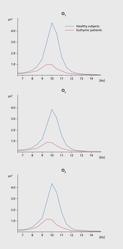
Power spectral analysis of spontaneous EEG activity is one of the most successfully applied methods for identifying biomarkers. (). shows the grand averages of power spectra in 18 bipolar euthymic subjects (red) and 18 healthy controls (black) in the alpha frequency range for the eyes closed recording session from occipital locations (O1, Oz, and O2): the power spectrum in the euthymic subjects ranged up to 1 μV2 across all electrodes but up to 4.8 μV2 for O1, 4 μV2 for Oz, and 4.5 μV2 for O2 in the controls.
Event-related spectra in the alpha frequency range are also drastically reduced in BD.Citation25 Only the marked decrease in alpha power shown in (). could possibly serve as a neurophysiologic marker in BD.
Phase locking in the gamma band in healthy subjects
In inter-trial coherence plots of EROs (), the general time course and frequency composition are completely changed.
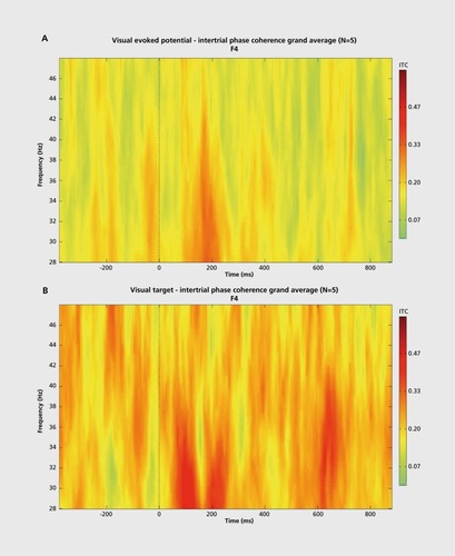
1) a) At the O2 location there are phase-locked components at 400 ms and 600 ms in addition to phase locking at around 100 ms. b) Moreover, the frequencies of phase-locked oscillations increase to over 40 Hz (200 ms periodicity), indicating superposition with the 5 Hz frequency band.
There are abundant phase-locked response components in comparison to sensory evoked responses in Figure 2b.
2) Responses at the F4 location are similar to those at O2. There is 10 Hz periodicity at 100-200 ms with lower frequencies around 30 Hz, whereas at around 600 ms we find solid phase locking (0.45) with a frequency higher than 40 Hz.
Differentiated changes in target responses in bipolar disorder
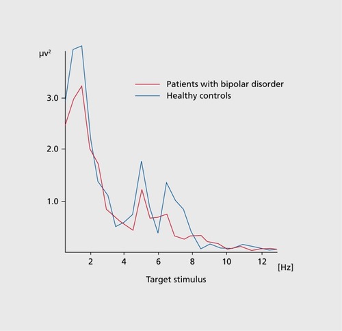
Evoked and event-related slow and fast theta oscillations in response to an auditory stimulus were studied in 22 euthymic drug-free patients with BD I (n =19) or BE) II (n =3). A two-tone oddball task was used, with frequent 1600-Hz target tones, and infrequent 1500-Hz non-target tones. The tones were presented in a random sequence at 3-7 second intervals. The subjects were instructed to keep a mental count of the number of 1600 Hz target tones. A FFT was applied to the 0-800 ms period after stimulus onset.
Slow (4-6 Hz) and fast (6-8 Hz) theta responses behaved differently during the oddball paradigm in euthymic BP patients. Fast theta responses (6-8 Hz) almost disappearedCitation26 ().
Application of digital filters to the analysis of neuropsychiatry patients requires refinement using adaptive filters chosen according to the cutoff frequency in power spectra instead of rigid filters in the conventional frequency ranges. Sometimes a peak is missed or else it shifts to other frequencies in patients, especially after drug administration.
Selective connectivity deficit
There are several forms of connection between different structures in the brain. The connectivity that can be measured using wavelet coherence function in healthy subjects is well defined, in contrast to the deficit in selective connectivity displayed by patients whose substructures are anatomically or physiologically disrupted.
An important brain mechanism underlying cognitive processes is the exchange of information between brain areas.Citation27-Citation28
Decreased event-related gamma coherence in euthymic bipolar patients
Ozerdem et alCitation29 studied cortico-cortical connectivity by examining sensory-evoked coherence and event-related coherence values for the gamma frequency band during simple light stimulation and visual oddball paradigm in 20 euthymic drug-free BD patients and 20 sex- and age-matched healthy controls. The coherence values of the left and right intra-hemispheric electrode pairs (F3-T3, F3-TP7, F3-P3, F3-O1), and F4 -T4, F4-TP4, F4-P4, F4-O2, respectively) were compared between the groups. BD patients showed bilaterally diminished long-distance gamma coherence between frontal and temporal as well as between frontal and temporo-parietal regions compared with healthy controls. The reductions in gamma coherence between the electrode pairs were statistically significant.
However, the patient group showed no significant reduction in sensory-evoked coherence compared with the healthy controls. The decrease in event-related coherence differed topologicaly and ranged from 29% (right fronto-temporal location) to 44% (left fronto-temporo parietal location). (). depict the grand average of visual event-related coherence in the gamma frequency band (28-48 Hz) in response to target stimuli between the right (F4-T8) and left (F3-T7) fronto-temporal electrode pairs in euthymic bipolar patients (n =20) compared with healthy controls (n =20).Citation29
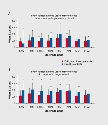
Oscillatory responses to both target and non-target stimuli are manifestations of working memory-processes. Therefore, the decrease in coherence in response to both stimuli points to an inadequacy of connectivity between different parts of the brain under cognitive load that in patients with cognitive impairment is greater than when they are processing purely sensory-signals.
Signal analysis results
The preceding analysis prompts a number of hypotheses, conclusions and lines of further enquiry:
1. Intrinsic oscillatory activity by single neurons forms the basis of the natural frequencies of neural assemblies. These natural frequencies, classified as alpha, beta, gamma, theta and delta, are the brain's real responses.Citation30-Citation32
2. Morphologically different neurons or neural networks respond to sensory-cognitive stimuli in the same frequency ranges of EEG oscillations. The type of neuronal assembly does not play a major role in the frequency tuning of oscillatory networks. Research has shown that neural populations in the cerebral cortex, hippocampus, and cerebellum are all tuned to the very same frequency ranges, although these structures have completely different neural organizations.Citation21,Citation33-Citation37 It is therefore suggested that whole-brain networks communicate via the same set of EEG oscillation frequency codes.
3. The brain has response susceptibilities that mostly originate from its intrinsic (ie, spontaneous) rhythmic activity.Citation15,Citation38-Citation41 A brain system responds to external or internal stimuli with those rhythms or frequency components that are among its intrinsic (natural) rhythms. Accordingly, if a given frequency range does not exist in its spontaneous activity, it will also be absent from its evoked activity. Conversely, if activity in a given frequency range does not exist in evoked activity, it will also be absent from spontaneous activity.
4. There is an inverse relationship between the EEG and ERR. EEG amplitude thus serves as a control parameter for brain responsiveness in the form of EP or ERE.Citation33,Citation42-Citation45
5. Combined with the concept of response susceptibility, this characteristic leads to the conclusion that EEG oscillatory-activity governs most general transfer functions in the brain.Citation46
6. Oscillatory neural tissue selectively distributed throughout the brain is activated by sensory-cognitive input. Such oscillatory activity can be described by a number of response parameters — enhancement (amplitude), delay (latency), blocking or desynchronization, prolongation (duration), degree of coherence between different oscillations, degree of entropy — that are differently configured depending on the particular task and the functions which that task elicits.Citation47-Citation60 In other words, the brain uses the same frequency range to perform not just one but multiple functions.
7. The number of oscillations and the ensemble of parameters that are obtained under given conditions increase as the complexity of the stimulus increases, or as recognition of the stimulus becomes difficult.Citation15,Citation21,Citation61-Citation62
8. Each function is represented in the brain by the superposition of oscillations in various frequency ranges. The values of the oscillations vary across a number of response parameters. The comparative polarity and phase angle of different oscillations are decisive in producing function-specific configurations. Neuron assemblies do not obey the all-or-none rule of single neurons.Citation63-Citation67
9. The superposition principle indicates synergy between alpha, beta, gamma, theta, and delta oscillations during the performance of sensory-cognitive tasks. Integrative brain function operates through the combined action of multiple oscillations.
10. Our results strongly support the recommendation to use several methods in multiple frequency windows when attempting to identify biomarkers that will differentiate between cognitive diseases.
Conclusions and perspectives
Research scientists in neuropsychiatry tend to be nonselective and non concept-oriented in using only one of the methods from the ensemble of oscillatory analyses. Consequently, brain oscillations often fail to provide useful and effective results in terms of biomarker identification or cognitive deficiency screening. This paper has suggested ways of improving such analyses by emphasizing the complementarity - between the different methods of brain oscillation investigation and the overriding need to use them in combination with one another.
Selected abbreviations and acronyms
| AFC | = | amplitude frequency characteristics |
| EEG | = | electroencephalogram |
| EP | = | evoked potential |
| ERP | = | event-related potential |
| ERO | = | event-related oscillation |
| FFT | = | fast Fourier transform |
| JMRI | = | functional magnetic resonance imaging |
| MEF | = | magnetic evoked field |
| MEG | = | magnetoencephalography |
| PET | = | positron emission tomography |
REFERENCES
- BaşarE.GüntekinB.Atagüni.Turp-GölbaşiB.TülayE.ÖzerdemA.Brain alpha activity is highly reduced in euthymic bipolar disorder patients.Cogn Neurodyn.20126112023372616
- Başar-EroğluC.Schmiedt-FehrC.MathesB.Auditory evoked alpha oscillations imply reduced anterior and increased posterior amplitudes in schizophrenia.Clin Neurophysiol.201262121129
- YenerGG.BajarE.Brain oscillations as biomarkers in neuropsychiatric disorders: following an interactive panel discussion and synopsis.Clin Neurophysiol.201262237257
- ÖzerdemA.GüntekinB.AtagünMi.BaşarE.Brain oscillations in bipolar disorder in search of new biomarkers.Clin Neurophysiol.201262207221
- VecchioF.BabiloniC.LizioR.et alResting state cortical EEG rhythms in Alzheimer's disease: towards EEG markers for clinical applications. A review.Clin Neurophysiol.201262223236
- YenerGG.BaşarE.Biomarkers in Alzheimer's disease with a special emphasis on event-related oscillatory responses.Clin Neurophysiol.201262325360
- MountcastleVB.Perceptual Neuroscience: The Cerebral Cortex. Cambridge, MA: Harvard University Press;1998
- EcclesJC.The Understanding of the Brain. New York, NY: McGraw-Hill.1973
- AdeyWR.DunlopCW.HendrixCE.Hippocampal slow waves. Distribution and phase relationships in the course of approach learning.Arch Neurol.19603749013791882
- AdeyWR.Neurophysiological correlates of information transaction and storage in brain tissue. In: Stellar E, Sprague JM, eds.Progress in Physiological Psychology. New York, NY: Academic Press;1966143
- AdeyWR.Cell membranes, electromagnetic fields, and intercellular communication. In: Başar E, Bullock TH, eds.Brain Dynamics-Progress and Perspectives. New York; NY: Springer;19892642
- FreemanWJ.Foreword. In: Basar E, eds. Brain Function and Oscillations: II. Integrative Brain Function.Neurophysiology and Cognitive Processes. Berlin, Heidelberg, Germany: Springer;1999
- KarakaşS.KarakasH.ErzenginOU.Early sensory gamma represents the integration of bottom-up and top-down processing.Int J Psychophysiol.20024539
- KarakaşS.BekçiB.ErzenginOU.Early gamma response in human neuroelectric activity is correlated with neuropsychological test scores.Neurosci Lett.2003340374012648753
- BaşarE.EEG-Brain Dynamics.Relation between EEG and Brain Evoked Potentials. Amsterdam, the Netherlands: Elsevier;1980
- YordanovaJ.KolevV.Event-related alpha oscillations are functionally associated with P300 during information processing.Neuroreport.199814315931649831444
- KlimeshW.DoppelmayrM.PachingerT.RipperB.Brain oscillations and human memory performance: EEG correlates in the upper alpha and theta bands.Neurosci Lett.19972389129464642
- BaşarE.BullockTH.eds.Induced Rhythms in the Brain. Boston, MA: Birkhäuser;1992
- GurtubayIG.AlegreM.LabargaA.MalandaA.ArtiedaJ.Gamma band responses to target and non-target auditory stimuli in humans.Neurosci Lett.20043676915308286
- BuszakiG.Rhythms of the Brain. New York, NY: Oxford University Press;2006
- BaşarE.Brain function and oscillations II: integrative brain function.Neurophysiology and cognitive processes. Heidelberg, Germany: Springer;1999
- RöschkeJ.MannK.RiemannD.FrankC.FellJ.Sequential analysis of the brain's transfer properties during consecutive REM episodes.Electroencephalogram Neurophysiol.199596390397
- YordanovaJ.KolevV.Developmental changes in the event-related EEG theta response and P300.Electroencephalogr Clin Neurophysiol.19971044184309344078
- SolodovnikovVV.Introduction to the Statistical Dynamics of Automatic Control Systems. New York: Dover;1960
- BasarE.GüntekinB.AtagünMi.TurpGölbaşi B.TülayE.ÖzerdemA.Brain's alpha activity is highly reduced in euthymic bipolar disorder patients.Cogn Neurodyn.20126112023372616
- AtagünMi.GüntekinB.ÖzerdemA.TülayE.BaşarE.Online first article. Decrease of theta response in euthymic bipolar patients during an oddball paradigm.Cogn Neurodyn.2013DOI 10. 1007/s11571-012-9228-7
- BaşarE.GüntekinB.TülayE.YenerGG.Evoked and event-related coherence of alzheimer patients manifest differentiation of sensory-cognitive networks.Brain Res.20101357799020732310
- GïntekinB.SaatçiE.YenerG.Decrease of evoked delta, theta and alpha coherences in Alzheimer patients during a visual oddball paradigm.Neurosci Lett.20084249499
- ÖzerdemA.GüntekinB.AtagünMi.TurpB.BaşarE.Reduced long distance gamma (28-48 Hz) coherence in euthymic patients with bipolar disorder.J Affect Disord.201113232533221459454
- BaşarE.Başar-EroğluC.KarakaşS.SchürmannM.Gamma, alpha, delta, and theta oscillations govern cognitive processes,int J Psychophysiol.20013924124811163901
- BaşarE.ÖzgörenM.KarakaşS.A brain theory based on neural assemblies and superbinding. In: Reuter H, Schwab P, Gniech KD, eds.Wahrnehmen und Erkennen. Lengerich, Germany: Pabst Science Pub; 20011124
- BaşarE.SchürmannM.SakowitzO.The selectively distributed theta system: functions,int J Psychophysiol.20013919721211163897
- BasarE.Brain Function and Oscillations I: Principles and Approaches. Berlin, Heidelberg, Germany: Springer-Publishers;1998
- EckhornR.BauerR.JordanR.et alCoherent oscillations: a mechanism of feature linking in the visual cortex?Biol Cybern.1988601211303228555
- LlinasRR.The intrinsic electrophysiological properties of mammalian neurons: insights into central nervous system function.Science.1988242165416643059497
- SingerW.The brain, a self-organizing system. In: Klivington KA, ed.The Science of Mind. Cambridge, MA: MIT Press;1989174179
- SteriadeM.Corro-DossiR.PareD.Mesopontine cholinergic system suppress slow rhythms and induce fast oscillations in thalamocortical circuits. In: Başar E, Bullock TH, eds.Induced Rhythm in the Brain. Boston, MA: Birkhauser;1992251268
- BasarE.Toward a physical approach to integrative physiology: I. Brain dynamics and physical causality.Am J Physiol.198314510533
- BasarE.Synergetics of neuronal populations. A survey on experiments. In: Basar E, Flohr H, Haken H, Mandell A, eds.Synergetics of the Brain. Berlin, Germany: Springer;1983183200
- BaşarE.DosselO.FuchsM.RahnE.SaermarkK.SchürmannM.Evoked alpha response from frontal-temporal areas in multichannel SQUID systems. Paper presented at: Proceedings of the IEEE Satellite Symposium on Neuroscienceand Technology; 1992; Lyon: Nov 28-33.
- NariciL.PizzellaV.RomaniGL.TorrioliG.TraversaR.RossiniPM.Evoked alpha and mu rhythms in humans: a neuromagnetic study.Brain Res.19905202222312207633
- BarryRJ.De PascalisV.HodderD.ClarkeAR.JohnstoneSJ.Preferred EEG brain states at stimulus onset in a fixed interstimulus interval auditory oddball task, and their effects on ERP components.Int J Psychophysiol.20034718719812663064
- BaşarE.ÖzgörenM.Başar-EroğluC.KarakaşS.Superbinding: spatiotemporal oscillatory dynamics.Theory Biosci.2003121370385
- RahnE.BasarE.Prestimulus EEG activity strongly influences the auditory evoked vertex responses: a new method for selective averaging.Int J Neurosci.1993692072208083007
- JansenBH.ZouridakisG.BrandtME.A neurophysiologically-based mathematical model of flash visual evoked potentials.Biol Cybern.1993682752838452897
- BaşarE.Chaos in Brain Function. Berlin, Germany: Springer;1990
- BaşarE.Memory and Brain Dynamics: Oscillations Integrating Attention, Perception, Learning and Memory. Florida: CRC Press;2004
- BaşarE.Başar-EroğluC.KarakaşS.SchurmannM.Are cognitive processes manifested in event-related gamma, alpha, theta and delta oscillations in the EEG?Neurosci Lett.199925916516810025584
- BaşarE.Başar-EroğluC.KarakaşS.SchurmannM.Oscillatory brain theory: a new trend in neuroscience.IEEE Eng Med Biol Mag.199918566610337564
- KocsisB.Di PriscoGV.VertesRP.Theta synchronization in the limbic system: the role of Gudden's tegmental nuclei.Eur J Neurosci.20011338138811168543
- MiltnerWH.BraunC.ArnoldM.WitteH.TaubE.Coherence of gammaband EEG activity as a basis for associative learning.Nature.19993974344369989409
- PfurtschellerG.EEG event-related desynchronization (ERD) and synchronization (ERS).Electroencephalogr Clin Neurophysiol.199710326
- PfurtschellerG.Functional brain imaging based on ERD/ERS.Vision Res.2001411257126011322970
- PfurtschellerG.NeuperC.RamoserH.Müller-GerkingJ.Visually guided motor activates sensorimotor areas in humans.Neurosci Lett.1999 26915315610454155
- PfurtschellerG.BrunnerC.SchlöglA.Lopesda Silva.FHMu rhythm, (de)synchronization and EEG single-trial classification of different motor imagery tasks.Neuroimage.20063115315916443377
- NeuperC.PfurtschellerG.15 event-related de synchronization (ERD) and synchronization (ERS) of rolandic EEG rhythms during motor behavior.Int J Psy Psychophysiol.19983078
- NeuperC.PfurtschellerG.134 ERD/ERS based brain computer interface (BCI): effects of motor imagery on sensorimotor rhythms.Int J Psychophysiol.1998305354
- RossoOA.BlancoS.YordanovaJ.KolevV.FigliolaA.SchurmannM.BaşarE.Wavelet entropy: a new tool for analysis of short duration brain electrical signals.J Neurosci Methods.2001105657511166367
- RossoOA.MartinMT.PlastinoA.Brain electrical activity analysis using wavelet-based information tools I.Physics.2002313587608
- SchürmannM.DemiralpT.BaşarE.Başar-EroğluC.Electroencephalogram alpha (8-15 Hz) responses to visual stimuli in cat cortex, thalamus, and hippocampus: a distributed alpha network?Neurosci Lett.200029217517811018305
- BaşarE.Başar-EroğluC.KarakaşS.SchürmannM.Brain oscillations in perception and memory.Int J Psychophysiol.2000359512410677641
- BaşarE.Başar-EroğluC.KarakaşS.SchürmannM.Gamma, alpha, delta, and theta oscillations govern cognitive processes.Int J Psychophysiol.20013924124811163901
- ChenAC.HerrmannCS.Perception of pain coincides with the spatial expansion of electroencephalographic dynamics in human subjects.Neurosci Lett.200129718318611137758
- KarakaşS.ErzenginOU.BaşarE.The genesis of human event related responses explained through the theory of oscillatory neural assemblies.Neurosci Lett.2000285454810788704
- KarakaşS.ErzenginOU.BaşarE.A new strategy involving multiple cognitive paradigms demonstrates that ERP components are determined by the superposition of oscillatory responses.Clin Neurophysiol.20001111719173211018485
- KlimeschW.DoppelmayrM.RöhmD.PöllhuberD.StadlerW.Simultaneous desynchronization and synchronization of different alpha responses in the human electroencephalograph: a neglected paradox?Neurosci Lett.20002849710010771171
- KlimeschW.DoppelmayrM.SchwaigerJ.WinklerT.GruberW.Theta oscillations and the ERP old/new effect: independent phenomena?Clin Neurophysiol.200011178179310802447