
Abstract
Background
To date, the most efficient and robust method for isolating avian influenza A viruses (IAVs) is using embryonated chicken eggs (ECEs). It is known that low-pathogenic avian IAVs undergo rapid genetic changes when introduced to poultry holdings, but the factors driving mutagenesis are not well understood. Despite this, there is limited data on the effects of the standard method of virus isolation of avian-derived viruses, that is, whether isolation in ECEs causes adaptive changes in avian IAVs. Eggs from a homologous species could potentially offer an isolation vessel less prone to induce adaptive changes.
Methods
We performed eight serial passages of two avian IAVs isolated from fecal samples of wild Mallards in both ECEs and embryonated Mallard eggs, and hemagglutination assay titers and hemagglutinin sequences were compared.
Results
There was no obvious difference in titers between ECEs and embryonated Mallard eggs. Sequence analyses of the isolates showed no apparent difference in the rate of introduction of amino acid substitutions in the hemagglutinin gene (three substitutions in total in embryonated Mallard eggs and two substitutions in ECEs).
Conclusion
Embryonated Mallard eggs seem to be good isolation vessels for avian IAVs but carry some practical problems such as limited availability and short egg-laying season of Mallards. Our study finds isolation of Mallard-derived avian IAVs in ECEs non-inferior to isolation in embryonated Mallard eggs, but more research in the area may be warranted as this is a small-scale study.
Isolation of avian influenza viruses (IAVs) is traditionally performed by inoculation of a material containing viral particles into embryonated chicken eggs (ECEs), and this method has been the gold standard since it was introduced in the 1930s (Citation1). Isolation in ECEs can be successfully applied at different scales, and it is used industrially to amplify large quantities of attenuated IAV vaccine strains, and it is also the recommended method for the isolation of influenza A virus from samples in national surveillance programs of domestic and wild animals (Citation2, Citation3). Research on IAV has increased dramatically in the last decade, and careful conclusions must be drawn from experimental research in this field if it is unknown whether an isolate has retained its wild-type characteristics after being passaged in eggs. Many studies have compared virus isolation on different cell lines and ECEs, but there have been few comparative studies of eggs from different species of birds as isolation vessels, and none addressing selection and mutations (Citation4, (Citation5). Waterfowl, and particularly ducks of the genus Anas, is considered the natural reservoir of avian IAVs (Citation6. When an IAV crosses host species barriers, it is exposed to a new growth environment, which likely promotes a new selection. Differences in diet, body structure, distribution of cell types, immune system, and receptor expression could all influence selection pressures exerted on the virus in its new host. Spread to other bird species, particularly domestic chickens, has been associated with an increase in mutations and a higher pathogenicity (Citation7–Citation9). In the case of IAVs adapted to humans and birds, the tropism is largely dependent on the linkage between cell-bound receptors with terminal sialic acid (SA) to galactose (Gal). IAVs from humans show a preference for α2,6 linked SA and Gal, while avian IAVs have a higher affinity for receptors with an α2,3 linkage (Citation10). Thus, when a human-adapted virus is inoculated in an ECE, there is a pressure that selects for quasi-species with higher affinity to α2,3 SA. IAV adapted to humans has been shown to respond to the change in selection pressure when isolated on ECEs by amino acid substitutions in the receptor binding pocket on the apical surface of the hemagglutinin protein (Citation11–Citation15). However, although low-pathogenic avian IAVs from the natural waterfowl reservoir have been known to develop new characteristics upon interspecies transmission to domestic fowl, there are no data on amino acid substitution in avian viruses due to virus isolation in embryonated eggs from a different bird species (Citation7, Citation9) (Citation16).
In this study, we first isolated and then serially passaged two Mallard-derived IAVs both in standard ECEs method and in embryonated eggs from Mallards (Anas platyrhynchos). The hemagglutinin gene of one isolate from each passage and egg species was sequenced and analyzed with regard to mutations and resulting amino acid substitutions. Each passage from the different species was compared to determine whether one species was more prone than the other to select for a mutated hemagglutinin protein. As the Mallard is considered the natural host of avian IAVs, it was postulated that traditional isolation methods would apply a different selection pressure on the virus and possibly result in more amino acid substitutions than isolation in eggs from a homologous species.
Materials and methods
Viruses
Material for virus isolation was obtained from an ongoing IAV surveillance in southeastern Sweden, where approximately 3,000–5,000 fecal samples are collected each year (Citation17). All samples are swabs stored in Hank's balanced salt solution containing 0.5% lactalbumin, 10% glycerol, 200 U/mL penicillin, 200 µg/mL streptomycin, 100 U/mL polymyxin B sulfate, 250 µg/mL gentamycin, and 50 U/mL nystatin (Sigma, Stockholm, Sweden). Two viruses, A/Mallard/Sweden/79947/2008/H5N2 and A/Mallard/Sweden/80863/2008/H11N6 from recently collected fecal samples, were used for the experiment. These IAVs were selected on the basis of signal strength in real-time, reverse-transcription polymerase chain reaction (PCR) screening assay used for surveillance.
Eggs
Mallard eggs were obtained from a commercial Mallard breeder in Sweden (Schedewij Säteri, Flen, Sweden). Specific pathogen free (SPF) chicken eggs were acquired from Lohmann Tierzucht GmbH (Cuxhaven, Germany).
Passaging
For the first passage, the original fecal sample from a wild Mallard was diluted 10× and treated with penicillin/streptomycin (Sigma). Two hundred microliters of the resulting virus solution was inoculated in the allantoic cavity of 5-, 11-, and 12-day-old ECEs and Mallard eggs, respectively, and harvested after 2 days. Virus growth was confirmed by hemagglutination assay (HA) of SPF chicken erythrocytes (0.5%), and HA titers were calculated in accordance with the WHO manual (Citation2). Subsequent passaging of isolates was performed after diluting the positive isolate grown from the highest dilution of the previous passage at 10−6, 10−7, and 10−8. In case of two positive isolates from the highest dilution, the isolate with the highest HA titer was chosen. The dilutions were treated with penicillin/streptomycin in accordance with the manufacturer's instructions for 30 min prior to inoculation. One to three eggs were inoculated with 200-µL diluted isolate each, and virus was allowed to grow for 2 days before harvest.
The setup with multiple passaging would have made the use of replicates at each passaging step extremely cumbersome. For example, using a triplicate of isolates at each passage step that would then be further passaged in triplicates and so on would result in 38=6,561 samples to analyze. Thus, rather than analyzing several isolates per passage, we chose to follow one viral population throughout the experiment. This means that we might have missed mutations that occurred in unanalyzed isolates, but the multiple passages would still allow for detection of mutations with a significantly higher fitness during egg propagation. Therefore, we found our approach reasonable for a small-scale study.
RNA extraction
Upon thawing, the tube containing original material from fecal sample (first passage) or viral isolate (subsequent passages) was thoroughly vortexed and 150 µL was removed and mixed with 450 µL Trizol reagent (Invitrogen, Paisley, England). In the case of the original material, 15 µL was diluted to 150 µL. Adding one hundred sixty microliters of pure chloroform, a 350-µL water phase containing RNA could be separated. RNA from the water phase was then extracted using RNeasy Mini kit (Qiagen, Hilden, Germany) treating the water phase as homogenized animal cell lysate. The elution volume was 30 µL.
Reverse transcription PCR
RNA was transcribed to cDNA using Uni12 primers (Citation18) and an SSIII RT kit according to the manufacturer's specifications (Invitrogen). One microliter of RNA template was used from isolates, and 5 µL of RNA template was used from fecal samples. Amplification of the hemagglutinin gene was performed by PCR, using Taq Platinum HiFi (Invitrogen) and published primers (Citation18).
PCR cycling settings: activation at 95°C for 2 min; cycling 40× at 95° for 30 s, at 60°C for 1 min, and at 68°C for 3 min. Two microliters of cDNA was used as the template in all PCRs. The PCR products were separated using gel electrophoresis on 1% agarose/TBE gel over 80 V for 80 min. Bands of correct size, that is, 1.8 kb were cut from the gel and purified using illustra GFX PCR and Gel Band Purification Kit (GE Healthcare, Waukesha, WI) according to the manufacturer's specifications.
Sequencing
Sanger sequencing of purified PCR product was performed with BigDye v.3.1 (Applied Biosystems, Carlsbad, CA). Custom primers were designed for each virus to cover the hemagglutinin gene (shown in ).
Table 1 Custom primers used for sequencing of the hemagglutinin gene
Sequence analysis
Gene sequences were analyzed using SeqMan and MegAlign of the Lasergene software package (DNASTAR, Madison, WI) and BioEdit version 7.0.9.0 (Citation19).
Results
Titers in HA
IAVs grew to high titers in embryonated Mallard eggs; HA titers over 2,048 were recorded during passage. Primary isolation in ECEs did not yield an HA titer over 48, while Mallard eggs yielded HA titers of 1,024 and 384 for the two viruses, respectively (). The A/Mallard/Sweden/79947/2008/H5N2 did not grow well in ECEs in the first three passages; therefore, at passage 4 this virus was inoculated at lower dilutions to enable further propagation. After this, the virus grew better and was capable of growing at higher dilutions at subsequent passages (). This phenomenon was not observed when the same virus was passaged in Mallard eggs, where a markedly higher HA titer was observed for the first three passages (). HA titers are not directly translatable to viral infectivity but as the same viral strains are compared, the HA titers should correlate to the relative amount of virus present in each isolate.
Table 2 Overview of the HA titers obtained from each embryonated egg species (chicken/Mallard) and virus
The virus A/Mallard/Sweden/79947/2008/H5N2 was lost after five passages in Mallard eggs. At the end of the egg laying period of Mallards, the quality and survival of the eggs decrease and we assume that this was the reason for the non-completion of eight passages for this virus.
Amino acid substitutions in the hemagglutinin
In total, five mutations that gave rise to amino acid substitutions in the hemagglutinin protein could be detected in viruses from both tested species of embryonated eggs. In Mallard eggs, two substitutions were detected in A/Mallard/Sweden/80863/2008/H11N6 and one substitution in A/Mallard/Sweden/79947/2008/H5N2. Both viruses changed immediately after primary isolation and further passaging did not induce more mutations. Isolating and passaging in ECEs yielded one substitution in each virus. A/Mallard/Sweden/79947/2008/H5N2 switched immediately after primary isolation, as after isolation of the same virus in Mallard eggs. A/Mallard/Sweden/80863/2008/H11N6 did not acquire any amino acid changes until after the third passage; then one change was established ().
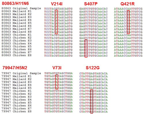
Fig. 1. Parts of aligned sequences of the hemagglutinin gene where mutations were found giving rise to amino acid substitutions. Mallard=virus passaged in embryonated Mallard eggs. Chicken=virus passaged in ECEs. V214I: the codon GTA (valine) changed to ATA (isoleucine) in Mallard E1–8. S407P: the codon TCT (serine) changed to CCT (proline) in chicken E3–8. Q421R: the codon CAA (glutamine) changed to CGA (arginine) in Mallard E1–8. V73I: the codon GTA (valine) changed to ATA (isoleucine) in Mallard E1–5 (virus was lost after E5). S122G: the codon AGT (serine) changed to GGT (glycine) in chicken E1–8.
No other mutations at the nucleotide level were seen in any of the hemagglutinin gene sequences, that is, there were no silent mutations during the passaging of either virus strain in embryonated eggs from either species.
Discussion
Research on viruses is commonly performed with isolates, as wild-type material is rarely available in the quantities needed for most techniques. Thus, in the design of a project, it is imperative to take precautions to make sure the isolate is indeed phenotypically as similar as possible to the wild-type virus. If one cannot be sure that the isolate has maintained its wild-type characteristics, the investigator has to be careful when extrapolating results and conclusions to the wild-type virus. This study compared isolation and passage of IAV using the traditional method (ECEs) and eggs from a homologous species (Mallards) with regard to HA titers and mutations leading to amino acid substitutions in the hemagglutinin gene. Our results demonstrate that embryonated Mallard eggs were suitable for the propagation of IAV. In most aspects, the Mallard eggs performed equally or better than ECEs (with a higher titer at initial isolation, a lower dilution had to be used at one point in ECEs for one of the strains to allow for further propagation, and in the case of A/Mallard/Sweden/79947/2008/H5N2 we were able to dilute the virus more and still obtain higher titers during the first three passages using Mallard eggs). The Mallard eggs performed worse than ECEs in one aspect; one of the strains was lost after five passages. Probably, this was due to logistical problems with Mallard eggs; the quality of the eggs decrease toward the end of the egg-laying period of Mallards (corresponding to the later part of the experiment), and the eggs are difficult to store. However, we cannot exclude that the loss of the virus indicates impaired growth of certain IAVs in Mallard eggs after multiple passages for other reasons. Our results indicate that Mallard eggs can be favorable to ECEs in some cases, but whether Mallard eggs are generally better for isolation in terms of virus titer cannot be conclusively answered on the basis of this study. A similar experiment has also found contradictory results (Citation4). The use of Mallard eggs carry some practical problems such as lower availability than ECEs, difficulties to obtain SPF Mallard eggs, and the tendency of Mallard eggs to decrease in quality later in the egg-laying season.
A tendency toward lower virus titers after several passages was seen in both species, although no mutations in the hemagglutinin could be coupled to this phenomenon. In terms of selection pressure and mutation rate, and whether one species was to be preferred over the other, no conclusive evidence was found for any one alternative. In both species, amino acid substitutions were introduced after the primary isolation and only ECEs managed to maintain the wild-type sequence for one of the two tested viruses, and then for passages 1 and 2 only before an amino acid substitution was introduced. Unrestricted RNA replication has been reported to incorporate mismatching nucleotides at a rate of 1/103 to 1/104 nucleotides (Citation20, Citation21). In this experiment, a non-linear mutation rate was recorded, where the first passage introduced one and two mutations, respectively, for the different viruses in Mallard embryonated eggs. ECEs introduced one mutation in passage 1 for A/Mallard/Sweden/79947/2008/H5N2 and one mutation in passage 3 for A/Mallard/Sweden/80863/2008/H11N6. The number of mutations found in passage 1 correlates well with the expected rate, whereas mutations in the following passages fall well below the random mismatch rate. However, when interpreting the results it is important to bear in mind that the present study was performed at a small scale with only two virus strains and one analyzed isolate per passage and egg species, making the results difficult to generalize.
All substitutions but one were homologous, with little biological significance (). Only the substitution that occurred in ECEs for A/Mallard/Sweden/80863/2008/H11N6 was of major importance, changing a serine to proline. Proline is a rigid amino acid with a clear function to bend a peptide chain, and such a substitution will reasonably have some effect on the biological properties of the protein. The location, however, at position 407 of H0, or 79 of H2, is not in close vicinity of either the cleavage site or the binding pocket. Nor were any of the other mutations detected in this experiment located at the binding site where the SA is bound, or at the H0 cleavage site. As the glycosylation of the SA is similar in both species with an α2,3 linkage, it is not unexpected that the binding pocket remains unchanged. What is less understood is the exact mechanism in the interaction between host and virus before and during fusion of the membranes of the virus and the host lysosome. Even though the observed mutations are not directly connected to the binding site, fusion peptide, or the cleavage site of H0, there could be other factors that differ between the species when H undergoes its conformational change and secondary cleavage inside the lysosome (Citation22). Changes in quasi-species structure over time have not been assessed in this study, but it seems reasonable to assume that any mutation with a significant selective advantage would be noticeable also in Sanger sequencing after eight passages. Although hemagglutinin is the gene where host adaptation-related mutations are most likely, there could also be mutations in other segments of the genome that contribute to host adaptation. For example, changes in the neuraminidase could cause an altered hemagglutinin/neuraminidase activity balance, resulting in altered adhesion. Also, changes in internal genes could contribute via more indirect mechanisms such as differences in polymerase activity/replication ability or differences in interactions with the innate immune response of the host. To assess these questions, a larger study using a next-generation sequencing approach is necessary.
In conclusion, our results demonstrate that embryonated Mallard eggs are good isolation vessels for IAVs. However, we find no obvious advantages as compared to the standard method of ECEs; neither with regard to virus titers nor to the amount of amino acid substitutions in the hemagglutinin.
Conflict of interest and funding
This study was supported by grants from the Swedish Research Council (Vetenskapsrådet) and the Swedish Research Council FORMAS.
References
- Bull DR, Burnet FM. Changes in influenza virus associated with adaptation to passage in chick embryos. Aust J Exp Biol Med Sci. 1943; 21: 55–69.
- Stöhr K, Cox NJ, Webster RG . WHO Manual on Animal Influenza Diagnosis and Surveillance. 2002; Geneva, Switzerland: WHO.
- Tosh PK, Jacobson RM, Poland GA. Influenza vaccines: from surveillance through production to protection. Mayo Clin Proc. 2010; 85: 257–73.
- Moresco KA, Stallknecht DE, Swayne DE. Evaluation and attempted optimization of avian embryos and cell culture methods for efficient isolation and propagation of low pathogenicity avian influenza viruses. Avian Dis. 2010; 54(1 Suppl): 622–6.
- Munster VJ, Baas C, Lexmond P, Bestebroer TM, Guldemeester J, Beyer WEP, etal. Practical considerations for high-throughputinfluenza A virus surveillance studies of wild birds by use of molecular diagnostic tests. J Clin Microbiol. 2009; 47: 666–73.
- Olsen B, Munster VJ, Wallensten A, Waldenström J, Osterhaus AD, Fouchier RA. Global patterns of influenza a virus in wild birds. Science. 2006; 312: 384–8.
- Banks J, Speidel ES, Moore E, Plowright L, Piccirillo A, Capua I, etal. Changes in the haemagglutinin and the neuraminidase genes prior to the emergence of highly pathogenic H7N1 avian influenza viruses in Italy. Arch Virol. 2001; 146: 963–73.
- Alexander DJ. A review of avian influenza in different bird species. Vet Microbiol. 2000; 74: 3–13.
- Capua I, Marangon S. The avian influenza epidemic in Italy, 1999–2000: a review. Avian Pathol. 2000; 29: 289–94.
- Rogers GN, Paulson JC. Receptor determinants of human and animal influenza virus isolates: differences in receptor specificity of the H3 hemagglutinin based on species of origin. Virology. 1983; 127: 361–73.
- Rocha EP, Xu X, Hall HE, Allen JR, Regnery HL, Cox NJ. Comparison of 10 influenza A (H1N1 and H3N2) haemagglutinin sequences obtained directly from clinical specimens to those of MDCK cell- and egg grown viruses. J Gen Virol. 1993; 74: 2513–18.
- Azzi A, Bartolomei-Corsi O, Zakrzewska K, Corcoran T, Newman R, Robertson JS, etal. The haemagglutinins of influenza A (H1N1) viruses in the ‘O’ or ‘D’ phases exhibit biological and antigenic differences. Epidemiol Infect. 1993; 111: 135–42.
- Katz JM, Naeve CW, Webster RG. Host cell-mediated variation in H3N2 influenza viruses. Virology. 1987; 156: 386–95.
- Widjaja L, Ilyushina N, Webster RG, Webby RJ. Molecular changes associated with adaptation of human influenza A virus in embryonated chicken eggs. Virology. 2006; 350: 137–45.
- Rogers GN, Paulson JC, Daniels RS, Shekel JJ, Wilson IA, Wiley DC. Single amino acid substitutions in influenza haemagglutinin change receptor binding specificity. Nature. 1983; 304: 76–8.
- Alexander DJ, Brown IH. Recent zoonoses caused by influenza A viruses. Rev Sci Tech. 2000; 19: 197–225.
- Latorre-Margalef N, Gunnarsson G, Munster VJ, Fouchier RAM, Osterhaus ADME, Elmberg J, etal. Effects of influenza A virus infection on migrating mallard ducks. Proc Biol Sci. 2009; 276: 1029–36.
- Hoffmann E, Stech J, Guan Y, Webster RG, Perez DR. Universal primer set for the full-length amplification of all influenza A viruses. Arch Virol. 2001; 146: 2275–89.
- Hall TA. BioEdit: a user-friendly biological sequence alignment editor and analysis program for Windows 95/98/NT. Nucl Acids Symp Ser. 1999; 41: 95–8.
- Holland J, Spindler K, Horodyski F, Grabau E, Nichol S, VandePol S. Rapid evolution of RNA genomes. Science. 1982; 215: 1577–85.
- Duffy S, Shackelton LA, Holmes EC. Rates of evolutionary change in viruses: patterns and determinants. Nat Rev Genet. 2008; 9: 267–76.
- Skehel JJ, Wiley DC. Receptor binding and membrane fusion in virus entry: the influenza hemagglutinin. Annu Rev Biochem. 2000; 69: 531–69.