We would like to report a 20-year-old female presented with gradually progressive left renal angle tenderness for two years. Examination revealed fullness in the left renal angle and the ultrasound scan showed severe left hydronephrosis with a normal caliber ureter.
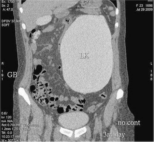
Abdominal CT scan was arranged and 40 cc of 350 mg I/mL of injection iohexol was injected intravenously prior to the scan (). In view of severe hydronephrosis, no contrast excretion was seen on the scanogram until after 4 hours and hence the patient was recalled after 24 hours for the scan. A 100 cc of injection iohexol (350 mg I/mL) was injected at a rate of 4 cc/sec and the patient was scanned in arterial phase from the level of diaphragm to the iliac crest (). In the middle of the scan, however, the patient moved on the table. Hence, a repeat scan was performed with additional 100 cc of injection iohexol (350 mg I/mL) in arterial and venous phases for the renal vessels (). As there was no contrast excretion in the hydronephrotic kidney, the patient was recalled after another 24 hours for an urographic scan (). At this time, a thin section plain scan was performed from the level of dome of diaphragm to the symphysis pubis and it was observed that there was gall bladder and colonic opacification with contrast, with similar Hounsfield values to excreted contrast in hydronephrotic left kidney (). There was no evidence of contrast-induced nephropathy.
Figure 1. First scan: axial CT scan with 40 cc intravenous contrast.
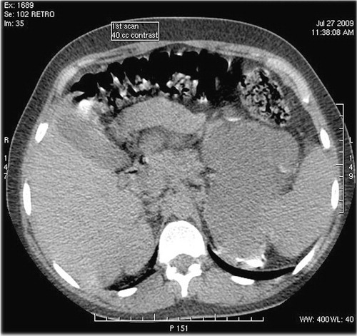
Figure 2. Second scan: 24 hours. After first scan. Arterial phase axial CT scan with 100 cc I.V. contrast.
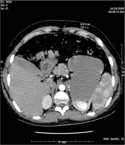
Figure 3. Third scan: additional 100 cc I.V. contrast after second scan.
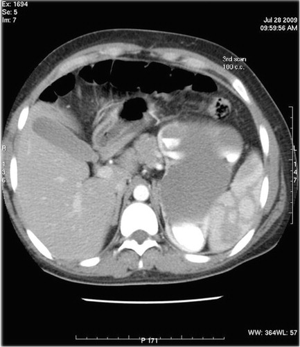
Figure 4. 48 hours delayed scan shows contrast in gall bladder and colon besides hydronephrotic left kidney (marked with oval).
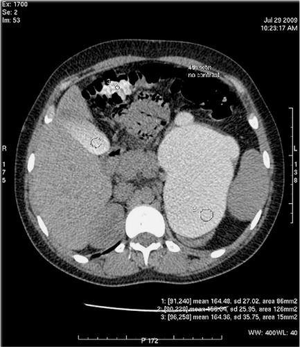
Figure 5. Coronal reformat of delayed scan showing opacified gall bladder and ascending colon besides opacified hydronephrotic left kidney.
Abdominal imaging has undergone a major change with introduction of specialized, dedicated protocols for organ-specific imaging thereby creating a huge demand for higher volumes of contrast. In our center, we use injection iohexol in strength of 300 and 350 mg I/mL as CT scan contrast. It is a non-ionic, monomeric tri-iodinated, water soluble X-ray contrast medium. The dose of contrast ranges from 50 to 150 cc depending on the type of examination, age, weight, and general condition of the patient.
Yamazaki et al. found no significant correlation between delayed gall bladder opacification and contrast associated nephropathy as in our case Citation1. Kaizu et al. and Tajima et al. attributed gall bladder opacification to the important role of hepatobiliary tract in the excretion of ioxaglate Citation2Citation3. However, iohexol is excreted completely almost unchanged through the kidneys within 24 hours in patients with normal renal function with half life of 2 hours with no detectable metabolites. The protein binding of iohexol is so low (<2%) that it has no clinical relevance, and can therefore be neglected (information from product leaflet published by Amersham Health, Norway, December 2003).
We conclude that gall bladder and colonic opacification may not be as uncommon as is generally thought, but their timing and location in most of the cases does not allow making this observation.
Anuj Mishra
Department of Radiology
National Organ Transplant Program
Tripoli Central Hospital, Tripoli, Libya
Ehtuish Farag Ehtuish
Department of Surgery
AlFatah University of Medical Sciences
Tripoli, Libya
Dallia Abukhair
Department of Hepatology
National Organ Transplant Program
Tripoli Central Hospital, Tripoli, Libya
References
- Yamazaki H, Oi H, Matsushita M, Kim T, El-Baradie M, Inoue T, et al.. Lack of correlation between gallbladder opacification in delayed CT and contrast-associated nephropathy. Eur Radiol. 1997; 7: 1328–31.
- Kaizu T, Tajima H, Ichikawa T, Kumazaki T. Nippon Igaku Hoshasen Gakkai Zasshi [Hepato-biliary excretion of water-soluble iodinated contrast medium shortly after abdominal angiography]. 1995; 55: 1038–41.
- Tajima H, Kaizu T, Ichikawa T, Kumazaki T. Gallbladder visualization on CT shortly after angiography with ioxaglate. Acta Radiol. 1994; 35: 634–5.