Abstract
Objective: This study was carried out to determine the reliability of sex determination from teeth pulp tissue.
Patients and methods: This study was carried on 60 maxillary and mandibular premolars and permanent molars (30 male teeth and 30 female teeth) which were indicated for extraction. The teeth were categorized into three groups of 20 each (10 from males and 10 from females).Group 1-pulp tissue from teeth examined immediately after extraction. Group 2- and Group 3-pulp tissue examined from teeth one and five month after extraction, respectively. Teeth was sectioned and pulpal cells were stained with quinacrine dihydrochloride. The cells were observed with fluorescent microscope for fluorescent body. Gender was determined by identification of Y chromosome fluorescence in dental pulp.
Results: Freshly extracted teeth and for those examined one month later, sensitivity, specificity, positive predictive value, negative predictive value, and efficiency were all 100%.
Conclusion: The fluorescent Y body test is shown to be a reliable, simple, and cost-effective technique for gender identification in the immediate postmortem period up to one month.
Introduction
Human identification is a mainstay of civilization and identification of unknown individuals has always been of paramount importance to society Citation1. Identification in the medico-legal sense refers to the determination of the individuality of a person which may be complete or partial Citation2. Dental identification has long been considered a reliable method when other methods fail because of critical body conditions or unavailability of body parts Citation3.
The most reliable means of identification include fingerprints, dental comparisons, and biological methods such as DNA profiling. In some cases, however, fingerprints are not available from the deceased or antemortem prints cannot be obtained. Although the techniques of postmortem fingerprinting have improved dramatically, cases of extreme decomposition, incineration or skeletonization preclude accurate finger printing Citation1.
The use of biological evidence for identification of an individual is a relatively recent development. Biological evidence generally means the comparison of genetic material such as DNA. But DNA analysis can be expensive and time consuming Citation4. Teeth are the most durable organs in the body and can be heated to temperatures of 1,600°C without appreciable loss of microstructure. Teeth can survive long after soft and skeletal tissues have been destroyed Citation1. Determination of sex can prove refractory after catastrophic events, such as fires, high impact crashes, and explosions. Tooth pulp is embedded in a hard tissue casting that protects it from the detrimental effects of impact, trauma, and heat. The present study was conducted to determine the reliability of sex determination from tooth pulp during the five months after tooth extraction using a fluorescent body test.
2 Material and methods
This study was carried out in the Department of Oral Medicine and Radiology, Oral Pathology and Microbiology, Bapuji Dental College & Hospital, Davangere, India. This study was approved by the Head of the Ethics Committee of the Institution and informed consent was obtained from all the patients who came for extraction, who were informed of the procedure details regarding tooth extraction and about the research.
This study was done on 60 maxillary and mandibular premolars and permanent molars that were free of dental caries, but were indicated for extraction because they were periodontally compromised. One hundred and thirty patients aged 18–74 years came for extraction of teeth during three days (70 males and 60 females). Of these, 50 males and 40 females were found to be eligible for the study. Thirty male teeth and 30 female teeth found to be eligible for the study were selected by consecutive sampling. The teeth were categorised into three groups of 20 each (10 from males and 10 from females), for examination immediately after extraction (Group 1), one month after extraction (Group 2), and five months after extraction (Group 3). The teeth examined after one month or after five months were kept at room temperature without any preservation. Determination of gender by Y chromosome fluorescence in dental pulp was carried out in the Department of Oral pathology and Microbiology, Bapuji Dental College & Hospital, Davangere, India.
2.1 Examination of patients and extraction of teeth
The patients were made to sit comfortably on a dental chair with artificial illumination. Sterile hand gloves and a mouth mask were worn when the patients were examined. The clinical examination was carried out by adopting the method of Kerr et al. Citation5. Teeth were extracted according to method described by Kruger Citation6.
2.2 Sectioning of teeth
Modeling wax was folded and made into a block. The tooth was embedded on the modeling block. To free the pulp, the crown was separated longitudinally by using a carborundum disc at 30,000 rpm. Similarly, the root was split for pulp removal. As described by Malaver and Yunis Citation7, the pulp was removed with a sterile curette and kept dry in a sterile test tube.
2.3 Staining dental pulp cells
The pulp tissue was transferred into small mortar with a needle and 0.5 ml of 20% acetic acid was used to soften the dental pulp, which was then crushed to separate the cells. Again 2 ml of 20% acetic acid was added and the suspension was stirred well. Two drops of the suspension were placed in a cytofunnel clamped to a slide holder, which was then placed into the cytospin (Shandon Inc, USA) and spun at 2,000 rpm for 5 minutes to obtain a monolayer of cells on a fluorescence microscope glass slide. After drying at room temperature, a few drops of absolute methanol were added to fix the material. After natural evaporation of the methanol, the material was stained with 0.5% quinacrine dihydrochloride for 20 minutes. The slide was then washed with double distilled water and kept in Mcllvaine's buffer (0.1 M citric acid, 0.2 M dibasic sodium phosphate, pH 5.5) for 3 minutes. The slide was then washed with 0.4 g/L magnesium chloride for 10 minutes, and then a drop of glycerol was added and a cover slip was placed on top.
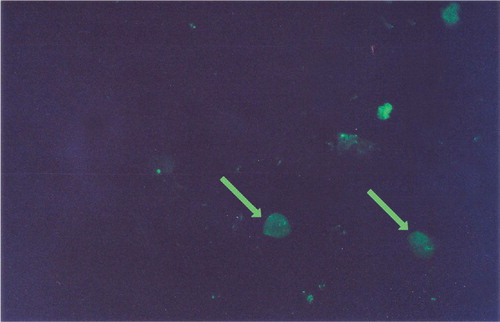
The specimen was observed with a Leica DMR fluorescent microscope (Leica Microsystems, USA) under oil immersion in dark field at an objective of 40×by BV exciting method (emitting a blue-violet colour mainly at 4.047 and 4.038 Å). Gender was identified as male if one fluorescent spot was observed in the nucleus (F-body, seen in the Y chromosome) and as female if no spot was observed ( & ).
Figure 1. Nucleus of male cell with fluoresent ‘Y’ chromosome.
Figure 2. Nucleus of female cell with faint fluorescence of the whole nucleus.
3 Statistics
This study was done to evaluate the use of the fluorescent body test as a diagnostic test for sex determination. Sensitivity, specificity, positive predictive value, negative predictive value, and efficiency were calculated for each of the three groups of teeth(). The descriptive data included mean, standard deviation, and range(). The median was used as the cut-off value.
Table 1. Diagnostic utility of fluorescent body in identifying gender
Table 2. The mean, standard deviation, and range of the number of fluorescent bodies observed in freshly extracted teeth and one month and five months after extraction
4 Results and discussion
We often encounter cases of murder or injury from assault where a single tooth is the sole material left behind at the scene of crime. In such cases even a single tooth might prove to be an important factor for identifying the individual.
Sex determination from skulls is highly unreliable and there is also much conflicting evidence as to the reliability of estimates from tooth size Citation8. Although a number of workers have described differences in the size of male and female teeth, such measurements seem unlikely to be used to determine the sex of an individual.
A technique has been developed that might offer a solution whenever soft tissue parts cannot be recovered. It has been shown that when chromosomes are stained with quinacrine mustard, they fluoresce differentially along their length when viewed under ultraviolet light and that the human Y chromosome fluoresces more brightly than the other chromosomes Citation8.
It has been demonstrated that the Y chromosome can be seen not only during mitosis but also at interphase. The reason for the bright fluorescence of the Y chromosome is not entirely clear. Caspersson et al. Citation9 suggested that alkylating agents such as quinacrine accumulate in DNA regions rich in guanine. This technique has been used in forensic science for sex determination from dried blood stains, saliva, and hair Citation8. Thus this technique might be applicable to sex determination from tooth pulp some time after death because the teeth are a stable part of the skeleton and the pulp tissue is well protected. In 1972, Seno and Ishizu Citation10 carried out the detection of Y chromosome in the nuclei of dental pulp. In this study we examined dental pulp tissue one and five months after extraction.
In this study we observed in males 44–71 fluorescent Y bodies (mean 57.7 bodies) in fresh samples, 19–49 bodies (mean 30.6) one month after extraction, and 5–22 bodies (mean 12) five months after extraction (Tables ). Seno and Ishizu Citation9 showed a range of fluorescent bodies of 56–72 after one month and 30–47 after five months.
Table 3. Correct and incorrect identification of gender from freshly extracted teeth
Table 4. Correct and incorrect identification of gender one month after extraction of teeth
Table 5. Correct and incorrect identification of gender five months after extraction of teeth
The present study showed pseudo Y body in females in the range of 0–13 (mean 4.4), 4–14 (mean 9.4), and 2–14 (mean 9) for fresh samples, one month and five months after extraction, respectively (Tables ). Seno and Ishizu Citation10 attributed the appearance of fluorescent bodies in females to artifacts and Caspersson et al. Citation9 attributed it to the presence of fluorescent debris. Age and the disease for which the teeth were extracted were not factors in assessment of fluorescent Y body as the aim of the study was to obtain the gender characteristic of individuals from fluorescent body assessment.
In the study by Whitaker, assessment of gender was correct in 30–100% of instances. Correct assessment was common in tissues putrefying up to four weeks after extraction, but accuracy was reduced in specimens left for 6–10 weeks, and it was concluded that it was necessary to smear pulp cells thinly to prevent masking the fluorescent Y chromosomes by fluorescent debris. Y chromosomes were usually readily visible in thin smears Citation8.
We show that for freshly extracted teeth and those examined one month later, sensitivity, specificity, positive predictive value, negative predictive value, and efficiency were all 100% ().
The discrepancy between the present and previous studies could be attributed to variation in sample size, random inclusion of carious and non-carious teeth, or lack of bias in double blind trials such as the study conducted by Whittaker and Llewellyn (8). A similar study by Adachi (11) demonstrated the appearance of Y chromatin in the dental pulp stained by quinacrine mustard.
A decrease in the Y positive cells and decreased reliability of the study as the tooth ages was noted in the male teeth, which is in accord with the work done on dried blood stains by Phillips and Gaten Citation12, and on dental pulp cells by Seno and IshizuCitation10, Whittaker and LlewellynCitation8, and Adachi Citation11.
Future developments in isolation of pulpal cells and staining procedure might yield better results. In the present study, the fluorescent Y body test is shown to be a reliable, simple, and cost-effective technique for gender identification in the immediate postmortem period up to one month after death.
5 Conflict of interest and funding
The authors have not received any funding or benefits from industry to conduct this study.
Acknowledgements
We thank the staff of the Department of Oral Pathology and Microbiology of Bapuji Dental College and the College of Dental Sciences, Davangere, India, for their help in the investigative procedures.
References
- Rothwell BR. Principles of dental identification. Dent Clin North Am. 2001; 45: 2253–70.
- Pillay VV. MKR Krishnan's hand book of forensic medicine and toxicology12th ed. Paras Publications. HyderabadIndia, 2001; 34.
- Bailoor DN, Nagesh KS. Fundamentals of oral medicine and radiology2nd ed. Jaypee Publishers. New DelhiIndia, 2001; 333.
- Ramenzoni LL, Line SRP. Automated biometrics-based personal identification of the Hunter Schreger bands of dental enamel. Proc Biol Sci. 2006; 273: 1155–58.
- Kerr DA, Ash MM, Millard HD. Oral diagnosis6th ed. C.V. Mosby Company. St. Louis MO, 1983; 82.
- Kruger OG. Text book of oral and maxillofacial surgery6th ed. Jaypee Publishers. New DelhiIndia, 1989; 52.
- Malaver PC, Yunis JJ. Different dental tissues as source of DNA for human identification in forensic cases. Croat Med J. 2003; 44: 306–9.
- Whittaker DK, Llewellyn DR. Sex determination from necrotic pulp tissue. Br Dent J. 1975; 139: 403–5.
- Caspersson T, Zech L, Johansson C, Modest EJ. Identification of human chromosomes by DNA binding fluorescent agents. Chromosoma. 1970; 30: 215–7.
- Seno M, Ishizu H. Sex identification of human tooth. Int J Forensic Dent. 1973; 1: 8–11.
- Adachi H. (1) Studies on sex determination using human dental pulp; (2) Sex determination of teeth left in a room. Nippon Hoigaku Zasshi. 1989; 43: 27–39.
- Phillips AP, Gaten E. Y chromosome fluorescence in blood stains. Lancet. 1971; 298: 371–2.