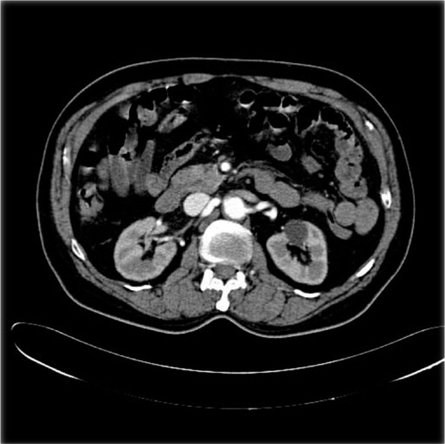
A 75-year-old male was admitted with sudden onset of unilateral left lower limb swelling and pain. He was a hypertensive on treatment for the past 10 years and a chronic smoker. On examination, the patient's blood pressure was 220/130 mm Hg and he had palpable abdominal pulsations. A bruit was audible over the patient's lower abdomen. An ultrasound showed a retroperitoneal cystic mass and CT scan confirmed the presence of large aneurysm involving the left common iliac artery () which was compressing upon the ipsilateral common iliac vein (, white arrow). A fistulous communication was found between the left common iliac artery and the vein (, black arrow) with early contrast opacification of the common iliac vein and the inferior vena cava in the arterial phase (). The patient developed chest pain and breathlessness and eventually expired due to cardiorespiratory arrest.
Fig. 1. Coronal reformatted CT scan image showing a large aneurysm arising from the left common iliac artery.
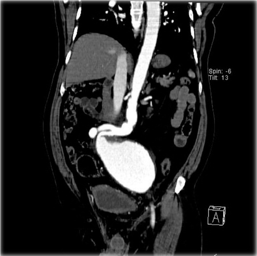
Fig. 2. Axial CT scan image showing the left common iliac vein (white arrow) is compressed by the aneurysm with formation of a fistulous communication (black arrow).
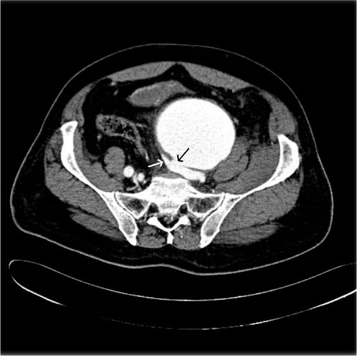
Fig. 3. Axial CT scan showing early opacification of the inferior vena cava in the arterial phase.
Isolated iliac artery aneurysm is a rare entity with reported incidence of 0.03%. They tend to occur predominantly in male patients (5:1) with a mean age of presentation of 75 years. Iliac artery aneurysms often go undetected for years as the patients usually remain asymptomatic unless complications supervene. In addition, the relative rarity of these aneurysms lead to low index of suspicion in patients with chronic pelvic pain or an overt vascular emergency. The rate of rupture of common iliac artery aneurysms varies from 14 to 75%. The aneurysm can rupture into any of the adjacent organs including the bowel, bladder, ureter or very rarely into the adjacent common iliac vein resulting in an arteriovenous fistula. Patients with arteriovenous fistula present with a distinct triad of findings described by McAuley et al. Citation1: high-output cardiac failure of precipitous onset, a pulsatile abdominal mass accompanied by a thrill and bruit, and unilateral lower-extremity ischemia or venous engorgement. This triad of clinical findings should raise a suspicion of arteriovenous fistula and guide the further investigations.
Learning points
In a patient presenting with sudden onset lower limb edema, abdominal bruit and high-output cardiac failure, a possibility of an arteriovenous fistula should be considered in the differential diagnosis.
CT scan is the modality of choice for investigation as it can exquisitely demonstrate the site of the fistula and can provide a further roadmap for endovascular treatment.
References
- McAuley CE, Peitzman AB, deVries EJ, Silver MR, Steed DL, Webster MW. The syndrome of spontaneous iliac arteriovenous fistula: a distinct clinical and pathophysiologic entity. Surgery. 1986; 99: 373–7.