Abstract
Background : Over-the-counter (OTC) feminine hygiene products come with little warning about possible side effects. This study evaluates in-vitro their effects on Lactobacillus crispatus, which is dominant in the normal vaginal microbiota and helps maintain a healthy mucosal barrier essential for normal reproductive function and prevention of sexually transmitted infections and gynecologic cancer.
Methods : A feminine moisturizer (Vagisil), personal lubricant, and douche were purchased OTC. A topical spermicide (nonoxynol-9) known to alter the vaginal immune barrier was used as a control. L. crispatus was incubated with each product for 2 and 24h and then seeded on agar for colony forming units (CFU). Human vaginal epithelial cells were exposed to products in the presence or absence of L. crispatus for 24h, followed by epithelium-associated CFU enumeration. Interleukin-8 was immunoassayed and ANOVA was used for statistical evaluation.
Results : Nonoxynol-9 and Vagisil suppressed Lactobacillus growth at 2h and killed all bacteria at 24h. The lubricant decreased bacterial growth insignificantly at 2h but killed all at 24h. The douche did not have a significant effect. At full strength, all products suppressed epithelial viability and all, except the douche, suppressed epithelial-associated CFU. When applied at non-toxic dose in the absence of bacteria, the douche and moisturizer induced an increase of IL-8, suggesting a potential to initiate inflammatory reaction. In the presence of L. crispatus, the proinflammatory effects of the douche and moisturizer were countered, and IL-8 production was inhibited in the presence of the other products.
Conclusion : Some OTC vaginal products may be harmful to L. crispatus and alter the vaginal immune environment. Illustrated through these results, L. crispatus is essential in the preservation of the function of vaginal epithelial cells in the presence of some feminine hygiene products. More research should be invested toward these products before they are placed on the market.
Introduction
Numerous women use feminine hygiene products every day; for some, it is part of their daily cleansing. Feminine hygiene products or methods may upset the normal pH level of ~4.5 in the vagina, which is important for maintaining the healthy vaginal immune barrier environment Citation1. Through the change of pH or through direct bactericidal properties, such products and practices may affect the composition of the normal vaginal microbiome, which is essential for the healthy mucosal environment and protection against yeast infection or other sexually transmitted pathogens. Vaginal lactobacilli are also important for having a healthy pregnancy and may prevent premature delivery and illness in the newborns Citation2. The disturbance of the normal microbiome, a condition called bacterial vaginosis (BV), is associated with significant risks for women's health as well as inflammatory complications in the newborn Citation3, while the presence of Lactobacillus has been associated with lower risk of inflammation Citation2. Lactobacilli and, especially, L. crispatus are among the bacteria that are most common in healthy women and characteristic for the healthy vaginal environment Citation4 Citation5. Lactobacilli are anaerobic bacteria that convert lactose and other sugars into lactic acid, which has a role in preventing infections Citation4. Lactobacillus is found in the gut and vagina, where it has anti-inflammatory and anti-cancer activities. In the industry, it is used to make yogurt and cheese, and it can also be used as a biotherapeutic. Because of its beneficial properties to the host, some Lactobacillus species have been used as probiotics available over the counter to aid mucosal health, for example, in the digestive tract. Lactobacillus products are also under investigation to restore vaginal health after prolonged use of antibiotics or to cure BV and prevent urinary tract infections Citation6 Citation7. L. crispatus has been found to effectively help inhibit the growth of harmful pathogenic microorganisms such as Neisseria gonorrhoeae Citation8. This study applied an experimental system for safety evaluation of vaginal products by testing their effects on L. crispatus growth and survival in-vitro. The evaluation was carried out by comparing products representing over-the-counter (OTC) douche kits, moisturizers, and lubricants to cell culture medium that maintains the growth of Lactobacilli in-vitro and nonoxynol-9 (N-9), a spermicide known to upset the vaginal immune barrier Citation9–Citation11.
Materials and methods
Test products
The test articles listed in (Vagisil Feminine Moisturizer, personal lubricant and moisturizer, and CareOne Douche) were purchased over the counter and were stored completely sealed, in their original box, at room temperature. The selection of products was made based on availability and public knowledge on each of the products. Once purchased, each product was screened to check for sterility. This was done by seeding each product undiluted over agar plates. None of the products showed any bacterial growth in this test. Phosphate-buffered saline (PBS) (Invitrogen by Life Technologies, Carlsbad, CA) and keratinocyte serum-free medium (KSFM) (Invitrogen, Carlsbad, CA) supplemented with epidermal growth factor, bovine pituitary extract, and CaCl2 as described Citation12 were obtained from Invitrogen, Carlsbad, CA. Nonoxynol-9 (N-9) was obtained from Personal Products Company (Skillman, NJ) Citation13 and diluted to 2% with KSFM. For assessing their effect on bacterial growth, all products were applied at 1:1 ratio over the bacterial suspensions.
Table 1. pH level along with ingredients listed on OTC product package
Preparation of bacterial suspension and treatment
L. crispatus was originally isolated from vaginal swab samples from healthy women participating in a vaginal microflora research study and then characterized and expanded in the laboratory of Dr. A. Onderdonk, Brigham and Women's Hospital Citation14. Once the volume needed was calculated, the frozen bacterial stocks were thawed in a room temperature water bath. The desired volume was transferred into a 50 mL tube and centrifuged down at 5000g× for 10 min at 25°C. After discarding the supernant, the bacterial pellet was resuspended in KSFM cell culture medium to the concentration of 7 × 10⁶ CFU/mL. Hundred microliter of the bacterial suspension was then added over 100 microliter undiluted product in 96-well tissue culture plates (Becton Dickson and Company, Franklin Lakes, NJ). The designated plates were then incubated on an orbital shaker in an anaerobic chamber (AnaeroPack System, PML Microbiologicals, Wilsonville, OR) at 35°C for 24 h or 2 h. From the 200 µl of test products along with bacterial suspension, three replicates within the same tissue culture plate were made. Each replicate contained 50 µl of the mixture of product and bacteria, along with 50µl of 50% glycerol, and was cryopreserved at −80°C.
Dilution of test plates
To assess colony forming units, the bacterial suspensions in KSFM or in test product collected at each time point were serially diluted using 1× PBS. The first row of the 96-well plate contained L. crispatus along with the product. The following rows contained the desired amount of dilution in PBS achieved by transferring desired volume from one row to another using multichannel pipettes, and when the product had high viscosity, we used positive displacement pipettes (Gilson Medical Electronics through Fisher Scientific, Pittsburgh, PA).
Seeding bacteria over agar plates
Brucella Agar Plates with 5% sheep blood, hemin, and vitamin K1 (BD, Franklin Lakes, NJ) were brought out from refrigerator to adjust to room temperature for 10 minutes. A pipette tip was used to mark either halves or thirds or the agar surface. After the serial dilutions of L. crispatus plus product were made, 30 µl was taken from a single well and was spread out over the agar, using a Steriloop (Fisher Scientific, Pittsburgh, PA). The plates were then incubated in an anaerobic chamber for 48–72 h, or until there were visible colonies.
Colony forming units counts
Bacterial colonies were counted in the agar plates. The data was then entered into Excel, multiplied by the dilution factor, and transformed logarithmically (log10). The average log10 data from all dilutions that generated countable colony-forming units (CFU) for each culture in each experiment were used for statistical analysis of the triplicate cultures conducted in at least two independent seeding experiments for each time point and product.
Preparation of vaginal epithelial cells and seeding over 96-well plates
We utilized for this study an immortalized vaginal keratinocyte cell line (Vk2/E6E7), which maintains the differentiation patterns and phenotypic characteristics of its normal human tissue of origin Citation12 Citation15 Citation16. Vaginal cells were grown in a T75 flask using KSFM medium. Once grown to confluence, the cells were trypsinized. After decanting the old medium, 5 mL of warm trypsin was added and the flask was tipped back and forth to wash away any dead cells. After removing the trypsin, 10 mL of fresh trypsin was added and incubated for 10 min at 37°C. After seven minutes, detachment of cells was checked under a microscope. Once enough cells were detached, equal volume of neutralization medium was added on top of the trypsin. The cell suspensions were then spun down at 500 g for 10 min. Next, the supernatant was decanted, and the cells were resuspended in 2 mL of antibiotic-free medium (AB-) and counted. The cell density was adjusted to 3 × 10⁵/mL and the suspension transferred to a sterile reservoir. Hundred microliter of suspension was transferred per a well into a 96-well plate. The plate was then incubated for at least 24 h before adding test products or bacterial coculture.
Epithelial cell viability and IL-8 measurement
Following 24h incubation with undiluted and diluted test products, epithelial cell viability was assessed by MTT assay as described Citation17. IL-8 levels were measured in cell culture supernatants using a Meso Scale Discovery (MSD) small-spot immunoassay and Sector Imager 2400 (Meso Scale Discovery, Gaithersburg, MD).
Bacterial-epithelial co-culture and epithelial-associated CFU counts
Details on the bacterial colonization model used in this study are published elsewhere Citation17. In brief, once the vaginal keratinocytes reached 100% confluence, medium was removed and replaced by 50 µL of bacterial suspension and 50 µL of test product. The plate was then placed in an anaerobic jar with one AnaeroPack inside and incubated for 24 h. After 24 h incubation, supernants were collected and frozen for cytokine assays. Using 200 µL of (1×) PBS, cell layers were washed in original plate twice with a multichannel pipette. Cells were then lysed by adding 100 µl of Hypure water and incubated at room temperature for 15 minutes followed by adding 100µl of (2×) PBS over the Hypure water and mixing well. Lysates were serially diluted in PBS and seeded over agar plates with a sterile loop. Agar plates were incubated under anaerobic conditions for 48–72 h, followed by CFU counting.
Statistical analysis
Two-tail ANOVA analysis of variance and Prism (GraphPad Software Inc, San Diego, CA) were used to compare the CFU values, viability, and IL-8 levels obtained for test product and the control medium.
Results
Effect of products on planktonic L. crispatus culture
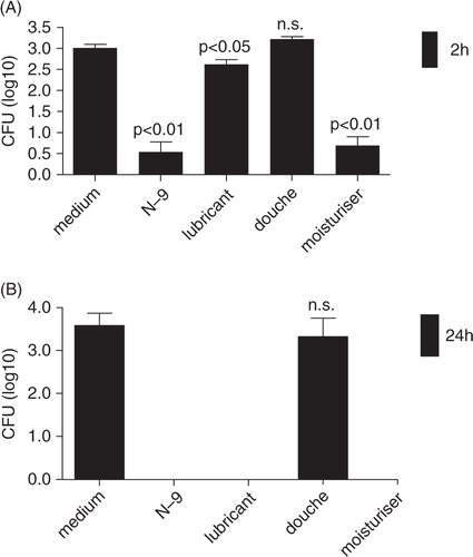
To simulate a naturally thriving environment of the natural microbiota of the vagina, L. crispatus was incubated at 2 and 24 h with KSFM medium, which supports the growth of human vaginal epithelial cells Citation12. Acting as a control, it was expected that under these conditions the bacteria would grow to a sufficient number of colonies. These plates were used as a maximum threshold to compare the serial dilutions done in the absence or presence of OTC vaginal products. The results from the series of incubations and dilutions concurred with the expectations for good support of bacterial growth demonstrating recovery of CFU input counts ( columns one). Acting as a bactericidal control, N-9 was incubated along with L. crispatus for both 2 and 24 h. As expected, N-9 had a negative effect on the bacteria. At 24 h N-9 completely killed the bacteria, while at 2 h, N-9 significantly (p<0.01) decreased the number of bacterial colonies (). The personal lubricant explored during this study decreased number of bacterial colonies by less than 1 log at 2 h incubation period (p<0.05) (A); however, at 24 h, it completely prevented bacterial growth and no colonies were visible from seeding of either diluted or undiluted bacterial suspensions (B). Vagisil significantly suppressed Lactobacillus growth even at a short 2 h incubation time (A) and at 24 h completely abolished bacterial colonies (B). The vaginal douche tested in this study did not show any significant effect on L. crispatus as compared to the KSFM control (A and B).
Fig. 1. Composite effect of vaginal products on L. crispatus CFU. Bars represent logarithmically transformed means and standard errors of the mean from triplicate CFU measurements at (A) 2 h and (B) 24 h for each vaginal product exposed to bacteria twice. P values <0.05 and <0.01 show significant differences from the medium control. n.s. = no significant difference from control.
Effect of products on bacterial-epithelial interactions
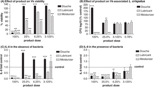
All products tested were toxic to the vaginal keratinocytes when applied undiluted (at 100% dose) (A). The douche was the least toxic and the moisturizer most toxic over a set of dilutions (A). While the undiluted douche stimulated bacterial growth (increasing recovery of epithelial cell-associated CFU to ~200%), the other products suppressed bacterial growth, with the moisturizer being suppressive up to a 4-fold dilution (at 25% dose) (B). The products had differential effects on the production of IL-8, which is an important proinflammatory innate immunity mediator produced by the vaginal epithelium to attract leukocytes to the mucosal surface in response to infection Citation18 Citation19. In the absence of bacteria the douche triggered increased IL-8 production (C). L. crispatus counteracted this effect. In the presence of L. crispatus, all products suppressed IL-8 production (D).
Fig. 2. (A) Effect of product on vaginal keratinocyte (Vk) viability assessed by MTT assay, (B) L. crispatus colonization assessed by Vk-associated CFU, and Vk IL-8 production in (C) the absence or (D) the presence of L. crispatus. Data represent means and SEM of biological triplicates. * = p<0.05, ** = p<0.01, *** = p<0.001, values in product-treated cultures lower than medium control; + = p<0.05, + + + = p<0.001, values in product-treated cultures higher than medium control.
Discussion
The mucosal lining of the vagina provides essential help in maintaining a healthy vaginal environment. The results from this experimental study demonstrate that some vaginal products may be harmful to the Lactobacillus bacteria and therefore should be used with caution. Referring to , both the lubricant and N-9 showed to have a significant effect on the growth of L. crispatus. At 24 h (), both the lubricant and N-9 completely inhibited bacteria growth in our experimental system.
The results from this experimental study confirm the hypothesis that some feminine hygiene products may alter the vaginal immune barrier by having a negative effect on the epithelial cell integrity (measured by MTT), survival of beneficial Lactobacillus species in the vaginal microenvironment (measured by epithelial-cell-associated CFU), and by changing the ability of the vaginal epithelial cells to produce protective or inflammatory immune mediators, for example, IL-8. The results confirm clinical findings of the harmful effects of certain vaginal hygiene practices on susceptibility to infections. The moisturizer and the lubricant were especially cytotoxic and negatively affecting L. crispatus survival. The douche, on the other hand, could promote a proinflammatory environment in the absence of Lactobacillus or when the microbiome is disturbed. More clinical studies are needed to test the findings and conclusions from these experiments, which if confirmed would argue for the benefit of using probiotics for vaginal health.
Our results provide experimental warning that women who have used nonoxynol-9, Vagisil, or lubricant may have a weakened vaginal barrier due to destroyed L. crispatus and perhaps other normal microflora species not assessed in our study, thus becoming at risk for the development of the syndrome of disturbed vaginal microbiome Citation20. Women using such products on a regular basis should take measures such as regular use of condoms to prevent sexually transmitted infections and should be evaluated for BV, which can sometime be asymptomatic and thus remain unnoticed by the vaginal product user. Although the particular douche kit tested did not show to have a negative effect on L. crispatus growth in our experimental system, it must be noted that some douching products have been associated with B streptococci and different Candida species Citation21. Douching has also been associated with a non-regression of low-grade squamous intraepithelial lesions Citation21. The use of vaginal spermicides and vaginal medications has been known to onset an unstable microflora and BV Citation22. It has to also be noted that the effects of vaginal products on bacterial growth can be selective and can vary among commercial brands, as suggested by another in-vitro study, which demonstrated differences among seven commercial douches tested against Lactobacillus isolates as well as BV associated bacteria Citation23. All three products tested were estimated to be fairly acidic (), although the pH varied within values typical for normal vaginal environment, except for the douche, which was more acidic.
Using the data that was amassed from this project, more research can hopefully go into vaginal health and development of probiotics for restoring and maintenance of a healthy vaginal microflora. Conventional health care wisdom has warned consumers against the use of douches a due to their stripping the vagina of its protective lining and altering its pH. Though the brand we tested did not show to inhibit the growth in-vitro of L. crispatus, frequent douching with this or other brands may throw the vagina's internal protective system off balance and promote non-sexually and sexually transmitted infections. Regardless of this, millions of females still continue to use it for personal and cultural reasoning. Hopefully this research may blossom into a new field in the biopharmaceutical market. Just as vaginal suppositories exist containing estrogen replacement therapies and anti-fungal agents to combat non-sexually transmitted infections such as candida (yeast) and BV, we may envision vaginal probiotic replacement therapies in the future in order to promote vaginal health and curb the frequency of reoccurring yeast infections and as a supplement available to women who continue to use these products. Preventative therapies are the path of modern medicine. Finding treatments for tomorrows’ disease may be the cloud with a silver lining for patients who are afflicted, but preventing them is golden.
Acknowledgements
The authors gratefully acknowledge the funding support and career mentorship from the Center for Community Health and Health Equity's Student Success Jobs Program (SSJP), a program of Brigham and Women's Hospital. The first author of this article, Bisiayo Fashemi, conceived the idea, conducted all experiments described here, performed literature search and wrote the manuscript. While conducting the study she was a student at Boston Latin Academy and intern supported by SSJP under the training, mentorship and guidance by Dr. Fichorova. Dr. Fichorova assisted with the experimental design and with editing the manuscript. M. Delaney isolated the primary L. crispatus strain and expanded and provided the bacterial stocks for these experiments. Dr. Onderdonk provided guidance for the culture of L. crispatus under anaerobic conditions, read the manuscript, and provided comments. The authors thank Hidemi Yamamoto, lab manager of Dr. Fichorova's laboratory, for her invaluable guidance and encouragement to the students.
Related Research Data
References
- Hainer BL, Gibson MV. Vaginitis: diagnosis and treatment. Am Fam Physician. 2011; 83: 807–15.
- Fichorova RN, Onderdonk AB, Yamamoto H, Delaney ML, Dubois AM, Allred E, et al. Maternal microbe-specific modulation of inflammatory response in extremely low-gestational-age newborns. mBio. 2011; 2: e00280–10.
- Larsson PG, Bergstrom M, Forsum U, Jacobsson B, Strand A, Wolner-Hanssen P. Bacterial vaginosis. Transmission, role in genital tract infection and pregnancy outcome: an enigma. APMIS. 2005; 113: 233–45. 10.3402/mehd.v24i0.19703.
- Antonio MA, Hawes SE, Hillier SL. The identification of vaginal Lactobacillus species and the demographic and microbiologic characteristics of women colonized by these species. J Infect Dis. 1999; 180: 1950–6. 10.3402/mehd.v24i0.19703.
- Verstraelen H, Verhelst R, Claeys G, De Backer E, Temmerman M, Vaneechoutte M. Longitudinal analysis of the vaginal microflora in pregnancy suggests that L. crispatus promotes the stability of the normal vaginal microflora and that L. gasseri and/or L. iners are more conducive to the occurrence of abnormal vaginal microflora. BMC Microbiol. 2009; 9: 116. 10.3402/mehd.v24i0.19703.
- Stapleton AE, Au-Yeung M, Hooton TM, Fredricks DN, Roberts PL, Czaja CA, et al. Randomized, placebo-controlled phase 2 trial of a Lactobacillus crispatus probiotic given intravaginally for prevention of recurrent urinary tract infection. Clin Infect Dis. 2011; 52: 1212–7. 10.3402/mehd.v24i0.19703.
- Senok AC, Verstraelen H, Temmerman M, Botta GA. Probiotics for the treatment of bacterial vaginosis. Cochrane database of systematic reviews (Online). 2009: CD006289.
- Graver MA, Wade JJ. The role of acidification in the inhibition of Neisseria gonorrhoeae by vaginal lactobacilli during anaerobic growth. Ann Clin Microbiol Antimicrob. 2011; 10: 8. 10.3402/mehd.v24i0.19703.
- Hillier SL, Moench T, Shattock R, Black R, Reichelderfer P, Veronese F. In vitro and in vivo: the story of nonoxynol 9. J Acquir Immune Defic Syndr. 2005; 39: 1–8. 10.3402/mehd.v24i0.19703.
- Fichorova RN, Tucker L, Anderson DJ. The molecular basis of nonoxynol-9 induced vaginal inflammation and its possible relevance to human immunodeficiency virus type 1 transmission. J Infect Dis. 2001; 184: 418–28. 10.3402/mehd.v24i0.19703.
- Trifonova RT, Pasicznyk JM, Fichorova RN. Biocompatibility of solid-dosage forms of anti-human immunodeficiency virus type 1 microbicides with the human cervicovaginal mucosa modeled ex vivo. Antimicrob Agents Chemother. 2006; 50: 4005–10. 10.3402/mehd.v24i0.19703.
- Fichorova RN, Rheinwald JG, Anderson DJ. Generation of papillomavirus-immortalized cell lines from normal human ectocervical, endocervical and vaginal epithelium that maintain expression of tissue-specific differentiation proteins. Biol Reprod. 1997; 57: 847–55. 10.3402/mehd.v24i0.19703.
- Fichorova RN, Bajpai M, Chandra N, Hsiu JG, Spangler M, Ratnam V, et al. Interleukin (IL)-1, IL-6, and IL-8 predict mucosal toxicity of vaginal microbicidal contraceptives. Biol Reprod. 2004; 71: 761–9. 10.3402/mehd.v24i0.19703.
- Onderdonk AB, Zamarchi GR, Rodriguez ML, Hirsch ML, Munoz A, Kass EH. Qualitative assessment of vaginal microflora during use of tampons of various compositions. Appl Environ Microbiol. 1987; 53: 2779–84.
- Fichorova RN, Cronin AO, Lien E, Anderson DJ, Ingalls RR. Response to Neisseria gonorrhoeae by cervicovaginal epithelial cells occurs in the absence of toll-like receptor 4-mediated signaling. J Immunol. 2002; 168: 2424–32.
- Fichorova RN, Anderson DJ. Differential expression of immunobiological mediators by immortalized human cervical and vaginal epithelial cells. Biol Reprod. 1999; 60: 508–14. 10.3402/mehd.v24i0.19703.
- Fichorova RN, Yamamoto HS, Delaney ML, Onderdonk AB, Doncel GF. Novel vaginal microflora colonization model providing new insight into microbicide mechanism of action. MBio. 2011; 2: e00168–11.
- Fichorova RN, Desai PJ, Gibson FC 3rd, Genco CA. Distinct proinflammatory host responses to Neisseria gonorrhoeae infection in immortalized human cervical and vaginal epithelial cells. Infect Immun. 2001; 69: 5840–8. 10.3402/mehd.v24i0.19703.
- Fichorova RN, Trifonova RT, Gilbert RO, Costello CE, Hayes GR, Lucas JJ, et al. Trichomonas vaginalis lipophosphoglycan triggers a selective upregulation of cytokines by human female reproductive tract epithelial cells. Infect Immun. 2006; 74: 5773–9. 10.3402/mehd.v24i0.19703.
- Koumans EH, Sternberg M, Bruce C, McQuillan G, Kendrick J, Sutton M, et al. The prevalence of bacterial vaginosis in the United States, 2001–2004; associations with symptoms, sexual behaviors, and reproductive health. Sex Transm Dis. 2007; 34: 864–9. 10.3402/mehd.v24i0.19703.
- Chu T-Y, Hsiung CA, Chen C-A, Chou H-H, Ho C-M, Chien T-Y, et al. Post-coital vaginal douching is risky for non-regression of low-grade squamous intraepithelial lesion of the cervix. Gynecol Oncol. 2010; 120: 449–53. 10.3402/mehd.v24i0.19703.
- Schwebke JR, Richey CM, Weiss HL. Correlation of behaviors with microbiological changes in vaginal flora. The Journal of infectious diseases. 1999; 180: 1632–6. 10.3402/mehd.v24i0.19703.
- Pavlova SI, Tao L. In vitro inhibition of commercial douche products against vaginal microflora. Infect Dis Obstetr Gynecol. 2000; 8: 99–104.