Abstract
Well-differentiated fetal adenocarcinoma (WDFA) is a rare pulmonary malignancy. Biomarkers of tumor biology has rarely been studied in WDFA. Here, we report two WDFA patients. Both patients had blood-streaked sputum or mild hemoptysis at presentation. They underwent lobectomy and systematic mediastinal lymphadenectomy. Expression of PDGFRα on the plasma membrane was demonstrated by immunohistochemistry (IHC) in the resected tumor specimens. Further IHC examination showed intense immunostaining of β-catenin in both patients but negative staining for TP53, CEA, CD56, EGFR, CK5/6, HER2, S-100, ER, PR, BCL2, and NSE. Both patients had no recurrence to date after more than 3 years of follow up. Herein, we reviewed this rare disease with special emphasis on the clinico-pathological features, treatment and potential role of PDGFRα.
Introduction
Well-differentiated fetal adenocarcinoma (WDFA) is a rare type of lung cancer. It is characteristically composed of fetal type glands and tubules. The normal stromal components lack sarcomatous feature, which is different from the conventional pulmonary blastoma.Citation1,Citation2 WDFA is considered a variant of invasive adenocarcinoma in modern classification of lung adenocarcinomas but usually with good prognosis.Citation3
The PDGFR-PDGF signaling system includes two cell surface receptors (PDGFRα and PDGFRβ) and several ligands (PDGFA, PDGFB, PDGFC, and PDGFD). Ligand binding induces receptor dimerization, enabling autophosphorylation of specific tyrosine residues and subsequent recruitment of signal transduction molecules.Citation4 PDGFRα, one of the isoforms of platelet-derived growth factor receptor (PDGFR), is a trans-membrane tyrosine kinase receptor with an important role in cancer growth in several tumor types and a small subset of NSCLCs.Citation5,Citation6 In this study, we report two WDFA patients with positive PDGFRα expression and discuss the clinico-pathological features, treatment and potential role of PDGFRα.
Results
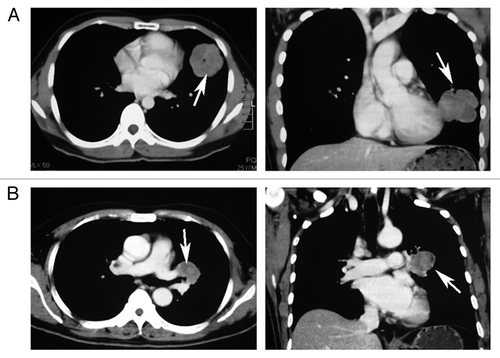
A 25-year-old male patient was referred to the Department of Thoracic Surgery, the First Affiliated Hospital of Sun Yat-sen University for reports mild left chest pain and expectoration of blood-streaked sputum. The patient had no respiratory distress, dyspnea, cough, or wheezing. There was no history of smoking, and the family history was noncontributory. Physical examination was unremarkable. CT of the chest showed a mass measuring 5.6 cm in its greatest diameter in the left lower lobe; there was no mediastinal lymphadenopathy, pleural effusion, or any signs of invasion to the chest wall (). CT of the head, abdomen and pelvis and radionuclide bone scintigraphy scan revealed no abnormal findings. The patient underwent lobectomy of left lower lobe and systemic mediastinal lymph node dissection in February 2009. The pathological stage was T2bN0M0 (stage IIA) according to the updated staging system of lung cancer.
Figure 1. Chest CT. (A) Case 1. There is a homogeneous peripheral mass in the left lower lobe of lung (white arrows). (B) Case 2. There is a 3.6-cm popcorn-like hilar mass located in the left upper lobe of the lung (white arrows).

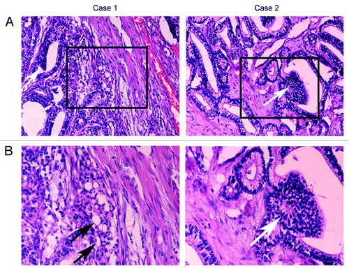
Microscopically, the tumor was composed of irregular ductal and papillary proliferation of cuboidal or columnar tumor cells which were predominantly arranged in large flat sheets, morphologically resembling the fetal lung (). The majority of the stromal cells were spindle-like cells without malignant findings. The nucleoli were indistinct, and the mitotic activity was very low. Some of the cells had prominent subnuclear and supranuclear cytoplasmic vacuoles. Squamoid-appearing morules were also identified, intimately admixed with the glandular component ().
Figure 2. H&E staining of two samples. (A) H&E stained section from the surgical resections showed well-differentiated glands and scant stroma. The glands were consisting of irregular tubular structures lined with pseudostratified columnar epithelium, and typical squamoid morules (White arrows) were found. (B) The stroma consisted of cytologically bland fibroblast-like spindle cell without sarcomatous features. The subunclear and supranuclear vacuolization was occasionally observed (Black arrows).

The other patient was a 51-year-old man, who admitted was to our hospital with reports of cough and mild hemoptysis. CT scan of the chest showed a solitary 3.6-cm mass in the left upper lobe of the lungs (). There was no pleural effusion, mediastinal or hilar lymphadenopathy. The patient underwent a left upper lobectomy with systematic mediastinal lymphadenectomy in November 2008, and the tumor was pathologically staged as T2aN0M0, stage IB. The resected tumor showed classic histologic features of WDFA, i.e., a well-differentiated glandular component showing perinuclear vacuoles and squamous morule formation, and a delicate fibrous stroma containing cytologically normal spindle cells ().
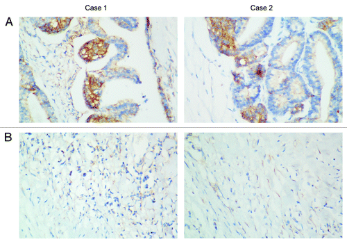
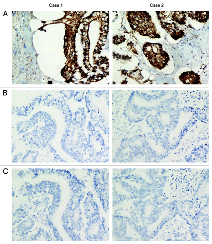
In normal lung tissues, PDGFRα is usually not detectable by immunohistochemistry, and the staining pattern of PDGFRα in lung tumors is at the plasma membrane and in the cytoplasm.Citation7 By immunohistochemistry (IHC), we found moderate staining for PDGFRα at the plasmalemma and cytoplasm of tumor cells in these two WDFA cases and negative expression of PDGFRα in the adjacent normal lung tissues (). The tumors also showed intense immunostaining for β-catenin but negative staining for TP53 and CEA (). Additionally, we performed immunostaining for CD56, EGFR, CK5/6, HER2, S-100, ER, PR, BCL2 and NSE. All these tumor markers showed negative staining (data not shown).
Figure 3. PDGFRα staining in two cases. (A) PDGFRα staining showed positive staining at the plasmalemma of the glandular cells. (B) Adjacent lung tissues of tumors in two cases showed negative expression of PDGFRα. Representative photomicrographs are shown at the original magnification × 400.

Figure 4. Immunohistochemical examination of two samples. (A) Both cases were strong positive for the β-catenin staining in the nuclei and cytoplasm. (B) CEA examination showed negative staining in two samples. (C) Negative staining of p53 in immunohistochemical examination. Representative photomicrographs are shown at the original magnification × 400.

Both patients had uneventful postoperative recovery. After more than three years of follow up, both patients have no recurrence to date.
Discussion
WDFA is a subtype of pulmonary blastoma, which was first described by Kradin et al. in 1982. It is a rare lung tumor with an incidence less than 0.1% of that for lung cancers.Citation1,Citation8 WDFA is defined by a monophasic carcinoma solely consisting of malignant epithelial components (squamoid morules) with subnuclear and supranuclear vacuoles, and a benign-appearing mesenchymal stroma.Citation1,Citation9,Citation10 Recently, WDFA is classified as a variant of invasive adenocarcinoma according to its predominant component the malignant glandular structures.Citation3 However, it is a low-grade fetal adenocarcinoma with a favorable clinical outcome.
PDGFs are a growth factor family; they exert their mitogenic effects through PDGFRα and PDGFRβ tyrosine kinase receptors. Activation and autophosphorylation of the tyrosine kinase domain of PDGFR recruits SH-2 domain-containing signal transduction proteins to activate downstream pathways through SRC, PI3K/AKT/mTORCitation11 and phospholipase CCitation12 and promote oncogenic transformation.Citation13 Recent studies revealed that high expression of PDGFRα was associated with shorter patient’s survival in gastroenteropancreatic neuroendocrine tumorsCitation14 and high metastatic potential of medulloblastoma cells.Citation15 However, the role and involvement of PDGFRα expression in WDFA as a prognostic factor has not been studied. In addition to its prognostic role, PDGFR is also an important target for chemotherapy treatment. Inhibition of PDGFRα signaling disrupts cancer cell survival in the subset of GISTs with activating PDGFRα mutations.Citation16,Citation17 The efficacy of targeted disruption of PDGFRα signaling using small molecular tyrosine kinase inhibitors in recurrent or metastatic WDFA expressing PDGFRα remains to be investigated in the future. Given the potential role of PDGFRα in the prognosis and targeted therapy in the above cancers, the value of PDGFRα in prognosis and guidance of therapy for WDFA should be further evaluated.
Little specific biomarker information has been reported for this rare tumor. Lineage-specific transcription factors (TTF-1 and GATA-6) were associated with fetal lung epithelial morphogenesis in WDFA.Citation18 Lori et al. reported that aberrant nuclear/cytoplasmic expression of β-catenin, which was involved in both cadherin-mediated cellular adhesion and intracellular signaling in the WNT pathway,Citation19,Citation20 had diagnostic utility for WDFA. The cases we reported also showed strong immunoreactivity with β-catenin. The molecular biomarkers reported for WDFA are summarized in .
Table 1. Summary of molecular biomarkers reported in WDFA
The standard treatment for WDFA is surgical resection, and most of the reported cases were in stage I with a 5-y mortality rate of 10–20% after complete resection.Citation2,Citation10 Postoperative chemotherapy or radiotherapy are selectively used for patients with advanced lung cancers, but the potential benefits in WDFA are unclear. We reviewed 9 cases of WDFA with complete resection, reported between 1995 and 2012, 6/9 were in staged I and II.Citation8,Citation21,Citation22 Among the 6 cases, 1/6 (IIB) received adjuvant chemotherapy (carboplatin and paclitaxel), 5/6 received no adjuvant therapy after complete resection, and all of them had a good prognosis.Citation8,Citation21,Citation23-Citation27 The two cases reported here were in stage IIA and IB, and no other treatment was performed after surgical resection. Up to now, no recurrence or metastasis has occurred. We speculate that adjuvant therapy may be unnecessary for patients with stage I and II WDFA after complete resection. Nevertheless, for WDFA patients with more advanced diseases or recurrent diseases, the expression and activation of PDGFRα may suggest that they may benefit from therapies targeting PDGFRα.
In summary, WDFA is a rare lung cancer with good prognosis, and complete surgical resection is still the primary therapy for this disease. The biomarkers, such as β-catenin, PDGFRα, need to be further investigated for potential contribution to preoperative diagnosis and targeted therapy in WDFA.
Patients and Methods
The two patients in this report were diagnosed of WDFA by pathologists in our hospital. The study was approved by the Committee for the Conduct of Human Research and Institutional Review Board of the First Affiliated Hospital of Sun Yat-Sen University, and informed consent was obtained from each patient.
Immunohistochemistry (IHC) was performed as previously described.Citation28 Formalin-fixed lung tissues and tumor tissues were embedded in paraffin and cut by microtome into tissue sections (4-μm thick) for immunostaining according to standard techniques. The primary antibodies were rabbit polyclonal antibody against PDGFRα (Santa Cruz Biotechnology, Inc.), monoclonal mouse antibody against β-catenin (Dako North America, Inc.), monoclonal rabbit antibody against p53 (Dako North America, Inc.) and monoclonal mouse antibody against CEA (Dako North America, Inc.). All other antibodies were from Dako Technology. As negative controls for non-specificity of the antibody for PDGFRα, immunostaining was performed for each specimen by omitting the primary antibody in the procedure. Subjective assessments of the intensity of staining were performed according to the manufacturer’s package insert.
Additional material
Download Zip (3.9 MB)Disclosure of Potential Conflicts of Interest
No potential conflicts of interest were disclosed.
Acknowledgments
No potential conflicts of interest were disclosed. The work was supported by grants from China National Natural Sciences Foundation (NO.30801136 to C. Cheng) and (No.81171125 to C. Cheng) and the Research and Development Program of Sun Yat-sen University (10YKPY09 to C. Cheng).
Supplemental Materials
Supplemental materials may be found here:
http://www.landesbioscience.com/journals/cbt/article/22253/
References
- Kradin RL, Young RH, Dickersin GR, Kirkham SE, Mark EJ. Pulmonary blastoma with argyrophil cells and lacking sarcomatous features (pulmonary endodermal tumor resembling fetal lung). Am J Surg Pathol 1982; 6:165 - 72; http://dx.doi.org/10.1097/00000478-198203000-00009; PMID: 6179430
- Sato S, Koike T, Yamato Y, Yoshiya K, Honma K, Tsukada H. Resected well-differentiated fetal pulmonary adenocarcinoma and summary of 25 cases reported in Japan. Jpn J Thorac Cardiovasc Surg 2006; 54:539 - 42; http://dx.doi.org/10.1007/s11748-006-0048-8; PMID: 17236658
- Travis WD, Brambilla E, Noguchi M, Nicholson AG, Geisinger KR, Yatabe Y, et al. International association for the study of lung cancer/american thoracic society/european respiratory society international multidisciplinary classification of lung adenocarcinoma. J Thorac Oncol 2011; 6:244 - 85; http://dx.doi.org/10.1097/JTO.0b013e318206a221; PMID: 21252716
- Valius M, Kazlauskas A. Phospholipase C-gamma 1 and phosphatidylinositol 3 kinase are the downstream mediators of the PDGF receptor’s mitogenic signal. Cell 1993; 73:321 - 34; http://dx.doi.org/10.1016/0092-8674(93)90232-F; PMID: 7682895
- Williams LT. Signal transduction by the platelet-derived growth factor receptor. Science 1989; 243:1564 - 70; http://dx.doi.org/10.1126/science.2538922; PMID: 2538922
- McDermott U, Ames RY, Iafrate AJ, Maheswaran S, Stubbs H, Greninger P, et al. Ligand-dependent platelet-derived growth factor receptor (PDGFR)-alpha activation sensitizes rare lung cancer and sarcoma cells to PDGFR kinase inhibitors. Cancer Res 2009; 69:3937 - 46; http://dx.doi.org/10.1158/0008-5472.CAN-08-4327; PMID: 19366796
- Zhang P, Gao WY, Turner S, Ducatman BS. Gleevec (STI-571) inhibits lung cancer cell growth (A549) and potentiates the cisplatin effect in vitro. Mol Cancer 2003; 2:1; http://dx.doi.org/10.1186/1476-4598-2-1; PMID: 12537587
- Zaidi A, Zamvar V, Macbeth F, Gibbs AR, Kulatilake N, Butchart EG. Pulmonary blastoma: medium-term results from a regional center. Ann Thorac Surg 2002; 73:1572 - 5; http://dx.doi.org/10.1016/S0003-4975(02)03494-X; PMID: 12022552
- Nakatani Y, Kitamura H, Inayama Y, Kamijo S, Nagashima Y, Shimoyama K, et al. Pulmonary adenocarcinomas of the fetal lung type: a clinicopathologic study indicating differences in histology, epidemiology, and natural history of low-grade and high-grade forms. Am J Surg Pathol 1998; 22:399 - 411; http://dx.doi.org/10.1097/00000478-199804000-00003; PMID: 9537466
- Koss MN, Hochholzer L, O’Leary T. Pulmonary blastomas. Cancer 1991; 67:2368 - 81; http://dx.doi.org/10.1002/1097-0142(19910501)67:9<2368::AID-CNCR2820670926>3.0.CO;2-G; PMID: 1849449
- Zhang H, Bajraszewski N, Wu E, Wang H, Moseman AP, Dabora SL, et al. PDGFRs are critical for PI3K/Akt activation and negatively regulated by mTOR. J Clin Invest 2007; 117:730 - 8; http://dx.doi.org/10.1172/JCI28984; PMID: 17290308
- Tallquist M, Kazlauskas A. PDGF signaling in cells and mice. Cytokine Growth Factor Rev 2004; 15:205 - 13; http://dx.doi.org/10.1016/j.cytogfr.2004.03.003; PMID: 15207812
- Gazit A, Igarashi H, Chiu IM, Srinivasan A, Yaniv A, Tronick SR, et al. Expression of the normal human sis/PDGF-2 coding sequence induces cellular transformation. Cell 1984; 39:89 - 97; http://dx.doi.org/10.1016/0092-8674(84)90194-6; PMID: 6091919
- Knösel T, Chen Y, Altendorf-Hofmann A, Danielczok C, Freesmeyer M, Settmacher U, et al. High KIT and PDGFRA are associated with shorter patients survival in gastroenteropancreatic neuroendocrine tumors, but mutations are a rare event. J Cancer Res Clin Oncol 2012; 138:397 - 403; http://dx.doi.org/10.1007/s00432-011-1107-9; PMID: 22160160
- MacDonald TJ, Brown KM, LaFleur B, Peterson K, Lawlor C, Chen Y, et al. Expression profiling of medulloblastoma: PDGFRA and the RAS/MAPK pathway as therapeutic targets for metastatic disease. Nat Genet 2001; 29:143 - 52; http://dx.doi.org/10.1038/ng731; PMID: 11544480
- Corless CL, Schroeder A, Griffith D, Town A, McGreevey L, Harrell P, et al. PDGFRA mutations in gastrointestinal stromal tumors: frequency, spectrum and in vitro sensitivity to imatinib. J Clin Oncol 2005; 23:5357 - 64; http://dx.doi.org/10.1200/JCO.2005.14.068; PMID: 15928335
- Dagher R, Cohen M, Williams G, Rothmann M, Gobburu J, Robbie G, et al. Approval summary: imatinib mesylate in the treatment of metastatic and/or unresectable malignant gastrointestinal stromal tumors. Clin Cancer Res 2002; 8:3034 - 8; PMID: 12374669
- Yamazaki K. Pulmonary well-differentiated fetal adenocarcinoma expressing lineage-specific transcription factors (TTF-1 and GATA-6) to respiratory epithelial differentiation: an immunohistochemical and ultrastructural study. Virchows Arch 2003; 442:393 - 9; PMID: 12684771
- Nakatani Y, Miyagi Y, Takemura T, Oka T, Yokoi T, Takagi M, et al. Aberrant nuclear/cytoplasmic localization and gene mutation of beta-catenin in classic pulmonary blastoma: beta-catenin immunostaining is useful for distinguishing between classic pulmonary blastoma and a blastomatoid variant of carcinosarcoma. Am J Surg Pathol 2004; 28:921 - 7; http://dx.doi.org/10.1097/00000478-200407000-00012; PMID: 15223963
- Heldin CH, Westermark B. Platelet-derived growth factor: three isoforms and two receptor types. Trends Genet 1989; 5:108 - 11; http://dx.doi.org/10.1016/0168-9525(89)90040-1; PMID: 2543106
- El Ouazzani H, Jniene A, Bouchikh M, Achachi L, El Ftouh M, Achir A, et al. Well-differentiated fetal adenocarcinoma: a very uncommon malignant lung tumor. Rev Port Pneumol 2012; 18:39 - 41; http://dx.doi.org/10.1016/j.rppneu.2011.06.003; PMID: 21778030
- Van Loo S, Boeykens E, Stappaerts I, Rutsaert R. Classic biphasic pulmonary blastoma: a case report and review of the literature. Lung Cancer 2011; 73:127 - 32; http://dx.doi.org/10.1016/j.lungcan.2011.03.018; PMID: 21513998
- Fujino S, Asada Y, Konishi T, Asakura S, Kato H, Mori A. Well-differentiated fetal adenocarcinoma of lung. Lung Cancer 1995; 13:311 - 6; http://dx.doi.org/10.1016/0169-5002(95)00489-0; PMID: 8719071
- Lee HJ, Goo JM, Kim KW, Im JG, Kim JH. Pulmonary blastoma: radiologic findings in five patients. Clin Imaging 2004; 28:113 - 8; http://dx.doi.org/10.1016/S0899-7071(03)00240-7; PMID: 15050223
- Pilla ES, Sanchez P, Mädke GR, Camargo S, Camargo JdeJ. Pulmonary blastoma: treatment through sleeve resection of the right upper lobe. J Bras Pneumol 2006; 32:75 - 7; http://dx.doi.org/10.1590/S1806-37132006000100014; PMID: 17273572
- Longo M, Levra MG, Capelletto E, Billè A, Ardissone F, Familiari U, et al. Fetal adenocarcinoma of the lung in a 25-year-old woman. J Thorac Oncol 2008; 3:441 - 3; http://dx.doi.org/10.1097/JTO.0b013e318169cd9a; PMID: 18379367
- Wang J, Sun H, Bai R, Liu H, Yu T. Pulmonary blastoma with endobronchial growth. J Thorac Oncol 2009; 4:543 - 4; http://dx.doi.org/10.1097/JTO.0b013e31819c8646; PMID: 19333073
- Cheng C, Liu ZG, Zhang H, Xie JD, Chen XG, Zhao XQ, et al. Enhancing chemosensitivity in abcb1- and abcg2-overexpressing cells and cancer stem-like cells by an aurora kinase inhibitor cct129202. Mol Pharm 2012; http://dx.doi.org/10.1021/mp2006714; PMID: 22632055