Abstract
Extragonadal germ cell tumors (EGCTs) in the lung are extremely rare and their pathogenesis is poorly understood. We report a case in a 48-year-old female which was very aggressive and stained positive for primoridial germ cell markers. Interestingly, there was chromosome 3 polysomy noted. To our knowledge this is the first chromosomal aberration noted in a primary germ cell tumor of the lung.
Keywords: :
Introduction
Oct3/4 (Octamer-binding transcription factor 3/4) is a transcription factor essential for the maintenance of totipotency in embryonic stem cells. It is expressed in germ cell tumors and has been labeled as the marker for primordial germ cells. During the process of differentiation this gene is downregulated.Citation1 Its interaction with the caudal-related homeobox gene (Cdx2) is responsible for the differentiation and maintenance of trophectoderm at the initial stages of development. The interaction between Oct3/4 and Cdx2 has been described as a reciprocal inhibitionCitation1 and both are markers for germ cell tumors. Oct3/4 along with a transcriptional cofactor expressed in embryonic stem cells called SRY (sex-determining region Y)-box 2(Sox-2) forms a regulatory complex needed for the maintenance of the pluripotency of primitive embryonic stem cells. The Sox-2 gene is located on chromosome 3 in humans and its upregulation is associated with aggressive nature of the tumor at least in murine lung cancer cell lines.Citation2
Extragonadal germ cell tumors in the lung are extremely rare tumors and quoted as “one of the rarest tumors encountered in pathology.”Citation3 The etiology of the tumor and possible mechanisms are still not completely understood. We report a case of an Oct3/4-positive, Cdx2-negative germ cell tumor in the lung with no evidence of gonadal lesion. The tumor showed resistance to cisplatin-based chemotherapy. We also report the findings from in situ hybridization studies that were done to analyze the chromosomal anomalies within the tumor cells. We believe that this is the first report of such unique findings and may contribute to our understanding of the pathogenesis of this extremely rare malignancy.
Case Report
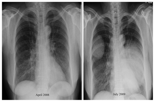
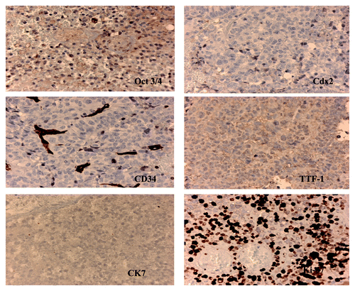
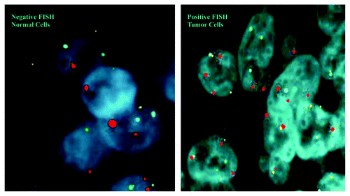
A 48-y-old woman with a past medical history significant for asthma and hypertension of 1 y duration was admitted to our hospital with a dry cough and occasional hemoptysis, which she had been experiencing for 3 mo before admission. There is a 35 pack/year history of cigarette smoking. On admission, the chest radiograph demonstrated a large soft tissue mass within the right upper lobe measuring 8 × 6 cm with additional soft tissue density measuring 4 × 2 cm along the right paratracheal region and a large soft tissue mass along the left lower lobe measuring 12 × 10 cm. These findings were compared with a previous radiograph done 15 mo previously, which was completely normal (). Chest CT scans with intravenous contrast confirmed chest radiograph findings. CT scan of the abdomen and pelvis was negative for involvement of any abdominal or pelvic organs. Pertinent labs included an α-fetoprotein (AFP) level of 254ng/ml and βHCG < 2 MIU/ml. CT-guided biopsy of one of the lung mass was reported to be negative for malignancy. The patient then underwent a left thoracotomy and left lobectomy for tissue diagnosis. The histopathology of the resected lung revealed a mixed anaplastic tumor with extensive necrosis, focal papillary feature and pseudo rosette formation. Histochemical studies showed focal positivity for α-fetoprotein, epithelial membrane antigen and HCG. Placental alkaline phosphatase (PLAP) and c-kit gene product (CD 117) were negative. The tumor was negative for thyroid transcription factor-1 (TTF-1), Cdx-2, cytokeratin (CK) 7 and 20. It was focally positive for Oct3/4 and strongly positive for the monoclonal antibody Ki-67. It was negative for the human hematopoietic progenitor cell antigen CD34 (). One of the three nodes showed focus of metastasis. Cytogenetic analysis was performed on the resected specimen using fluorescent in situ hybridization(FISH) technique for chromosome 3(red), 7(green), 9 (aqua) and 17(yellow) (). The tumor was staged IIIA based on Suster and Moran staging and chemotherapy with etoposide (100 mg/m2 per day on days 1–5) and cisplatin (30 mg/m2 per day on days 1–5) was started initially. The dose was increased later because of no radiological response and then she received six cycles of cisplatin at 30 mg/m2 and four cycles of Etoposide at 150 mg/day. After 6 mo of follow up, an increase in the size of the tumor on the right lung was noted, with no radiological recurrence noted on the site of prior resection. The AFP levels trended up to 1,454 ng/ml. The decision was made to perform a tumor resection with mediastinal lymph node sampling on the right lung. Surgery was performed on the right side, and 3 mo post-surgery follow-up the patient has no radiological tumor recurrence and the AFP levels are down to 153 ng/ml. The chromosome 3 polysomy was again noted on the second resected specimen (data not shown). The lymph node sampling was negative. Within a year of follow-up the patient died because of pneumonia.
Figure 1. Chest radiograph taken on admission compared with the one taken in 2008.The aggressive nature of the tumor is clearly evident by the rapid growth. Six month follow-up X Ray (January 2010) shows increase in the size of the tumor after three cycles of cisplatin and etoposide.

Discussion
An interesting point of discussion from this case is the cellular origin of the tumor. It appears that the tumor arose from primordial germ cells or embryonic stem cells because the tumor cells stained positive for Oct3/4. Although Oct 4 has been shown to be expressed in adenocarcinoma of the lung, the absence of CK7/20 staining suggests against the lung being the primary.Citation4,Citation5 Another possibility could be that the tumor arose from a cancer stem cell. Kim and colleagues had reported the identification of bronchioalveolar stem cells in normal and cancerous lungs.Citation6 However, those cells from their case report were positive for CD34, an accepted marker for hematopoietic stem cells. Pelosi and colleagues in their reported case of a pure yolk sac tumor of the lung showed that the tumor had diffuse Cdx2 immunoreactivity.Citation7 Our case was completely negative for Cdx2 but showed diffuse Oct3/4 immunostaining. Oct 4 is an accepted marker for identifying germ cell origin (ie, seminoma or embryonal carcinoma) in the work-up of an undifferentiated neoplasm. AFP (focally positive in our case) is not a reliable marker for yolk sac tumor because of its low sensitivity. PLAP was negative in our case, which supports the case being a non-seminomatous germ cell tumor. Low levels of AFP, as seen in our case, could be due to the poor differentiation of the tumor, again suggesting a more primordial origin. Our case appears to have a mixed embryonal carcinoma and non-seminomatous picture.
The next question we address is that did Oct 3/4 expression lead to the aggressive nature of the tumor? We hypothesize that while Oct3/4 is expressed more in germ cell tumors of the lung that are more undifferentiated and hence aggressive, our case had a greater than 50 percent Ki-67 labeling index, suggesting the aggressive nature of the tumor. Whether or not the degree of differentiation affects the response to chemotherapy is subject to further testing, but studies have shown that Oct3/4 expression confers resistance to platinum-based chemotherapy and more tumor invasion in bladder carcinoma.Citation8
We also tried to determine what might have led to the development of tumorigenesis by studying the chromosomal aberrations commonly implicated in lung cancer. We selected chromosomes 3, 7, 9 and 17 as they have been commonly implicated in the tumor biology of lung cancer. FISH analysis showed consistent polysomy in chromosome 3 in all the cells studied (). The Sox-2 gene is located on chromosome 3 and its multiplication could mean maintenance of primitive embryonic cells along by forming a regulatory complex with Oct3/4. However, planned research studies are needed to quantify the mRNA and protein analysis before any cause and effect relationship could be established. However this is the first ever reported finding which opens up new areas for research.
We hypothesize that Oct3/4 is the marker for poorly differentiated germ cell tumors in the lung and chromosome 3 polysomy may be playing a role in the development of the tumor through the above discussed mechanism.
References
- Niwa H, Toyooka Y, Shimosato D, Strumpf D, Takahashi K, Yagi R, et al. Interaction between Oct3/4 and Cdx2 determines trophectoderm differentiation. Cell 2005; 123:917 - 29; http://dx.doi.org/10.1016/j.cell.2005.08.040; PMID: 16325584
- Xiang R, Liao D, Cheng T, Zhou H, Shi Q, Chuang TS, et al. Downregulation of transcription factor SOX2 in cancer stem cells suppresses growth and metastasis of lung cancer. Br J Cancer 2011; 104:1410 - 7; http://dx.doi.org/10.1038/bjc.2011.94; PMID: 21468047
- Pont J, Pridun N, Vesely N, Kienzer HR, Pont E, Spital FJ. Extragonadal malignant germ cell tumor of the lung. J Thorac Cardiovasc Surg 1994; 107:311 - 2; PMID: 8283903
- Chen Z, Wang T, Cai L, Su C, Zhong B, Lei Y, et al. Clinicopathological significance of non-small cell lung cancer with high prevalence of Oct-4 tumor cells. J Exp Clin Cancer Res 2012; 31:10; http://dx.doi.org/10.1186/1756-9966-31-10; PMID: 22300949
- Kummar S, Fogarasi M, Canova A, Mota A, Ciesielski T. Cytokeratin 7 and 20 staining for the diagnosis of lung and colorectal adenocarcinoma. Br J Cancer 2002; 86:1884 - 7; http://dx.doi.org/10.1038/sj.bjc.6600326; PMID: 12085180
- Kim CF, Jackson EL, Woolfenden AE, Lawrence S, Babar I, Vogel S, et al. Identification of bronchioalveolar stem cells in normal lung and lung cancer. Cell 2005; 121:823 - 35; http://dx.doi.org/10.1016/j.cell.2005.03.032; PMID: 15960971
- Pelosi G, Petrella F, Sandri MT, Spaggiari L, Galetta D, Viale G. A primary pure yolk sac tumor of the lung exhibiting CDX-2 immunoreactivity and increased serum levels of alkaline phosphatase intestinal isoenzyme. Int J Surg Pathol 2006; 14:247 - 51; http://dx.doi.org/10.1177/1066896906290657; PMID: 16959714
- Chang CC, Shieh GS, Wu P, Lin CC, Shiau AL, Wu CL. Oct-3/4 expression reflects tumor progression and regulates motility of bladder cancer cells. Cancer Res 2008; 68:6281 - 91; http://dx.doi.org/10.1158/0008-5472.CAN-08-0094; PMID: 18676852

