Abstract
Objectives: The epithelial-mesenchymal-transition (EMT) is an important step in the invasion and metastasis of cancer. A critical molecular feature of this process is the downregulation of E-cadherin expression, which is mainly controlled by Snail-related zinc-finger transcription factors (Snail and Slug). The aim of this study was to evaluate the prognostic impact of EMT-related protein (E-cadherin, Snail and Slug) expression in endometrial cancer.
Methods: An immunohistochemical analysis was conducted using tissue microarray samples of 354 primary tumors and 30 metastases of endometrial carcinomas, and the relationship between protein expression, clinicopathological features and outcomes were investigated.
Results: Reduced E-cadherin was seen in 39.8% of primary tumors. Reduced E-cadherin was seen in 19.5%, 40.8% and 72.7% of G1, G2 and G3 endometrioid adenocarcinomas, respectively. The nuclear expression of Snail and Slug were positive in 16.9% and 3.7% of primary tumors, respectively. EMT status, which was represented by both reduced E-cadherin and nuclear expression of Snail, was significantly associated with histological type, FIGO stage, myometrial invasion, positive peritoneal cytology and patient survival (p < 0.01). There was no difference in the rates of EMT status between the primary tumors and metastases. A multivariate analysis showed that EMT-positive status was a significant predictor for both the progression-free survival and overall survival (p < 0.01).
Conclusions: These data indicate that EMT status has a prognostic impact in endometrial cancer. Therefore, the clarification and control of EMT signaling is a promising molecular targeting therapy in endometrial cancer.
Introduction
Endometrial cancer is the most common gynecologic malignancy in Japan and the western world, where the incidence has dramatically increased over the past decade. The 5-y survival rate for endometrial carcinoma has increased to approximately 80%, which seems to be comparatively higher than those of other malignancies.Citation1 However, patients with deep myometrial invasion, poor differentiation, serous or clear cell histology or extension of disease to other organs or lymph nodes within the pelvic region are at higher risk for disease recurrence.Citation2,Citation3 Endometrial carcinomas can spread via different routes including direct invasion of the myometrium or cervix with, in some cases, subsequent involvement of the uterine serosa and hence potential peritoneal dissemination. Finally, endometrial carcinomas may metastasize to the pelvic and para-aortic lymph nodes, or to distant sites, via lymphatic and hematogeneous routes. The biology of metastasis remains unsolved. The process of tumor metastasis consists of multiple steps, all of which are required to achieve tumor spreading.Citation4,Citation5 Proteins involved in metastasis are natural candidate molecular markers that can be analyzed in archived surgical samples and correlated with tumor recurrence and mortality in retrospective studies. Molecular markers have been studied intensely in endometrial cancer in attempt to predict metastatic potential or clinical outcome,Citation6-Citation10 yet few translate into widespread clinical use. Additional prognostic indicators will contribute to the better detection of patients with a higher risk of relapse or death from disease.
The epithelial-mesenchymal-transition (EMT), meaning changes in cell phenotype from epithelial morphology to mesenchymal morphology, is an important step in the invasion and metastasis of cancer. The EMT has key roles in embryonic development, and the importance in the pathogenesis of cancer and other human diseases is increasingly recognized.Citation11-Citation14 This process of EMT is associated with the progressive redistribution or downregulation of the apical and basolateral epithelial cell-specific tight and adherens junction proteins such as E-cadherin and cytokeratin, and novel expression of mesenchymal molecules such as vimentin and N-cadherin.Citation15,Citation16 One of the key factors that regulates EMT program is the Snail-related zinc-finger transcription factors (Snail and Slug).Citation17,Citation18 Snail was first described in Drosophila melanogaster as a regulator of mesoderm formation.Citation19 Both Snail and Slug have been suggested to be involved in the acquisition of resistance to apoptosis thereby promoting tumor survival.Citation20-Citation22 Therefore, Snail and Slug are thought to be involved in the invasion and metastasis process of cancer cells by promoting an EMT.
Alternations in cellular adhesion molecules such as E-cadherin are important for the development of invasive and metastatic capacity in human cancers.Citation23,Citation24 Decreased E-cadherin expression is related to a more infiltrative growth pattern in a variety of cancers,Citation25-Citation27 and is an independent prognostic factor of endometrial cancers.Citation28,Citation29 The loss of E-cadherin expression is a hallmark of EMT. Other transcriptional factors (Zeb1/dEF-1, Zeb2/SIP1 and E12/E47) have also been shown to repress the activity of E-cadherin.Citation17,Citation30,Citation31 Recent work in hepatocellular carcinoma, oral squamous cell carcinoma and breast cancerCitation32-Citation35 suggest that the transcriptional factors of Snail and Slug are important effectors of the process of invasiveness of E-cadherin, a component of adherens junctions.Citation36 Moreover, Snail and Slug both play key roles in gynecologic malignancies and also have a prognostic impact.Citation37-Citation40 However, no study has so far clarified the prognostic impact of EMT-related protein (E-cadherin, Snails and Slugs) expression in endometrial cancer. Therefore, the current study hypothesized that the Snail and Slug expression is related to the E-cadherin suppression in endometrial cancers and investigated the clinical relevance and prognostic impact of the EMT status, based on both a reduced E-cadherin expression and the nuclear Snail or Slug expression in this type of tumor.
Material and methods
Tissue samples
Tissue samples were obtained from 354 Japanese patients who underwent surgical resection for primary endometrial carcinomas at Osaka Medical College. The Institutional Review Board approved this study and informed consent was obtained from all patients. These specimens were fixed in 10% formalin and embedded in paraffin. Serial sections cut out from paraffin-embedded blocks were used for routine histopathology. A 4 μm section was cut from a tissue microarray block and immunohistochemically analyzed for the expression of E-cadherin, Snail and Slug. The specimens of the primary tumor as well as the corresponding lymph node metastases from 30 cases were also analyzed.
Immunohistochemistry
Tumor samples were formalin-fixed and embedded in paraffin. Deparaffinized and rehydrated sections (4 μm) were autoclaved in 0.01 mol/l citrate buffer pH 6.0 for 15 min at 121°C for antigen retrieval. Endogenous peroxidase activity was blocked with 0.3% solution hydrogen peroxide in methanol for 30 min. Tumor sections were incubated at 4°C for 12 h with the E-cadherin-specific antibodies E-cadherin (24E10; 1:50 dilution; Cell signaling Technology), Snail antibody (N-term D24; 1:100 dilution; ABGENT) and Slug antibody (C19G7 1:50 dilution; Cell signaling Technology). The sections were washed with 1X phosphate-buffered saline (PBS) and incubated with Histofine simple stain MAX PO (multi; Nichirei) for 30 min at room temperature. Finally, the sections were washed with 1X PBS, signals and then were visualized by incubation with H2O2/diaminobenzidine substrate solution for 5 min. The sections were counterstained with hematoxylin prior to dehydration and mounting. Evaluation of the immunohistochemical data was performed by two independent pathologists who were blinded to the clinicopathological data. The expression of e-cadherin, Snail and Slug was assessed using a semiquantitative system that was defined as described by Blechscmidt et al.Citation37 Briefly, E-cadherin expression was scored as: 0 (no stain), 1+ (low intensity immunoreactivity in more than 10% of tumor cells), 2+ (medium intensity immunoreactivity of more than 10% of tumor cells), 3+ (high intensity immunoreactivity of more than 10% of tumor cells). These data were summarized into two groups; preserved E-cadherin (3+) and reduced E-cadherin (0, 1+, 2+). The Snail and Slug expressions were evaluated as positive only when nuclear staining was detectable: 0 (no stain), 1+ (immunoreactivity of more than 1% of tumor cells), 2+ (immunoreactivity of more than 2–5% of tumor cells), 3+ (immunoreactivity of more than 5% of tumor cells), then divided into two groups, negative (0), and positive (1+, 2+, 3+). Scoring was performed three times per slide for three distinct fields, and the three scores were averaged.
Statistical analysis
Statistical analyses in this study were performed with the StatView statistical software package (SAS Institute, Cary, NC, USA). Fisher’s exact probability test was used for evaluating correlations between immunohistochemical and clinical data. The end points investigated were the progression-free and overall survival (PFS and OS). The progression-free survival was defined as the time from the first day of treatment until either death from any cause or disease progression. Overall survival was defined as time from the first day of treatment to death from any cause. Univariate and multivariate analyses of the progression-free survival and overall survival were determined with the Kaplan- Meier method using a log-rank test and the Cox proportional hazards model, respectively. Differences with p values of less than 0.05 were considered to be statistically significant.
Results
E-cadherin expression
The clinical features of endometrial carcinomas are outlined in . We investigated the data of patient’s age, BMI (Body Mass Index), histology, FIGO stage, myometrial invasion, ascites status, lymph node metastasis and patient’s outcomes. The 354 endometrial tumors included 17 atypical endometrial hyperplasia, 252 endometrioid adenocarcinomas (G1: 159, G2: 49, G3: 44), 18 adenoacanthomas, 18 serous adenocarcinomas, 10 clear cell adenocarcinomas, 22 carcinosarcomas and 17 others. Seventeen, 155, 11, 26 and 4 of the 354 investigated patients, were categorized as Stage 0, I, II, III and IV, respectively. Representative examples of immunohistochemically stained sections are shown in . Reduced E-cadherin was seen in 39.8% of primary tumors. Reduced E-cadherin was seen in 19.5%, 40.8% and 72.7% in G1, G2 and G3 endometrioid adenocarcinoma tumors, respectively. The rates of reduced E-cadherin were significantly higher in the patients with serous, clear cell carcinoma and carcinosarcoma (62.2%, 100%, 95.5%, respectively) in comparison to those observed in other histological subtypes. The analysis of FIGO stage revealed the rates to be 0%, 29.5%, 47.6%, 64.9%, 81.8% in stage 0, I, II, III and IV, respectively. The rate of reduced E-cadherin in advanced cancer (stage III and IV) was significantly higher than that of early cancer (stage 0, I and II). Reduced E-cadherin expression was observed in 29.8% of the cases with less than 50% myometrial invasion, on the other hand, 63.2% in the cases more than a half of myometrial invasion. A reduced E-cadherin expression was observed in 80.8% of the patients with positive peritoneal cytology, in comparison to 32.8% in the patients with negative peritoneal cytology (p < 0.0001). In addition, the rates of the reduced E-cadherin expression were significantly higher in the patients with positive lymph node metastasis than that with negative metastasis (p = 0.0002).
Table 1. Results of immunohistochemistry
Figure 1. Representative examples of immunohistochemically sequential sections stained by E-cadherin, Snail and Slug in endometrioid adenocarcinoma G1 (A and B) and carcinosarcoma (C) (A, B, C 40X original magnification). (A), E-cadherin expression was preserved (positive) in G1endometrioid adenocarcinoma. Snail expression was detected mainly in the cytoplasm of tumor specimens because Snail is a transcription factor (negative), but no Slug expression was detected. Endometrioid adenocarcinoma G1 (B) sections showed reduced E-cadherin (negative) and nuclear expression of Snail (positive) but no Slug staining. Carcinosarcoma (C) showed the nuclear expression of Snail and Slug, in addition to reduced E-cadherin (positive). Scale bars represent 100 µm.
Snail expression
Snail expression was observed mainly in the cytoplasm; therefore, any cell with nuclear Snail staining was identified as positive. The cytoplasmic staining was unexpected, because Snail was originally identified as a transcription factor. Nuclear expression of Snail was detected in 16.9% of primary tumors. The rates of Snail expression were significantly higher in G3 endometrioid tumors than that of G1 and G2 tumors. Moreover, the rates of nuclear expression of Snail were significantly higher in the patients of serous, clear cell carcinoma and carcinosarcoma (33.3%, 40%, 63.6%, respectively) than that in other histological subtype. The analysis of the FIGO stage revealed that the rates of nuclear expression of Snail were 0%, 11.8%, 4.8%, 32.4%, 40.9% in stage 0, I, II, III and IV, respectively. The rate of Snail expression was significantly higher in advanced cancer (stage III and IV) than that of early cancer (stage 0, I and II). Snail positive cells were observed in 12.1% of cases with less than 50% myometrial invasion. On the other hand, positive cells were observed in 28.3% of the cases with more than 50% myometrial invasion. Snail-positive cells were observed in 40.4% of the patients with positive peritoneal cytology, in comparison to 12.9% in the patients with negative peritoneal cytology. In addition, the rates were 35.3% and 15.0% in lymph node-positive and -negative patients, respectively. Positive Snail immunoreactivity was also associated with all variables except BMI.
Slug expression
Slug expression was identified as positive if nuclear staining was observed, as described with Snail expression. The nuclear expression of Slug was seen in only 3.7% of primary tumors. Interestingly, the nuclear expression of Slug was frequently recognized in carcinosarcomas. The analysis of FIGO stage revealed that the rates of Slug positive were 0%, 1.8%, 0%, 9.5%, 9.1% in stage 0, I, II, III and IV, respectively. The rate of Slug-positive in advanced cancer (stage III and IV) was significantly higher than that of early cancer (stage 0, I and II). A univariate logistic regression analysis found a Slug-positive expression to be associated with the histology, FIGO stage, myometrial invasion and peritoneal cytology status, but not with age, BMI and lymph node metastasis.
Correlation of E-cadherin, Snail and Slug expression in primary tumors in comparison to the corresponding metastases
Thirty specimens of primary tumors and the corresponding metastases of endometrial carcinomas were examined for E-cadherin, Snail and Slug immunoreactivity. Preserved E-cadherin expression was defined as E-cadherin positive, and reduced E-cadherin as negative. The results are shown in . No association was observed between the data of the primary tumors and that of corresponding metastatic sites. The rates of reduced E-cadherin, nuclear expression of Snail and Slug in the primary site and metastatic site were similar.
Table 2. Results of immunohistochemistry
Prognostic impact of EMT status in endometrial carcinomas
Interestingly, the overexpression of Snail and Slug was closely associated with a reduced expression of E-cadherin in this study. These findings were particularly noticeable at the invasive front of tumor (), thus EMT status was represented by both reduced E-cadherin expression and nuclear Snail expression. The results of immunohistochemistry were compared with the patient survival. Overall survival and progression-free survival were stratified according to the EMT status with the Kaplan- Meier method using a log-rank test (). EMT status was significantly associated with patient survival (p < 0.05). A multivariate analysis using the Cox proportional hazards models was conducted to assess the predictive value of the tumor EMT status (). The analysis included the following prognostic variables: FIGO stage, histological types (endometrioid/non endometrioid), myometrial invasion (< 50%/ > 50%), peritoneal cytology (positive/negative), lymph node metastasis (positive/negative). EMT-positive status (95% CI, 0.249–0.791; p = 0.0059) along with the FIGO stage (95% CI, 1.180–6.964; p = 0.0201), the initial histological type (endometrioid/non endometrioid; 95% CI, 1.534–4.936; p = 0.0007), myometrial invasion (< 50%/ > 50%; 95% CI, 1.481–5.704; p = 0.0019) and peritoneal cytology (95% CI, 0.226–0.906; p = 0.0252) were found to be significant predictors of progression-free survival (). EMT-positive status (95% CI, 0.197–0.678; p = 0.0014) along with the initial histological type (endometrioid/non endometrioid; 95% CI, 1.564–5.485; p = 0.0008), myometrial invasion (< 50%/ > 50%;95% CI, 1.773–7.639; p = 0.0005) and peritoneal cytology (95% CI, 0.170–0.783; p = 0.0097) were identified to be significant predictors of overall survival (). The results of the multivariate survival analyses are also summarized in .
Figure 2. Survival curves generated by using the Kaplan-Meier method in 354 endometrial cancer patients. The progression-free survival and overall survival of patients were stratified according to the EMT status. An EMT status which was represented by both reduced E-cadherin expression and nuclear Snail expression was defined as positive. P values were calculated using the log-rank test.
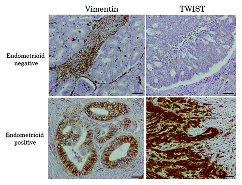
Table 3. Multivariate analysis of survival rates
Discussion
The current study revealed that the overexpression of Snail and Slug was closely associated with the reduced expression of E-cadherin in uterine endometrial cancer, and these findings were particularly noticeable at the invasive front of the tumor.
Malignant epithelial tumors can invade surrounding tissues through a variety of mechanisms.Citation41 EMT is considered to be an important means of tumor invasion and metastasis in many common cancers. The current study is the first report to demonstrate that the EMT status, as represented by both reduced E-cadherin expression and nuclear Snail expression, was identified to be an independent predictive factor of patient survival in endometrial cancer. Although we also evaluated the vimentin status, which was one of the mesenchymal markers in this tissue microarray sample to confirm the mesenchymal status, the rate of vimentin expression showed no substantial differences associated with the FIGO stage or in the histological subtypes, the rate of vimentin expression was not associated with myometrial invasion, peritoneal cytology and lymph node metastasis status (data not shown). These results were almost identical to those described in a previous report.Citation42 Therefore, we believe that the vimentin expression should be excluded as a marker of the EMT status in the current study. The transcription factor Snail is one of the repressors involved in E-cadherin downregulation. Snail is thought to be an important regulator of invasiveness during tumor progression.Citation43 The current study found a statistically significant inverse relationship between the Snail and E-cadherin expression in endometrial cancer. Specifically, the overexpression of Snail was closely associated with either the absence or a reduced expression of E-cadherin at the invasive front of tumors. Moreover, we showed the first report that EMT status, which was represented by both reduced E-cadherin expression and nuclear Snail expression, was associated with a significantly increased risk of death. An association was observed between the Snail expression and lymph node metastasis. However, there was no correlation between the Snail expression in primary tumors and their corresponding metastases, thus suggesting that the immunohistochemistry cannot directly demonstrate the status of invasive malignant cells. Further examination in vitro is therefore required to clarify whether Snail regulates the E-cadherin reduction, and Snail and E-cadherin regulate the invasiveness and metastasis of cancer cells in endometrial cancer.
The carcinosarcoma patients in the current series showed high expression rates of not only Snail but also Slug. Slug is overexpressed in numerous cancers,Citation44 including ovarian cancer, breast cancer, prostate cancer, esophageal cancer, gastric cancer, colorectal cancer, pancreatic cancer, lung cancer, leukemia, malignant mesothelioma, cholangiocarcinoma, hepatocellular carcinoma and glioma. An elevated expression of Slug is also associated with reduced E-cadherin expression, high histologic grade, lymph node metastasis, postoperative relapse and a shorter patient survival in a variety of cancers.Citation44-Citation46 In other words, Slug overexpression could contribute to downregulate the expression of E-cadherin and induce EMT in epithelial cells.Citation47 However, Snail and Slug may differ in their respective target genes. Forced overexpression of each of these transcription factors induces EMT in cancer cell lines.Citation48 Slug is also more relevant for generating breast cancer cells with cancer cell phenotype than Snail.Citation49 Slug overexpression was rare in endometrial cancer in the current study, but was selectively expressed in carcinosarcoma patients, and 10 of 13 positive cases were carcinosarcoma patients. Slug might thus be strongly involved in E-cadherin downregulation in carcinosarcoma, leading to the tumor cells acquiring the ability of metastasis and might be a more important regulator of the EMT in carcinosarcoma than that in endometrioid adenocarcinoma. These findings suggest that Slug might be a crucial factor that regulates the EMT program. However, the current study showed the immunoreactivity status of the EMT program in endometrial cancer. Further examination in vitro is necessary to clarify whether Slug regulates the reduction of E-cadherin, and whether Slug and E-cadherin regulate the cancer cells invasive and metastatic behavior in uterine carcinosarcoma.
In conclusion, the current study demonstrated that EMT status, which was represented by both reduced E-cadherin expression and nuclear Snail expression, was an independent predictor in endometrial cancer. Moreover, Slug might be the crucial factor that regulates EMT program in uterine carcinosarcoma. These results indicate that E-cadherin and Snail or Slug may therefore be potentially useful molecular targets in endometrial cancer.
Additional material
Download Zip (397.5 KB)Acknowledgments
This work was supported by a Grant-in-Aid for Scientific Research on Priority Areas, No.22591869 (to Y.T.) from the Ministry of Education, Culture, Sports, Science and Technology of Japan.
Disclosure of Potential Conflicts of Interest
No potential conflicts of interest were disclosed.
References
- Sankaranarayanan R, Ferlay J. Worldwide burden of gynaecological cancer: the size of the problem. Best Pract Res Clin Obstet Gynaecol 2006; 20:207 - 25; http://dx.doi.org/10.1016/j.bpobgyn.2005.10.007; PMID: 16359925
- Kadar N, Homesley HD, Malfetano JH. Prognostic factors in surgical stage III and IV carcinoma of the endometrium. Obstet Gynecol 1994; 84:983 - 6; PMID: 7970482
- Morrow CP, Bundy BN, Kurman RJ, Creasman WT, Heller P, Homesley HD, et al. Relationship between surgical-pathological risk factors and outcome in clinical stage I and II carcinoma of the endometrium: a Gynecologic Oncology Group study. Gynecol Oncol 1991; 40:55 - 65; http://dx.doi.org/10.1016/0090-8258(91)90086-K; PMID: 1989916
- Chambers AF, Groom AC, MacDonald IC. Dissemination and growth of cancer cells in metastatic sites. Nat Rev Cancer 2002; 2:563 - 72; http://dx.doi.org/10.1038/nrc865; PMID: 12154349
- Woodhouse EC, Chuaqui RF, Liotta LA. General mechanisms of metastasis. Cancer 1997; 80:Suppl 1529 - 37; http://dx.doi.org/10.1002/(SICI)1097-0142(19971015)80:8+<1529::AID-CNCR2>3.0.CO;2-F; PMID: 9362419
- Sivridis E, Giatromanolaki A, Gatter KC, Harris AL, Koukourakis MI, Tumor and Angiogenesis Research Group. Association of hypoxia-inducible factors 1alpha and 2alpha with activated angiogenic pathways and prognosis in patients with endometrial carcinoma. Cancer 2002; 95:1055 - 63; http://dx.doi.org/10.1002/cncr.10774; PMID: 12209691
- Inoue M. Current molecular aspects of the carcinogenesis of the uterine endometrium. Int J Gynecol Cancer 2001; 11:339 - 48; http://dx.doi.org/10.1046/j.1525-1438.2001.01046.x; PMID: 11737463
- Fanning J, Brown S, Phibbs G, Kramer T, Zaher A. Immunohistochemical evaluation is not prognostic for recurrence in fully staged high-risk endometrial cancer. Int J Gynecol Cancer 2002; 12:286 - 9; http://dx.doi.org/10.1046/j.1525-1438.2002.t01-1-01103.x; PMID: 12060450
- Salvesen HB, Das S, Akslen LA. Loss of nuclear p16 protein expression is not associated with promoter methylation but defines a subgroup of aggressive endometrial carcinomas with poor prognosis. Clin Cancer Res 2000; 6:153 - 9; PMID: 10656444
- Salvesen HB, Stefansson I, Kalvenes MB, Das S, Akslen LA. Loss of PTEN expression is associated with metastatic disease in patients with endometrial carcinoma. Cancer 2002; 94:2185 - 91; http://dx.doi.org/10.1002/cncr.10434; PMID: 12001116
- Thiery JP, Sleeman JP. Complex networks orchestrate epithelial-mesenchymal transitions. Nat Rev Mol Cell Biol 2006; 7:131 - 42; http://dx.doi.org/10.1038/nrm1835; PMID: 16493418
- Hugo H, Ackland ML, Blick T, Lawrence MG, Clements JA, Williams ED, et al. Epithelial--mesenchymal and mesenchymal--epithelial transitions in carcinoma progression. J Cell Physiol 2007; 213:374 - 83; http://dx.doi.org/10.1002/jcp.21223; PMID: 17680632
- Yang J, Weinberg RA. Epithelial-mesenchymal transition: at the crossroads of development and tumor metastasis. Dev Cell 2008; 14:818 - 29; http://dx.doi.org/10.1016/j.devcel.2008.05.009; PMID: 18539112
- Baum B, Settleman J, Quinlan MP. Transitions between epithelial and mesenchymal states in development and disease. Semin Cell Dev Biol 2008; 19:294 - 308; http://dx.doi.org/10.1016/j.semcdb.2008.02.001; PMID: 18343170
- Grünert S, Jechlinger M, Beug H. Diverse cellular and molecular mechanisms contribute to epithelial plasticity and metastasis. Nat Rev Mol Cell Biol 2003; 4:657 - 65; http://dx.doi.org/10.1038/nrm1175; PMID: 12923528
- Huber MA, Kraut N, Beug H. Molecular requirements for epithelial-mesenchymal transition during tumor progression. Curr Opin Cell Biol 2005; 17:548 - 58; http://dx.doi.org/10.1016/j.ceb.2005.08.001; PMID: 16098727
- Nieto MA. The snail superfamily of zinc-finger transcription factors. Nat Rev Mol Cell Biol 2002; 3:155 - 66; http://dx.doi.org/10.1038/nrm757; PMID: 11994736
- Hemavathy K, Ashraf SI, Ip YT. Snail/slug family of repressors: slowly going into the fast lane of development and cancer. Gene 2000; 257:1 - 12; http://dx.doi.org/10.1016/S0378-1119(00)00371-1; PMID: 11054563
- Grau Y, Carteret C, Simpson P. Mutations and Chromosomal Rearrangements Affecting the Expression of Snail, a Gene Involved in Embryonic Patterning in DROSOPHILA MELANOGASTER. Genetics 1984; 108:347 - 60; PMID: 17246230
- Valdés F, Alvarez AM, Locascio A, Vega S, Herrera B, Fernández M, et al. The epithelial mesenchymal transition confers resistance to the apoptotic effects of transforming growth factor Beta in fetal rat hepatocytes. Mol Cancer Res 2002; 1:68 - 78; PMID: 12496370
- Pérez-Losada J, Sánchez-Martín M, Pérez-Caro M, Pérez-Mancera PA, Sánchez-García I. The radioresistance biological function of the SCF/kit signaling pathway is mediated by the zinc-finger transcription factor Slug. Oncogene 2003; 22:4205 - 11; http://dx.doi.org/10.1038/sj.onc.1206467; PMID: 12833143
- Vega S, Morales AV, Ocaña OH, Valdés F, Fabregat I, Nieto MA. Snail blocks the cell cycle and confers resistance to cell death. Genes Dev 2004; 18:1131 - 43; http://dx.doi.org/10.1101/gad.294104; PMID: 15155580
- Takeichi M. Cadherins in cancer: implications for invasion and metastasis. Curr Opin Cell Biol 1993; 5:806 - 11; http://dx.doi.org/10.1016/0955-0674(93)90029-P; PMID: 8240824
- Wijnhoven BP, Dinjens WN, Pignatelli M. E-cadherin-catenin cell-cell adhesion complex and human cancer. Br J Surg 2000; 87:992 - 1005; http://dx.doi.org/10.1046/j.1365-2168.2000.01513.x; PMID: 10931041
- Sakuragi N, Nishiya M, Ikeda K, Ohkouch T, Furth EE, Hareyama H, et al. Decreased E-cadherin expression in endometrial carcinoma is associated with tumor dedifferentiation and deep myometrial invasion. Gynecol Oncol 1994; 53:183 - 9; http://dx.doi.org/10.1006/gyno.1994.1113; PMID: 8188077
- Cheng L, Nagabhushan M, Pretlow TP, Amini SB, Pretlow TG. Expression of E-cadherin in primary and metastatic prostate cancer. Am J Pathol 1996; 148:1375 - 80; PMID: 8623909
- Bremnes RM, Veve R, Gabrielson E, Hirsch FR, Baron A, Bemis L, et al. High-throughput tissue microarray analysis used to evaluate biology and prognostic significance of the E-cadherin pathway in non-small-cell lung cancer. J Clin Oncol 2002; 20:2417 - 28; http://dx.doi.org/10.1200/JCO.2002.08.159; PMID: 12011119
- Graff JR, Herman JG, Lapidus RG, Chopra H, Xu R, Jarrard DF, et al. E-cadherin expression is silenced by DNA hypermethylation in human breast and prostate carcinomas. Cancer Res 1995; 55:5195 - 9; PMID: 7585573
- Yoshiura K, Kanai Y, Ochiai A, Shimoyama Y, Sugimura T, Hirohashi S. Silencing of the E-cadherin invasion-suppressor gene by CpG methylation in human carcinomas. Proc Natl Acad Sci USA 1995; 92:7416 - 9; http://dx.doi.org/10.1073/pnas.92.16.7416; PMID: 7543680
- Grooteclaes ML, Frisch SM. Evidence for a function of CtBP in epithelial gene regulation and anoikis. Oncogene 2000; 19:3823 - 8; http://dx.doi.org/10.1038/sj.onc.1203721; PMID: 10949939
- Comijn J, Berx G, Vermassen P, Verschueren K, van Grunsven L, Bruyneel E, et al. The two-handed E box binding zinc finger protein SIP1 downregulates E-cadherin and induces invasion. Mol Cell 2001; 7:1267 - 78; http://dx.doi.org/10.1016/S1097-2765(01)00260-X; PMID: 11430829
- Blanco MJ, Moreno-Bueno G, Sarrio D, Locascio A, Cano A, Palacios J, et al. Correlation of Snail expression with histological grade and lymph node status in breast carcinomas. Oncogene 2002; 21:3241 - 6; http://dx.doi.org/10.1038/sj.onc.1205416; PMID: 12082640
- Grille SJ, Bellacosa A, Upson J, Klein-Szanto AJ, van Roy F, Lee-Kwon W, et al. The protein kinase Akt induces epithelial mesenchymal transition and promotes enhanced motility and invasiveness of squamous cell carcinoma lines. Cancer Res 2003; 63:2172 - 8; PMID: 12727836
- Hajra KM, Chen DY, Fearon ER. The SLUG zinc-finger protein represses E-cadherin in breast cancer. Cancer Res 2002; 62:1613 - 8; PMID: 11912130
- Sugimachi K, Tanaka S, Kameyama T, Taguchi K, Aishima S, Shimada M, et al. Transcriptional repressor snail and progression of human hepatocellular carcinoma. Clin Cancer Res 2003; 9:2657 - 64; PMID: 12855644
- Batlle E, Sancho E, Francí C, Domínguez D, Monfar M, Baulida J, et al. The transcription factor snail is a repressor of E-cadherin gene expression in epithelial tumour cells. Nat Cell Biol 2000; 2:84 - 9; http://dx.doi.org/10.1038/35000034; PMID: 10655587
- Blechschmidt K, Sassen S, Schmalfeldt B, Schuster T, Höfler H, Becker KF. The E-cadherin repressor Snail is associated with lower overall survival of ovarian cancer patients. Br J Cancer 2008; 98:489 - 95; http://dx.doi.org/10.1038/sj.bjc.6604115; PMID: 18026186
- Tuhkanen H, Soini Y, Kosma VM, Anttila M, Sironen R, Hämäläinen K, et al. Nuclear expression of Snail1 in borderline and malignant epithelial ovarian tumours is associated with tumour progression. BMC Cancer 2009; 9:289; http://dx.doi.org/10.1186/1471-2407-9-289; PMID: 19695091
- Kurrey NK, K A, Bapat SA. Snail and Slug are major determinants of ovarian cancer invasiveness at the transcription level. Gynecol Oncol 2005; 97:155 - 65; http://dx.doi.org/10.1016/j.ygyno.2004.12.043; PMID: 15790452
- Elloul S, Elstrand MB, Nesland JM, Tropé CG, Kvalheim G, Goldberg I, et al. Snail, Slug, and Smad-interacting protein 1 as novel parameters of disease aggressiveness in metastatic ovarian and breast carcinoma. Cancer 2005; 103:1631 - 43; http://dx.doi.org/10.1002/cncr.20946; PMID: 15742334
- Yilmaz M, Christofori G, Lehembre F. Distinct mechanisms of tumor invasion and metastasis. Trends Mol Med 2007; 13:535 - 41; http://dx.doi.org/10.1016/j.molmed.2007.10.004; PMID: 17981506
- Coppola D, Fu L, Nicosia SV, Kounelis S, Jones M. Prognostic significance of p53, bcl-2, vimentin, and S100 protein-positive Langerhans cells in endometrial carcinoma. Hum Pathol 1998; 29:455 - 62; http://dx.doi.org/10.1016/S0046-8177(98)90060-0; PMID: 9596268
- Cano A, Pérez-Moreno MA, Rodrigo I, Locascio A, Blanco MJ, del Barrio MG, et al. The transcription factor snail controls epithelial-mesenchymal transitions by repressing E-cadherin expression. Nat Cell Biol 2000; 2:76 - 83; http://dx.doi.org/10.1038/35000025; PMID: 10655586
- Alves CC, Carneiro F, Hoefler H, Becker KF. Role of the epithelial-mesenchymal transition regulator Slug in primary human cancers. Front Biosci 2009; 14:3035 - 50; http://dx.doi.org/10.2741/3433; PMID: 19273255
- Shih JY, Tsai MF, Chang TH, Chang YL, Yuan A, Yu CJ, et al. Transcription repressor slug promotes carcinoma invasion and predicts outcome of patients with lung adenocarcinoma. Clin Cancer Res 2005; 11:8070 - 8; http://dx.doi.org/10.1158/1078-0432.CCR-05-0687; PMID: 16299238
- Shioiri M, Shida T, Koda K, Oda K, Seike K, Nishimura M, et al. Slug expression is an independent prognostic parameter for poor survival in colorectal carcinoma patients. Br J Cancer 2006; 94:1816 - 22; http://dx.doi.org/10.1038/sj.bjc.6603193; PMID: 16773075
- Bolós V, Peinado H, Pérez-Moreno MA, Fraga MF, Esteller M, Cano A. The transcription factor Slug represses E-cadherin expression and induces epithelial to mesenchymal transitions: a comparison with Snail and E47 repressors. J Cell Sci 2003; 116:499 - 511; http://dx.doi.org/10.1242/jcs.00224; PMID: 12508111
- Kurrey NK, Jalgaonkar SP, Joglekar AV, Ghanate AD, Chaskar PD, Doiphode RY, et al. Snail and slug mediate radioresistance and chemoresistance by antagonizing p53-mediated apoptosis and acquiring a stem-like phenotype in ovarian cancer cells. Stem Cells 2009; 27:2059 - 68; http://dx.doi.org/10.1002/stem.154; PMID: 19544473
- Bhat-Nakshatri P, Appaiah H, Ballas C, Pick-Franke P, Goulet R Jr., Badve S, et al. SLUG/SNAI2 and tumor necrosis factor generate breast cells with CD44+/CD24- phenotype. BMC Cancer 2010; 10:411; http://dx.doi.org/10.1186/1471-2407-10-411; PMID: 20691079