Recent studies have demonstrated that gene expression is regulated not only by protein-coding genes, but also by non-protein-coding RNA molecules, including microRNAs (miRNAs). miRNAs are a class of 21–25 nt single-stranded non-coding small RNAs dependent upon ribonuclease III enzyme Dicer for the processing of mature and functional miRNAs.Citation1 Therefore, the deletion of the enzyme Dicer provides a genetic test for the relevance of miRNAs in mammalian development and function. We and others previously reported that loss of miRNAs in bone marrow induced by Tie2-Cre or in thymus by CD4-Cre, through tissue-specific deletion of miRNA-dependent Dicer, mainly regulated the development and function of CD4+CD25+Foxp3+ regulatory T (Treg) cells and invariant natural killer T (iNKT) cells, but did not affect conventional CD4 and CD8 T cell development in the thymus.Citation2-Citation6 However, the peripheral CD4 and CD8 T cells did require miRNAs for maintaining their homeostasis, and loss of Dicer increases T cell apoptosis and decreases their proliferation. Furthermore, Dicer-deficient helper T cells preferentially expressed interferon-gamma, the hallmark effector cytokine of the Th1 lineage.Citation4 Most importantly, T cell-specific Dicer-deficiency led to spontaneous development of colitis,Citation3 suggesting that miRNA-dependent regulation is critical for preventing spontaneous inflammation and autoimmunity. Surprisingly, mice with loss of Dicer in bone marrow did not spontaneously develop autoimmune diabetes and had no significant infiltration in the pancreas (data not shown here).
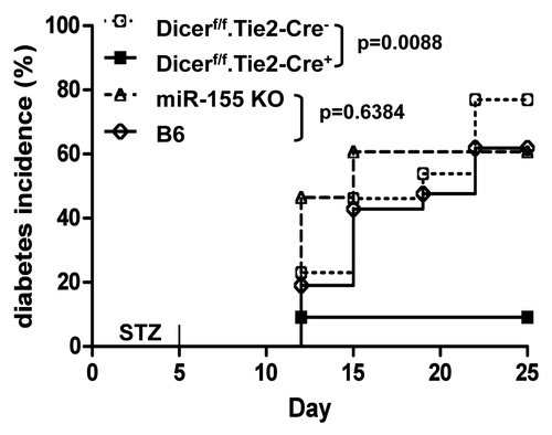
Multiple low dose streptozotocin (STZ) (MLDS)-induced autoimmune diabetes is an inflammatory disease of the pancreatic islets with clinical and histochemical features similar to those of human T1D. Given that defective iNKT cells and Treg cell number and function as well as increased Th1 cell differentiation are involved in MLDS-induced T1D, we hypothesized that loss of total miRNAs in T cells may enhance STZ-induced T1D. To test this, Dicerf/f.Tie2-cre+ knockout (KO) and Dicerf/f.Tie2-cre- wild-type (WT) miceCitation6 at 7–8 weeks of age were injected intraperitoneally with freshly dissolved STZ (50 mg/kg) for 5 consecutive days. Surprisingly, Dicer-KO mice showed a significantly decreased incidence of MLDS-induced diabetes compared with wild-type control mice. As shown in , less than 10% of male Dicer-KO mice developed diabetes at day 25 after STZ injection, while about 70% wild-type mice developed diabetes (p = 0.0088). Thus, our data suggest that loss of total miRNAs in hematopoiesis prevents STZ-induced autoimmune diabetes.
Figure 1. Deletion of microRNAs in bone marrow prevents streptozotocin-induced murine autoimmune diabetes, but deletion of miR-155 does not. Seven–9-week-old male Dicerf/f.Tie2-cre+ KO (■) and Dicerf/f.Tie2-cre- WT (○) mice, miR-155KO (∆) and B6 WT (◊) controls were injected intraperitoneally with freshly dissolved STZ (50 mg/kg) (Sigma) for 5 consecutive days. Development of glucosuria was monitored with Clinistix (Bayer Diagnostics). Clinical diabetes is defined by positive glucosuria and hyperglycemia (blood glucose levels > 250 mg/dl) on two consecutive days in nonfasting animals. Mice were followed until 25 d after the last injection of STZ. Data shown here is a combination (n = 11–28) of two to three experiments.
To identify the specific role of miRNAs in Treg cell development and function, three individual groups previously identified, almost simultaneously, the critical role of miRNAs in Treg function using mouse models with Foxp3Cre-mediated Dicer or Drosha deletion in the Treg cell lineage.Citation1 Disease onset was very early, with most mice dying within a few weeks of life. The fact that Treg-specific miRNA depletion led to a much more aggressive autoimmune phenotype than that from bone marrow (Tie2Cre)- or thymus (CD4Cre)-specific miRNA deletionCitation2-Citation5 indicates that the lack of an miRNA network in conventional T cells may delay the autoimmune process. More recent findings from Tian et al. further support this notion.Citation7 They found that the T cell miRNA network is essential for a robust IFN-g response in vivo in the absence of TGFβ, which is different from previous in vitro polarization experiments, where T cells lacking Dicer were preferential in Th1 induction.Citation4 The pathology of most organs affected by TGFβ signaling deficiency was either fully or partially rescued with a strong suppression of T cell activation by additional loss of Dicer, with the colon being the sole exception.Citation7 Even though some miRNAs may have dominant effects on autoimmunity, other miRNA are more likely to have a tolerance function, and overall, the net effect of miRNA function in T cells may reveal a systemic pro-autoimmune function, as loss of the entire miRNA network resulted in a profound reduction of autoimmune pathology in our current data plus a recent report in other organs.Citation7
As loss of miRNAs reduced both immunogenic capacity of effector T cellsCitation7 and tolerogeneic capacity of Treg and iNKT cells, the outcome from specific organs could depend on the balance of power between these two populations. miRNA miR-155 is widely expressed in T cells, B cells and DCs and is functionally involved in different aspects of the adaptive immune system. miR-155 is functionally relevant to Treg function, where its expression is regulated by Foxp3,Citation8 as well as to iNKT cell development and function (unpublished data). miR-155KO mice had defective Treg cell homeostasis but displayed a defective Th17 differentiation and bias toward Th2 differentiation. Most interestingly, miR-155KO mice were resistant to experimental autoimmune encephalomyelitis (EAE) due to defective Th17 and Th1 development, which supports a role for miR-155 in the inflammatory response.Citation9 We therefore further investigated the role of miR-155 in STZ-mediated T1D. As indicated in , surprisingly, the diabetes incidence was comparable between miR-155KO and wild-type B6 mice (p = 0.6384). Thus, unlike prevention in other autoimmune diseases, including EAE, colitis and arthritis, loss of miR-155 did not prevent MLDS-mediated T1D development. Our current data suggest that the same miRNA may play different roles in different organs and conditions.
The discovery of miRNAs has represented one of the most important mechanisms in gene regulation. Accumulated studies of miRNAs performed in the last decade have allowed the better understanding of the mechanisms of miRNA regulation in the disease development. Our current data plus recent reports suggest that the net systemic effect of miRNA on T cell function is dominantly immunogenic for T1D, implicating for the potential exploitation of miRNA as a therapeutic option for T1D. Inhibition of the entire miRNA network by targeting Dicer or other biogenesis proteins would prevent T1D development. For the individual miRNA, given that each miRNA can target and repress many different genes, and, vice versa, one gene can be regulated by multiple miRNAs, the complexity of miRNA regulation network may be linked to a redundancy system. Most experiments performed thus far have reported specific targeting of a specific mRNA at most times. Conversely, it is more likely that a few miRNAs may serve as ”key” regulators for gene expression in certain tissues or specific autoimmune diseases. Much work in these areas still needs to be done. Undoubtedly, defining key miRNAs related to autoimmunity in T1D will not only uncover new mechanisms, but will also help us to develop therapeutic targets or biomarkers for T1D.
| Abbreviation: | ||
| T1D | = | type 1 diabetes |
| miRNAs | = | microRNAs |
| MLDS | = | multiple low dose streptozotocin |
| STZ | = | streptozotocin |
| KO | = | knockout |
Acknowledgments
This study was supported in part by Juvenile Diabetes Research Foundation International Grants (1–2007–039, 5–2006–403, and 17–2012–678), Henry Ford Stubnitz Grant (J80005) and Henry Ford Immunology Program start-up (T71016 and T71017).
Disclosure of Potential Conflicts of Interest
No potential conflicts of interest were disclosed.
References
- Zhou L, Park JJ, Zheng Q, Dong Z, Mi Q. MicroRNAs are key regulators controlling iNKT and regulatory T-cell development and function. Cell Mol Immunol 2011; 8:380 - 7; http://dx.doi.org/10.1038/cmi.2011.27; PMID: 21822298
- Zhou L, Seo KH, Wong HK, Mi QS. MicroRNAs and immune regulatory T cells. Int Immunopharmacol 2009; 9:524 - 7; http://dx.doi.org/10.1016/j.intimp.2009.01.017; PMID: 19539573
- Cobb BS, Hertweck A, Smith J, O’Connor E, Graf D, Cook T, et al. A role for Dicer in immune regulation. J Exp Med 2006; 203:2519 - 27; http://dx.doi.org/10.1084/jem.20061692; PMID: 17060477
- Muljo SA, Ansel KM, Kanellopoulou C, Livingston DM, Rao A, Rajewsky K. Aberrant T cell differentiation in the absence of Dicer. J Exp Med 2005; 202:261 - 9; http://dx.doi.org/10.1084/jem.20050678; PMID: 16009718
- Seo KH, Zhou L, Meng D, Xu J, Dong Z, Mi QS. Loss of microRNAs in thymus perturbs invariant NKT cell development and function. Cell Mol Immunol 2010; 7:447 - 53; http://dx.doi.org/10.1038/cmi.2010.49; PMID: 20852654
- Zhou L, Seo KH, He HZ, Pacholczyk R, Meng DM, Li CG, et al. Tie2cre-induced inactivation of the miRNA-processing enzyme Dicer disrupts invariant NKT cell development. Proc Natl Acad Sci U S A 2009; 106:10266 - 71; http://dx.doi.org/10.1073/pnas.0811119106; PMID: 19509335
- Tian L, De Hertogh G, Fedeli M, Staats KA, Schonefeldt S, Humblet-Baron S, et al. Loss of T cell microRNA provides systemic protection against autoimmune pathology in mice. J Autoimmun 2012; 38:39 - 48; http://dx.doi.org/10.1016/j.jaut.2011.12.004; PMID: 22225602
- Kohlhaas S, Garden OA, Scudamore C, Turner M, Okkenhaug K, Vigorito E. Cutting edge: the Foxp3 target miR-155 contributes to the development of regulatory T cells. J Immunol 2009; 182:2578 - 82; http://dx.doi.org/10.4049/jimmunol.0803162; PMID: 19234151
- O’Connell RM, Kahn D, Gibson WSJ, Round JL, Scholz RL, Chaudhuri AA, et al. MicroRNA-155 promotes autoimmune inflammation by enhancing inflammatory T cell development. Immunity 2010; 33:607 - 19; http://dx.doi.org/10.1016/j.immuni.2010.09.009; PMID: 20888269