Abstract
When one type of sensory system is disrupted, other intact remaining sensory function can be improved. Although this form of plasticity, cross-modal plasticity, is widely known, the molecular and cellular mechanisms underlying it are poorly understood. In a recent study, we demonstrated that visual deprivation increases extracellular serotonin in the juvenile rat barrel cortex and resulted in facilitation of synaptic delivery of AMPA-type glutamate receptors (AMPARs) at layer4-2/3 synapses in the barrel cortex via the activation of serotonin 5HT2A/2C receptors and ERK. This caused sharpening of functional whisker-barrel map at layer2/3 of the barrel cortex. Thus, sensory dysfunction of one modality leads to improvement of remaining modalities by the refinement of cortical organization through serotonin signaling-mediated facilitation of synaptic AMPARs delivery.
Loss of one sensory system can induce improved function of other intact sensory systems. For example, blind individuals compensate their lack of visual input with improved somatosensory or auditory functions (e.g., increased sensitivity and accuracy). Although this type of neuronal plasticity, cross-modal plasticity, is well known, the molecular and cellular mechanisms are poorly understood.Citation1–Citation3
One of the best characterized synaptic plasticity is long-term potentiation (LTP). LTP has been suggested to play a crucial role in experience-dependent neuronal plasticity such as learning and memory.Citation4 Synaptic trafficking of AMPA (α-amino-3-hydroxy-5-methyl-4-isoxazole propionic acid)-type glutamate receptors (AMPARs) appears to be a major mechanism regulating expression of LTP.Citation5–Citation8 AMPARs forms tetramers comprised of a combination of four subunits (GluR1–4).Citation9 LTP-inducing stimuli in vitro drive GluR1-containing AMPARs into synapses.Citation6 In animals, sensory experience early in development (postnatal day 12–14) delivers GluR1 into layer4-2/3 synapses of the barrel cortex.Citation10,Citation11 Further, synaptic GluR1 delivery at thalamo-amygdala synapses induced by fear conditioning is required for the acquisition of fear memory.Citation12
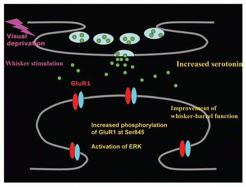
In our recent study, we reported (1) visual deprivation (VD) for 2 days drives GluR1 into layer4-2/3 synapses of the rat barrel cortex at the age (postnatal day 21–23) when natural whisker experience no longer delivers GluR1 at these synapses, (2) VD increases extracellular serotonin, (3) application of ketanserin, a 5HT2A/2C receptor antagonist, into layer2/3 of the barrel cortex prevents VD-induced synaptic GluR1 delivery, (4) knocking down of the expression of 5HT2A receptors with short hairpin RNA blocks VD-induced synaptic potentiation, (5) DOI (2,5-dimethoxy-4-iodoamphetamine), a 5HT2A/2C receptor agonist, facilitates synaptic GluR1 delivery in vivo as well as induction of LTP in vitro, (6) VD-induced activation of serotonergic system activates ERK and increases phosphorylation of GluR1 at Ser845, a critical site for synaptic GluR1 delivery, (7) VD sharpens functional whisker-barrel map ().Citation13 These results demonstrate that serotonin mediates VD-induced facilitation of synaptic GluR1 delivery in the barrel cortex and improvement of whisker-barrel function via activation of serotonin 5HT2A/2C receptors and potential down stream kinase, ERK. Phosphorylation of Ser845 has been demonstrated to increase surface expression of GluR1,Citation14 and synaptic incorporation of GluR1 is mainly mediated by lateral diffusion from extrasynaptic sites.Citation15 Therefore, VD could facilitate synaptic GluR1 trafficking by increasing the extrasynaptic pool of GluR1 through serotonin signaling-mediated increase of phosphorylation at Ser845. It will be interesting to test if VD enhances surface expression of GluR1.
Specific strengthening of layer4-2/3 synapses within a cortical column in the barrel cortex should increase response of each barrel column at layer2/3 to the principal whisker without enhancing response to surrounding whiskers and result in the sharpening of functional whisker-barrel map at layer2/3. While our study demonstrated that 2 days of VD strengthens layer4-2/3 synapses by synaptic delivery of GluR1 in the barrel cortex, leading to sharpening of whisker-barrel function,Citation13 previous study reported that 7 days of VD decreases AMPARs-mediated miniature EPSCs (mEPSC) at layer2/3 of the barrel cortex.Citation2 Consistent with these results, we found that the ratio of AMPARs-mediated to NMDARs-mediated synaptic transmission (A/N ratio), a measurement of synaptic AMPARs content, increases 2 days after VD but returns to the base line level 7 days after VD. However, we observed that whisker-dependent behavior kept improved even after 7 days of VD (Jitsuki S, et al. unpublished data). Thus, although AMPARs-mediated synaptic transmission which is enhanced 2 days after VD decreases to the basal level presumably by homeostatic mechanisms 7 days after VD, sharpened whisker-barrel function at layer2/3 likely to be retained for a week after VD. Lateral inhibition may be enhanced by selective acute synaptic strengthening at layer4-2/3 synapses within a cortical column in the barrel cortex after 2 days of VD, and increased lateral inhibition can be sustained for a week after the beginning of VD.
Cross-modal plasticity is an example of brain function to compensate for the loss of neuronal function. Since similar mechanism can be utilized for enhancement of remaining function after certain neuronal dysfunction, understanding the molecular and cellular mechanism underlying cross-modal plasticity may lead to effective treatment to enhance rehabilitation.
Figures and Tables
Figure 1 Visual deprivation increases extracellular serotonin at layer2/3 of juvenile rat barrel cortex. This leads to the facilitation of synaptic GluR1 delivery at layer4-2/3 synapses of the barrel cortex by increasing the phosphorylation of GluR1 at Ser845 via activation of serotonin 5HT2A/2C receptors and ERK. Furthermore, this results in the improvement whisker-barrel function.

Addendum to:
References
- Bavelier D, Neville HJ. Cross-modal plasticity: where and how?. Nat Rev Neurosci 2002; 3:443 - 452
- Goel A, Jiang B, Xu LW, Song L, Kirkwood A, Lee HK. Cross-modal regulation of synaptic AMPA receptors in primary sensory cortices by visual experience. Nat Neurosci 2006; 9:1001 - 1003
- Rauschecker JP, Tian B, Korte M, Egert U. Crossmodal changes in the somatosensory vibrissa/barrel system of visually deprived animals. Proc Natl Acad Sci USA 1992; 89:5063 - 5067
- Whitlock JR, Heynen AJ, Shuler MG, Bear MF. Learning induces long-term potentiation in the hippocampus. Science 2006; 313:1093 - 1097
- Malinow R, Malenka RC. AMPA receptor trafficking and synaptic plasticity. Annu Rev Neurosci 2002; 25:103 - 126
- Hayashi Y, Shi SH, Esteban JA, Piccini A, Poncer JC, Malinow R. Driving AMPA receptors into synapses by LTP and CaMKII: requirement for GluR1 and PDZ domain interaction. Science 2000; 287:2262 - 2267
- Bredt DS, Nicoll RA. AMPA receptor trafficking at excitatory synapses. Neuron 2003; 40:361 - 379
- Scannevin RH, Huganir RL. Postsynaptic organization and regulation of excitatory synapses. Nat Rev Neurosci 2000; 1:133 - 141
- Wenthold RJ, Petralia RS, Blahos J II, Niedzielski AS. Evidence for multiple AMPA receptor complexes in hippocampal CA1/CA2 neurons. J Neurosci 1996; 16:1982 - 1989
- Takahashi T, Svoboda K, Malinow R. Experience strengthening transmission by driving AMPA receptors into synapses. Science 2003; 299:1585 - 1588
- Clem RL, Barth A. Pathway-specific trafficking of native AMPARs by in vivo experience. Neuron 2006; 49:663 - 670
- Rumpel S, LeDoux J, Zador A, Malinow R. Postsynaptic receptor trafficking underlying a form of associative learning. Science 2005; 308:83 - 88
- Jitsuki S, Takemoto K, Kawasaki T, Tada H, Takahashi A, Becamel C, et al. Serotonin mediates cross-modal reorganization of cortical circuits. Neuron 2011; 69:780 - 792
- Derkach VA, Oh MC, Guire ES, Soderling TR. Regulatory mechanisms of AMPA receptors in synaptic plasticity. Nat Rev Neurosci 2007; 8:101 - 103
- Makino H, Malinow R. AMPA receptor incorporation into synapses during LTP: the role of lateral movement and exocytosis. Neuron 2009; 64:381 - 390