Abstract
The development of disseminated superficial porokeratosis is occasionally observed in association with renal transplant, autoimmune diseases, and various hematological disorders, suggesting a certain immunosuppression may trigger a wide-spread abnormal keratinization. Here we report a case of sudden onset disseminated superficial porokeratosis associated with an exacerbation of diabetes mellitus due to an anti-insulin antibody formation. To our knowledge, this is the first report of disseminated superficial porokeratosis in a patient with severe diabetes mellitus.
SIR, Porokeratosis is a disorder of epidermal keratinization that is characterized by variably sized plaques with elevated borders, which show cornoid lamellae on histologic examination.Citation1 Usually, porokeratosis exhibits single or few lesions, and is occasionally inherited in an autosomal dominant fashion. Disseminated superficial porokeratosis (DSP) is another rare type of porokeratosis. Although entire pathogenesis of DSP still remains unknown, it has been suggested that certain factors, including ultraviolet radiation and immunosuppression, may activate abnormal clones of keratinocytes, leading to the characteristic clinical and histological appearances of DSP. In fact, the development of DSP has been reported in renal transplant recipients,Citation1,Citation2 in a systemic lupus erythematosus (SLE) patient receiving long-term corticosteroid treatment,Citation3 and in individuals affected by various hematological disorders such as Hodgkin's disease and B-cell lymphoma.Citation4 Here we report a case of sudden onset DSP associated with an exacerbation of diabetes mellitus due to an anti-insulin antibody formation.
Case Report
A 75-year-old was referred to our hospital for evaluation of multiple, eruptive, itching annular lesions. One year before the initial visit to our hospital, the patient began to receive an insulin therapy with long-acting human insulin, detemir, for his type II diabetes mellitus. Six months after the initiation of insulin therapy, the patient felt an abrupt general fatigue and noticed to have itching skin eruptions on the extremeties and trunk. Blood examination revealed a high titer, more than 5,000 nU/ml, of anti-insulin antibodies (normal, less than 125 nU/ml) and high HbA1c (8.8%; normal, 4.3–5.8%). The patient stopped injection of this long-acting insulin and started a NPH insulin therapy with an intermediate duration of action. The blood glucose level became well controlled with a new insulin therapy, however, his eruptions persisted.
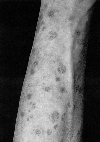
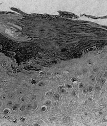
Physical examination revealed an approximately hundred number of discrete plaques, 0.5–2 cm in diameter, with elevated borders on the trunk and extremeties (). A biopsy specimen obtained from the right forearm showed parakeratotic columns, cornoid lamellae, with invaginations of the underlying granular layer (). These clinical and histological findings led us to the diagnosis of DSP. Plaques were treated with a topical 20% urea cream, resulting in a gradual resolution of pruritis and clearing of the lesions over several weeks.
Discussion
Patients with diabetes mellitus develop a wide variety of skin manifestations, such as gangrene, scleredema, xanthoma, necrobiosis lipiodica, disseminated granuloma annulare and Dupuytren contracture. Recently, a case of acquired ichthyosis associated with diabetes mellitus was reported.Citation5 Excessive glycation and/or glycoxidation of proteins in keratinocytes in diabetic patients might lead to an abnormal proliferation and differentiation of keratinocytes resulting in acquired ichthyosis and DSP. The simultaneous deterioration of diabetes mellitus and development of DSP provide a possibility that excessive glycation of proteins in keratinocytes contribute to the formation of DSP in our case. In addition, the patient began to produce antibodies against the injected insulin. As several immunological disorders often underlie the pathogenesis of DSP, the immunocomplex of insulin and antibodies might also affect the proliferation of keratinocytes.
Abbreviations
| DSP | = | disseminated superficial porokeratosis |
References
- Knoell KA, Patterson JW, Wilson BB. Sudden onset of disseminated porokeratosis of Mibelli in a renal transplant patient. J Am Acad Dermatol 1999; 41:830 - 832
- Robak E, Wozniacka A, Sysa-Jedrezejowska A, Biernat W, Robak T. Disseminated superficial actinic porokeratosis in a patient with systemic lupus erythematosus. Br J Dermatol 1999; 141:759 - 761
- Herranz P, Pizarro A, De Lucas R, et al. High incidence of porokeratosis in renal transpolant recipients. Br J Dermatol 1997; 136:176 - 179
- Diluvio L, Campione E, Paterno EJ, et al. Acute onset disseminated superficial porokeratosis heralding diffuse large B-cell lymphoma. Eur J Dermatol 2008; 18:349 - 350
- Sanli H, Akay BN, Sen BB, Kocak AY, Emral R, Bostanci S. Acquired ichthyosis associated with type 1 diabetes mellitus. Dermatoendocrinol 2009; 1:34 - 36

