Abstract
We present herein a case of right axillary accessory breast associated with galactorrhea in an adolescent girl. A 14-year-old Japanese girl presented with an 11-month history of a tender, subcutaneous lesion in the right axillary fossa. Seven months later, she experienced menarche. Subsequently, the patient noticed pressure-induced galactorrhea from both nipples. Physical examination revealed an elastic, firm and well-demarcated subcutaneous tumor 3 × 2 cm in size. A biopsy specimen showed proliferation of mammary gland tissue in the stroma located below the subcutaneous fat tissue. On the basis of these findings the patient was diagnosed with an accessory breast. Interestingly, the galactorrhea ceased after surgical removal of the accessory breast.
Accessory breast has been shown to be associated with several conditions such as kidney and urinary tract malformations and malignancies.Citation1 However, there are no reported cases of accessory breast associated with galactorrhea. We herein present a case of accessory breast associated with galactorrhea in an adolescent girl.
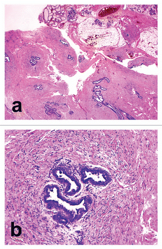
A 14-year-old Japanese girl presented with an 11-month history of a tender subcutaneous lesion in the right axillary fossa. Seven months after recognition of the subcutaneous lesion, she experienced menarche. Subsequently, the patient noticed a pressure-induced, white discharge from both nipples. The discharge was milky and not serous or hemorrhagic. She had not taken any medications. Physical examination of the right axilla revealed an elastic, firm and well-demarcated subcutaneous lesion 3 × 2 cm in size. The lesion was fixed to the underlying tissue. The surface skin was normal without the presence of a nipple, areola or hollow. A biopsy specimen showed groups of proliferating and branching ductal structures embedded in a fibrous connective tissue stroma in the subcutaneous fat tissue ( and B). The connective tissue stroma was sparse around the ducts (). The walls of the ducts were lined by 1 to 2 layers of columnar cells. Some of these ducts contained faint eosinophilic secreted materials. The epidermis was normal without hyperpigmentation in the basal layer. On the basis of these findings, the patient was diagnosed as having an accessory breast. Pituitary hormone levels including prolactin, leutenizing hormone (LH), growth hormone (GH), somatomedin C, thyroid stimulating hormone (TSH) and follicle-stimulating hormone (FSH) were all within normal limits. Levels of estradiol and free T4 were also normal. The lesion was surgically removed. Immediately after the surgery the galactorrhea stopped.
Accessory breast is residual mammary glandular tissue that persists from normal embryologic development and is found in 2–6% of woman.Citation2 Accessory breast can present as a mass anywhere along the course of the embryologic mammary streak (milk line) but is most frequently found in the axilla.Citation2 In a young child, the accessory breast tissue is not recognized since the mass is small. During menarche, the mass may enlarge and cause symptoms such as discomfort, pain, milk secretion and local skin irritation in response to hormonal stimulation.Citation3 Accessory breast is classified into eight types based on the presence or absence of glandular tissue, nipple and areola.Citation4 Our present case corresponded to type 4 (glandular tissue (+), nipple (−), areola (−)).
Galactorrhea is a relatively common problem in women of childbearing age.Citation5 However, galactorrhea is a significant finding if it is associated with anovulation and unrelated to exogenous factors such as hyperprolactinemia and hypothyroidism.Citation6 The cause of galactorrhea in our case is unknown. The prolactin level in our patient was within normal limits, indicating that a cause other than hyperprolactinemia was responsible for the galactorrhea.
The striking feature of our case is that the galactorrhea was associated with accessory breast and the galactorrhea ceased after the surgical removal of the accessory breast tissue. Thus, we speculate that the accessory breast contributed to the galactorrhea, although we cannot exclude the possibility that accessory breast and galactorrhea were unrelated. Furthermore, since the patient was noted to be a right-handed Kendo (Japanese fencing) player and chest wall irritation is a cause of galactorrheaCitation6 we hypothesize that mechanical stress on the accessory breast (i.e., friction between right upper arm and accessory breast during Kendo) caused galactorrhea. As such, we believe that an accessory breast may be considered as one of the differential diagnoses for galactorrhea in adolescent girls, especially in the absence of hyperprolactinemia.
Figures and Tables
Figure 1 Histopathological findings. (A) Groups of proliferating and branching ductal structures embedded in a fibrous connective tissue stroma in the subcutaneous fat tissue (haematoxylin and eosin, original magnification x6.25). (B) Dilated ducts with intraluminal secretion are lined by cuboidal cells. The connective tissue stroma was sparse around the ducts (haematoxylin and eosin, original magnification x100).

References
- Lewis EJ, Crutchfield CE, Prawer SE 3rd. Accessory nipples and associated conditions. Pediatr Dermatol 1997; 14:333 - 334
- Lesavoy MA, Gomez-Garcia A, Nejdl R, Yospur G, Syiau TJ, Chang P. Axillary breast tissue: clinical presentation and surgical treatment. Ann Plast Surg 1995; 35:356 - 360
- Loukas M, Clarke P, Tubbs RS. Accessory breasts: a historical and current perspective. Am Surg 2007; 73:525 - 528
- Kajava Y. The proportion of supernumerary nipples in Finnish population. Duodecim 1915; 31:143 - 170
- Buckman MT, Peake GT. Prolactin in clinical practice. JAMA 1976; 236:871 - 874
- Peña KS, Rosenfeld JA. Evaluation and treatment of galactorrhea. Am Fam Physician 2001; 63:1763 - 1770