Abstract
To our knowledge there have been no reports of scleroderma-like skin changes, not affecting the hand in prepubertal patients with Type I Diabetes Mellitus (T1DM). We report a prepubertal caucasian male with T1DM, and early morphea-type skin changes of the trunk and extremities, not involving the hand.
Introduction
Scleroderma-like skin changes as part of “diabetic hand limited joint mobility syndrome” is a clinical entity reported in 10–50% of adolescents and adults with diabetes mellitus.Citation1 The skin changes may occur early in the disease. The assumption is that vessel and connective tissue alterations as well as the impairment of the immune system and other associated metabolic changes caused by diabetes play an important role.Citation2 Male and female patients are equally affected and there is no racial predilection.Citation1 Its frequency appears to be related to duration of diabetes and increasing age, and most studies have failed to show a relationship to glycemic control (HbA1c).Citation2 Its importance appears to lie in its association with a two-to threefold greater risk of microvascular complications such as retinopathy and nephropathy during the first 15–20 years of diabetes.Citation1 To our knowledge there is no report of scleroderma-like skin changes, not affecting the hand and joint in a prepubertal patient with T1DM.
Case Report
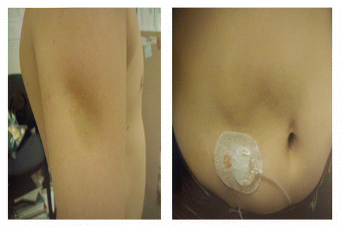
Patient is a 9-year-old boy diagnosed with T1DM at the age of 5 years. One year after diagnosis, he developed lipoatrophic skin changes specifically at insulin injection sites on his upper arms and thighs. His glycemic control was complicated by episodes of hypoglycemia and his hemoglobin A1C (HbA1C) was in the 6.5–7% range. The skin lesions were initially attributed to the insulin type, and he was switched from NPH insulin and regular insulin to Humalog insulin, delivered by an insulin pump. He subsequently developed similar lesions on is abdomen at sites not limited to the insulin infusion ().
On physical examination, weight was 37.6 kg (45%) and height 141 cm (20%) and normal pulse and blood pressure. He has painless, indurated, discolored, indentations of the skin of both upper extremities, thighs and lower abdomen, both at the sites of and far from insulin infusion (). There was no evidence of hand involvement such as thickening and induration of the skin of the dorsum and proximal interphalangeal joints. There was full range of motion in the joints, no flexion contractures, no trigger finger, Raynaud's phenomenon or telangiectasia. Other laboratory tests included ACA (anti-centromere antibody), ANA (anti-nuclear antibody), Scl-70 and celiac panel were all negative.
Six years later, the lesions continue progressing with the old ones enlarging and new ones developing at sites remote from insulin injections. He had new scleroderma lesions on his shoulder, abdomen and thigh. Recent follow up visit at the age of 17 years and 6 months and again noted to have stable scleroderma like changes on his abdomen and still there is no hand and joint involvement. He has fair diabetes control (HbA1C 7.8%) and no evidence of nephropathy or retinopathy.
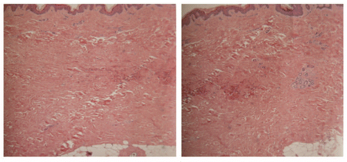
A biopsy from the affected areas revealed attenuated epidermis, dermis expanded with fibrous bands, atrophy of the adnexal structures (). There is no evidence of specific injury or inflammation of the subcutaneous fat. All of the above findings are consistent with the diagnosis of sclerodermal-like skin changes. Mucin stained had been performed on the skin biopsy specimen. The histologic features suggestive of a “scleroderma-like” disorder. However, histologic features are not diagnostic of scleroderma.”
Discussion
This is the first case report of a prepubertal male with T1DM and extensive scleroderma-like skin changes without evidence of the diabetic hand and joint syndrome whether this finding could be a variant of milder form of diabetic hand syndrome was unclear. This presentation is not consistent with linear scleroderma or diffuse morphic especially since the lesion originated in the areas of the insulin injections. In those patients with diabetic hand syndrome, the development of skin manifestation is influenced by the duration of diabetes and association with development of diabetic microvascular complications. Defective collagen formation may be due to the accumulation of advanced glycosylation end products has been assumed as the underlying pathogenic process.Citation1 In one study by Garza-Elizondo et al. it as noted that patients had scleroderma-like skin changes involving the PIP only and the juvenile patients had diabetes for 6 or more years and the difference in the disease duration between those with hand changes and those without was significant.Citation4 Therefore, most reports of scleroderma-like changes are noted in the hands of patients with T1DM. This skin changed initially was thought to be a lipohypothropy related to insulin injection. However the pathology report showed the change in the dermis not subcutaneous defect. There is also a case report of sclerderma and type I diabetes.Citation5 However the pathogenesis of these two co-existing conditions was unclear. It has been thought that interferons (IFNs) could play a major role. IFNs are well-known immunomodulators and inhibitors of collagen production. Beside their immunomodulatory action, IFNs can also be linked to autoimmune diseases.Citation6
It is noted that factors such as stratum corneum adhesion and accelerated aging of the skin may be implicated in the development of ichtyosiform skin changes in Type I DM patients and it could be explained by structural changes in skin proteins due to advance glycosylations.Citation3 There is strong correlation between ichthyoiform skin changes and diabetic retinopathy suggesting microvascular involvement in the pathogenesis of skin changes.Citation3,Citation7 Further observation will be needed in order to evaluate whether these lesions will progress to the full spectrum of the Limited Joint Mobility syndrome and represent an early risk marker for the development of diabetes complications.
Disclosure of Potential Conflicts of Interest
No potential conflicts of interest were disclosed.
Abbreviations
| T1DM | = | type I diabetes mellitus |
| HbA1C | = | hemoglobin A1C |
Figures and Tables
Acknowledgements
P.P. is currently on the speakers bureau for EMD Serono, Genetech, Inc., Endo phamaceutical and Novo Nordisk and serves as a consultant/advisor to EMD Serono. A.R. is currently on the speakers bureau for Amgen, Abbott and Pfizer and serves as a consultant/advisor to Amgen, Abbott, Pfizer, Merck and Novatis.
References
- Infante JR, Rosenbloom Al. Changes in frequency of limited joint mobility in children with Type 1 DM between 1976–78 and 1998. J Pediatr 2001; 138:33 - 37
- Yosipovitch G, Chuan Loh K. Medical Pearl: Scleroderma-like skin changes in patients with Diabetes Mellitus. J Am Acad Dermatology 2003; 49:1
- Yosipovitch G, Hodak E. The prevalence of cutaneous manifestations in IDDM patients and their association with diabetes risk factors and microvascular complications. Diabetes Care 1998; 21:506 - 509
- Garza-Elizondo MA, Diaz-Jouanene E. Joint Contractures and Scleroderma-like skin changes in the hands of insulin-dependent juvenile diabetics. J Rheumatol 1983; 10:797 - 800
- Polak M, Le Luyer B, Rybojab M, Czernichow P. Case report: insulin-dependent diabetes mellitus in childhood associated with scleroderma. Diabetes Metab 1996; 22:192 - 196
- Jimenez S, Derk C. Following the molecular pathways toward an understanding of the pathogenesis of systemic sclerosis. Ann Intern Med 2004; 37 - 50
- Sanli H, Akay BN, Sen BB, Kocak AY, Emral R, Bostanci S. Acquired ichthyosis associated with type 1 diabetes mellitus. Dermatoendocrinol 2009; 1:34 - 36

