Abstract
Background: Thyroid dysfunction is classically associated with alopecia. Studies focusing on manual thyroid examinations, with ultrasonography of palpable abnormalities, in alopecia patients are lacking.
Objective: To examine the clinical utility of manual and sonographic evaluation of the thyroid in alopecia patients.
Methods: A retrospective chart review was performed among patients diagnosed with alopecia.
Results: We found that 20.2% (74/367) of manual thyroid exams performed were deemed abnormal and 78.8% (41/52) of patients who had an ultrasound had an abnormal finding. Twenty two of the 74 patients did not obtain the requested ultrasound. Non-scarring alopecia was associated with 36 of 41 patients with abnormal ultrasounds (Telogen effluvium 29.3%, Androgenetic alopecia 27.8%, Alopecia areata 24.4%, and Traction alopecia 9.8%). No one specific structural abnormality was associated with a specific hair loss type. Of note, 78% (32/41) of patients with an abnormal ultrasound exam had normal thyroid function tests and only 9/41 (22%) patients had both.
Limitations: These include: a retrospective study design, small sample size, use of multiple sites for laboratory and sonographic thyroid evaluation, and a high attrition rate for ultrasound evaluation.
Conclusions: This study revealed that the manual examination of the thyroid in alopecia patients may identify additional thyroid abnormalities not detected with serologic evaluation alone. Further prospective studies are required to evaluate the necessity and significance of manual thyroid palpation and subsequent ultrasound studies in this patient population.
Introduction
The serum thyrotropin level (thyroid stimulating hormone or TSH) is a reliable initial evaluation for the detection of thyroid dysfunction,Citation1 but it does not detect the presence of structural abnormalities. Additionally, the majority of patients with a thyroid nodule and thyroid cancer are euthyroid.Citation2 Both manual palpation of the thyroid and ultrasound are employed to determine the size and presence of nodules however ultrasound is much more sensitive, especially in the detection of nonpalpable, sub centimeter nodules which have the potential to bear malignant disease.Citation3,Citation4
Serum thyroid function examinations are routinely ordered by the dermatologist when evaluating hair loss patients.Citation5,Citation6 However, the current literature discussing the necessity of thyroid palpation in this population is scarce and limited to patients with alopecia areata.Citation7,Citation8 A recent case at our institution's hair clinic, in which a thyroid malignancy in an alopecia areata patient was diagnosed after manual palpation and subsequent evaluation with ultrasound,Citation9 led to the addition of routine manual thyroid exams with subsequent ultrasound evaluation for perceived abnormal physical exam as part of the evaluation of alopecia. Retrospectively, we sought to investigate the utility of these manual and sonographic thyroid evaluations in our alopecia patients.
Results
Description of the study patients.
Demographics of the patients who received manual thyroid examinations are presented in . The average patient age was 44.1 years (standard deviation 16.7 years). The majority of patients were female (88.6%) and Caucasian (72.8%). In addition, 234 (63.8%) patients were white females. African American patients accounted for 13.6% of all exams. Non-scarring alopecia was the most prevalent type of hair loss in this study as 318 patients carried the diagnosis. The diagnosis of scarring alopecia was made for 49 patients.
Thyroid palpation and ultrasound evaluation.
The results of manual thyroid exams, TFTs, subsequent positive ultrasound evaluations and associated alopecia diagnoses are summarized in , and . Of the 367 patients examined, 74 (20.2%) were deemed to have an abnormal thyroid gland on palpation. Referrals for ultrasound evaluation were given to 69 of the 74 patients with an abnormal physical exam as 5 patients had a previous history of thyroid disease and ultrasound studies documented prior to our examine. A total of 22 patients did not obtain an ultrasound for various reasons: (1) the patient did not feel an additional exam was necessary despite the recommendation of a physician, (2) financial incapacity, and (3) the patient was unreachable (i.e. lost to follow-up). Not surprisingly, 9 (40.9%) of the 22 patients who never obtained a thyroid ultrasound also never obtained laboratory evaluations, therefore of the 350 patients who obtained TFTs 26 (7.2%) were observed to have serologic thyroid dysfunction. Approximately 41 of 52 (78.8%) of patients with abnormal manual thyroid exams had an abnormal finding on ultrasound. Only 8 of the 41 patients with an abnormal ultrasound also had abnormal serologic thyroid function. The most prevalent types of hair loss in patients () with an abnormal thyroid ultrasound were Telogen Effluvium (12/41), Androgenic Alopecia (10/41) and Alopecia Areata (10/41) followed by Traction Alopecia (4/41), Central Centrifugal Cicatricial Alopecia (3/41), Lichen Planopilaris (1/41) and Scarring Alopecia not otherwise specified (1/41). Of the 41 abnormal thyroid ultrasound evaluations 51 abnormalities were identified (). They included: 9 cysts (4/9 Colloid cysts), 8 adenopathy (1/8 found to have subacute thyroiditis and 2/8 found to have cervical lymphadenopathy), 18 sub-centimeter nodule(s), 5 >1cm nodule(s), 4 nodule with concerning features (i.e. taller than wide, increased vascularity, microcalcifications) with 1of the 4 being diagnosed with papillary thyroid cancer and 7 thyromegaly. No one type of alopecia was specifically associated with any one type of thyroid structural abnormalities ().
Methods
Subjects.
A retrospective analysis was performed on patients who were diagnosed with an alopecia disorder referred to the University of Pittsburgh Department of Dermatology Hair Clinic between January 1, 2008 and January 1, 2010. The Hair Clinic is conducted by one faculty dermatologist (JCE). Study approval was obtained from the University of Pittsburgh Institutional Review Board. A total of 367 patient records were reviewed. In order to maintain independence of the data, each patient was counted once, even in the event of multiple clinical visits. Each record was analyzed for demographic data, alopecia-spectrum diagnoses, serum thyroid function tests (TFTs), thyroid palpation, and results of ordered thyroid ultrasounds. Inter-examiner variability was eliminated as all thyroid physical exams included in this study were performed by the same faculty dermatologist (J.C.E). A total of 367 manual thyroid exams were performed within the study timeframe. The results were analyzed for the percentage of recorded abnormal examinations and in the event of subsequent referral for ultrasound evaluation the percentage of structural abnormalities identified. In addition, structural abnormalities were compared to alopecia spectrum diagnosis to evaluate for possible specific associations.
Statistical Analysis.
Limited statistical analysis was performed using the Microsoft Excel Software.
Comment
Physical examination of the thyroid is a safe, noninvasive and rapidly performed procedure that is commonly used to assess the thyroid for size, shape, consistency, tenderness and the presence of nodularity.Citation4,Citation10 While ultrasonography represents a more sensitive diagnostic tool, patients are unlikely to receive such testing unless clinical suspicion exists thus thyroid palpation represents a key method of reaching such suspicion.Citation4,Citation10 Ultrasonography is the recommended next step in the evaluation of thyromegaly detected on physical examination.Citation1
Thyroid nodules are found in approximate 5% of the general population and 95% of these nodules are benign in nature.Citation1,Citation2,Citation4,Citation11 Although thyroid carcinomas represent <2% of all cancers in the United States, approximately 37,000 new cases of thyroid carcinoma have been estimated to occur in 2010.Citation12 Although certain sonographic features of thyroid nodules have been associated with an increased risk of malignancy (i.e. predominantly solid nodule, hypoechogenicity, microcalcification, macrocalcification, ill-defined margins, intranodular vascularity, and taller-than-wide shape), no one feature is sensitive or specific enough to either exclude or diagnose malignancy.Citation13,Citation14 High-resolution ultrasonography has the capability to detect small, nonpalpable thyroid nodules, termed “incidentalomas” in the literature.Citation15,Citation16 Although most “incidentalomas” are benign, monitoring is required as the risk for malignancy in asymptomatic nodules found in non-irradiated glands ranges from 0.45% to 13%.Citation15,Citation16 Of note, a recent study suggests that the presence of benign-appearing enlarged cervical lymph nodes on ultrasound evaluation increases the predictive value in diagnosing thyroid cancer.Citation17 Biopsy via ultrasound guided fine needle aspiration is the gold standard to diagnosis thyroid cancer.Citation1,Citation2,Citation4,Citation14 Thyroid ultrasonography also helps to identify other potential thyroid disease. Diffuse thyroid echogenicity on sonography has been shown to predict patients with diffuse lymphocytic thyroiditis who are prone to develop hypothyroidism.Citation18
In this study, performance of a manual thyroid palpation was deemed sufficiently abnormal to consider sonographic evaluation in approximately one of every five patients, and over 75% of ultrasound assessments discovered evidence of an abnormality. The percentage of abnormal ultrasounds is likely under-representative as approximately 29.7% (22/74) of our population did not comply with our request for thyroid ultrasound. A true incidence of structural abnormalities in the study population cannot be determined. This is because the study was not prospective; all patients did not have the thyroid gland scanned and some structural abnormalities not detected on physical examination may have existed. Autopsy studies reveal that approximately 50% of patients 60 years of age have thyroid nodules.Citation19
Most concerning is that had the dermatologist obtained solely serum evaluation for thyroid function without manual palpation; several of these structural abnormalities may have been overlooked. The majority of patients with palpable thyromegaly and structural abnormalities were euthyroid (33/41). In our study, the majority of patients with thyromegaly and abnormal ultrasound evaluations had non-scarring types of alopecia. We observed that telogen effluvium and androgenetic alopecia patients had a slightly higher percentage of abnormal ultrasounds as that of alopecia areata patients. Upon further analysis no one specific structural abnormality was associated more frequently with a specific hair loss type ().
Several limitations are observed in this study. The retrospective design of this study prohibited the development of a standardized protocol for patient evaluation. The small sample size used in this study and bias of an academic setting in which it was carried out are additional limitations to our study. Ultrasound evaluations were performed and interpreted at multiple facilities by more than one radiologist may contribute to inter-observer variability. Our attrition rate for ultrasound evaluation was approximately 30%. Several factors likely contributed to this rate of noncompliance. In addition to financial limitations, patients may view alopecia as a self-contained disease and therefore feel that once initially treated, no further evaluation is necessary. Secondly, patients that we were unable to contact may have obtained further evaluation outside of our medical system. Therefore the percentage of structural thyroid abnormalities in our alopecia population with thyromegaly on physical examination may actually be higher than what was observed study.
In conclusion, the retrospective data obtained in this study suggests that patients with alopecia can have a wide range of structural abnormalities to the thyroid gland and the majority of these patients were euthryoid. A true pathologic cause and effect of structural thyroid abnormalities on alopecia in the setting of normal and abnormal thyroid function is unknown and further investigations will be needed to determine the significance of this data. Although the interaction between these two disease process is unknown, the literature on thyroid nodules suggest that when evaluating the thyroid one should perform a manual examine of the thyroid gland and obtaining sonographic imaging when thyromegaly is suspected. Future investigations with a prospective evaluation of a larger population with pan-ultrasound examinations of all alopecia subjects, comparing thyroid physical examination between dermatologists and endocrinologists and long-term follow-up to determine if alopecia patients with structural abnormalities have a greater risk of malignant transformation compared to the general population will further determine the significance of the our observation.
Disclosure of Potential Conflicts of Interest
No potential conflicts of interest were disclosed.
Figures and Tables
Figure 1 Results of manual thyroid exam, thyroid function tests and subsequent ultrasound evaluation flowchart. nl, normal; hx, history; abnl, abnormal; U/S, ultrasound; w/u, work-up; pts, patients; dz, disease; dx, diagnosis; TFT, thyroid function test (serum).
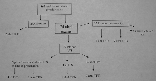
Table 1 Demographic features of the study population who received manual thyroid exam
Table 2 Type of hair loss in patients with abnormal ultrasound evaluations
Table 3 Results of abnormal ultrasound with associated alopecia
References
- Hegedüs L. Clinical practice. The thyroid nodule. N Engl J Med 2004; 351:1764 - 1771
- Sosa JA, Udelsman R. Papillary thyroid cancer. Surg Oncol Clin N Am 2006; 15:585 - 601
- Weist PW, Hartshorne MF, Inskip PD, Crooks LA, Vela BS, Telepak RJ, et al. Thyroid palpation versus high resolution-thyroid ultrasonography in the detection of nodules. J Ultrasound Med 1998; 17:487 - 496
- Dean DS, Gharib H. Epidemiology of thyroid nodules. Best Pract Res Clin Endocrinol Metab 2008; 22:901 - 911
- Shrivastava SB. Diffuse hair loss in an adult female: approach to diagnosis and management. Indian J Dermatol Venereol Leprol 2009; 75:20 - 27
- Artantas S, Gul U, Kilic A, Guler S. Skin findings in thyroid disease. Eur J Intern Med 2009; 20:158 - 161
- Kurtev A, Iliev E. Thyroid autoimmunity in children and adolescents with alopecia areata. Int J Dermatol 2005; 44:457 - 461
- Lutz G, Biersack HJ, Bauer R, Kreysel HW. Value of pathologic thyroid gland findings in alopecia areata. Z Hautkr 1987; 62:1253 - 1261
- McGuire S, English JC 3rd. Papillary thyroid cancer: An indication for thyroid palpation by the dermatologist. Arch Dermatol 2010; 146:1056 - 1057
- Slater S. Palpation of the thyroid gland. South Med J 1993; 86:1001 - 1003
- Miller MC. The patient with a thyroid nodule. Med Clin North Am 2010; 94:1003 - 1015
- Jemal A, Siegel R, Xu J, Ward E. Cancer statistics, 2010. CA Cancer J Clin 2010; 60:277 - 300
- Bastin S, Bolland MJ, Croxson MS. Role of ultrasound in the assessment of nodular thyroid disease. J Med Imaging Radiat Oncol 2009; 53:177 - 187
- Lew JI, Rodgers SE, Solorzano CC. Developments in the use of ultrasound for thyroid cancer 2010; 22:11 - 16
- Pinchera A. Thyroid incidentalomas. Horm Res 2007; 68:199 - 201
- Tan GH, Gharib H. Thyroid incidentalomas: management approaches to nonpalpable nodules discovered incidentally on thyroid imaging. Ann Intern Med 1997; 126:226 - 231
- Hands KE, Cervera A, Fowler LJ. Enlarged benign-appearing cervical lymph nodes by ultrasonography are associated with increased likelihood of cancer somewhere within the thyroid in patients undergoing thyroid nodule evaluation. Thyroid 2010; 20:857 - 862
- Marcocci C, Vitti P, Catalano F, et al. Thryoid ultrasonography helps identify patients with diffuse lymphocytic thyroiditis who are prone to develop hypothyroidism. J Clin Endorinol Metab 1991; 72:209 - 213
- Hegedus L, Bonnema SJ, Bennedbaek FN. Management of simple nodular goiter;current status and future perspectives. Endocr Res 2003; 24:102 - 132