Abstract
Our previous study has demonstrated that Yersinia pestis Microtus 201 is a low virulent strain to the Chinese-origin rhesus macaques, Macaca mulatta, and can protect it against high dose of virulent Y. pestis challenge by subcutaneous route. To investigate whether the Y. pestis Microtus 201 can be used as a live attenuated vaccine candidate, in this study its intravenous virulence was determined and compared with the live attenuated vaccine strain EV in the Chinese-origin rhesus macaque model. The results showed that the Chinese-origin rhesus macaques can survive intravenous infection with approximately 109 CFU of the Y. pestis Microtus 201, but all the animals succumbed to 1010 CFU of intravenous infection. By contrast, all the animals survive intravenous infection with 1010 CFU of the vaccine EV. Post-mortem examination showed multiple areas of severe abscess in the lungs of the dead animals infected with 1010 CFU of the Y. pestis Microtus 201, whereas histopathology observation, microbiological examination and immunohistochemistry staining showed that the Y. pestis Microtus 201 also invaded hearts, livers, spleens, kidneys and lymph nodes and caused different degrees of pathological changes in these organs. These results indicated that the Y. pestis Microtus 201 is indeed low virulent to monkeys, but it is more virulent than the vaccine EV when administered by intravenous route. The Y. pestis Microtus 201 mainly attack the lungs when administered by intravenous infection, which may be the leading cause of animal death.
Introduction
Yersinia pestis is the causative agent of plague, which is one of the most influential infectious diseases in human history. Y. pestis is genetically very closely related to the enteric pathogen Yersinia pseudotuberculosis and is considered as a clone evolved from a serotype of O:1b strain of Y. pseudotuberculosis, despite the fact that these 2 pathogens cause remarkably different diseases.Citation1-2 Plague is a zoonotic disease, which is occasionally transmitted to humans from Y. pestis-infected rodents via the bite of an infected flea.Citation1 Historically, 3 plague pandemics have occurred, leading to millions of human deaths.Citation3-4 Animal plague still currently persists in various zoonotic foci in Asia, Africa, Euroasia, and North or South America. The improvement of public health control countermeasures and the utilization of antibiotics considerably reduced the mortality, morbidity, and incidence of human plague during the latter half of the 20th century. However, Y. pestis strains with naturally acquired plasmid-mediated antibiotic resistance have been isolated from human cases.Citation2 In addition, since the beginning of the 1990s, a sharp rise in the number of human plague cases has been reported in 25 countries to the World Health Organization (WHO).Citation3-4 Recently, plague was classified as a re-emerging infectious disease by WHO.Citation5 Y. pestis has attracted considerable attention because of its potential misuse as an agent of biological warfare or bioterrorism.Citation6
Historically, Y. pestis strains have been divided into 3 biovars, including antiqua, mediaevalis and orientalis, based on their ability to ferment glycerol and to reduce nitrate. Biovars Y. pestis antiqua and mediaevalis are once considered to be associated with the Justinian Plague and the Black Death, respectively. Biovar Y. pestis orientalis originally from southern China is associated with modern plague.Citation7 Recently, the fourth Y. pestis biovar Microtus was proposed based on their unique pathogenic, biochemical and molecular features.Citation8 Whole genomic sequencing of a biovar Microtus strain 91001 reveals that this strain is composed of one chromosome and 4 plasmids (pCD1, pMT1, pPCP1 and pCRY). The genomic constituents of the strain 91001 do not differ dramatically from those of strains CO92 and KIM. However, the strain 91001 has undergone a unique accumulation of gene loss and pseudogenes in its genome.Citation9 The genome contents of another Y. pestis Microtus 201 were identical to Y. pestis strain 91001 according to our previous DNA microarray based comparative genomic analysis. Both strains belong to the newly established Y. pestis biovar, Microtus.Citation8
Our recent study has demonstrated that the Y. pestis Microtus 201 is highly attenuated in the Chinese-origin rhesus macaque, Macaca mulatta model of infection by subcutaneous route. The Y. pestis Microtus 201 represented a good plague vaccine candidate based on its ability to generate strong cytokine-mediated Th1-type cellular immune response and Th2-type humoral immune response as well as its good protection against high dose of subcutaneous virulent Y. pestis 141 challenge.Citation10 Recently, a case of lethal septicemic plague caused by an attenuated pgm¯ mutant of Y. pestis was reported in an U. S. laboratory. In this case, hereditary hemochromatosis caused by a C282Y mutation appears to be the leading cause of death.Citation11 In addition, live attenuated vaccine EV retains virulence when administered by the intranasal (i.n.) or intravenous (i.v.) route in some non-human primates,Citation12-14 which made it not to be licensed for human use in Western countries.Citation15-17 Therefore, it is imperative to explore novel rationally attenuated Y. pestis mutants as vaccine candidates for use against plague. Although we have demonstrated that the Y. pestis Microtus 201 strain shows a good plague vaccine candidate, and has a low virulence by the s.c. route in the Chinese-origin rhesus macaque model,Citation10 its virulence has not been evaluated by the intranasal (i.n.) or intravenous (i.v.) route. In order to investigate whether the Y. pestis Microtus 201 can be used as a live attenuated vaccine candidate for further evaluation, in this study the virulence of the Y. pestis Microtus 201 was determined and compared with the vaccine strain EV by intravenous route in the Chinese-origin rhesus macaque model.
Results
Clinical signs and post-mortem observation
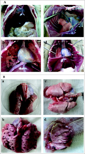
In our previous study, the Y. pestis Microtus 201 has been demonstrated to be highly attenuated in Chinese-origin rhesus macaque model of infection by subcutaneous route. All the animals survive the infection with the Y. pestis Microtus 201 (1.4 × 10¹⁰ CFU) or the EV vaccine (1.57 × 10¹⁰ CFU), and no systemic symptoms were observed in these 2 groups of animals.Citation10 This result seems to indicate that these 2 strains of Y. pestis had similar virulence by subcutaneous route. To further evaluate the virulence of these 2 strains of Y. pestis, 2 animals were first injected intravenously in a preliminary experiment to predict the virulence of the Y. pestis Microtus 201 and the vaccine EV. After intravenous infection, the animal infected with the vaccine EV (9.6 × 10⁸ CFU) eats less than normal animals from the second to third day, whereas the animal infected with the Y. pestis Microtus 201 (9 × 10⁸ CFU) has less than normal animals from the sixth to twentieth day. These two animals survive the infection with the Y. pestis Microtus 201 or the vaccine EV during the 21-day's post-infection observation. Post-mortem examination did not show any clear gross changes in the organs from the animal infected with the Y. pestis Microtus 201 (, a) or the vaccine EV (, c). This result seems to indicate that these 2 strains of Y. pestis might not cause serious damage to organs by intravenous route, and that the virulence difference between the Microtus Y. pestis 201 and the vaccine EV can not be distinguished at a level of 109 CFU by intravenous route.
Figure 1. Post-mortem examination of gross changes in the organs from the animal infected with the Y. pestis Microtus 201 or the vaccine EV. (A and B, a and c) Photos taken from the animals infected intravenously with approximately 109 CFU of the Y. pestis Microtus 201 and the vaccine EV. (A and B, b and d) Photos taken from the animals infected intravenously with approximately 1010 CFU of the Y. pestis Microtus 201 and the vaccine EV.

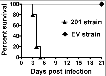
According to the results in the preliminary experiment, 2 groups of animals were infected intravenously with the Y. pestis Microtus 201 (1.28 × 10¹⁰ CFU) and the vaccine EV (1.5 × 10¹⁰ CFU), respectively, and then, closely observed for 21 d. After high-dose intravenous infection, all the animals infected with the Y. pestis Microtus 201 succumbed to the infection within 5 d, whereas all the animals infected with the vaccine EV survived the infection during the 21-day's post-infection observation, but their food intake decreased from the second to sixth days. Post-mortem examination did not show any gross changes in the organs of the animal infected with the vaccine EV (, d), whereas multiple areas of severe abscess were observed in the lungs of all the animals infected with the Y. pestis Microtus 201 (, b). This result indicates that the vaccine EV is still low virulent even if it is at a level of 1010 CFU by intravenous route, and is less virulent than the Microtus Y. pestis 201 by intravenous route. In addition, the result also indicates that bacteria attack mainly the lungs when administered by intravenous infection with up to 1010 CFU level of the Y. pestis Microtus 201.
Examination of Y. pestis within organs
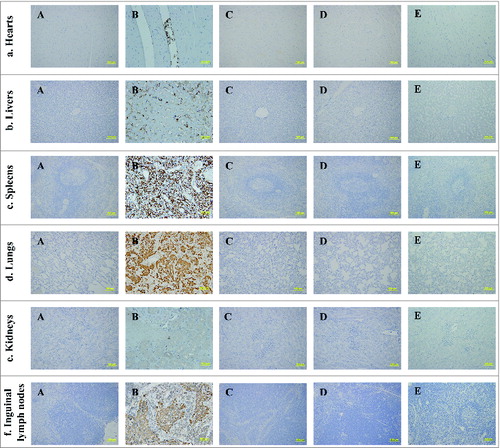
Microbiological examination and immunohistochemistry staining were used to investigate whether the Y. pestis Microtus 201 or the vaccine EV was completely eliminated from the infected animals. Tissues of the animals were harvested, homogenized, and then subjected to plate counts by using agar plates. The F1 antigen of the Y. pestis Microtus 201 or the EV vaccine was identified by immunohistochemistry staining in the formalin-fixed paraffin-embedded tissues. The tissues from one normal animal that was neither immunized with the Y. pestis Microtus 201 or the EV vaccine were used as the naïve controls. Bacteria could be visualized as those expressing the F1 antigen (brown and yellow stain). Microbiological examination () and immunohistochemistry staining () showed that no Y. pestis was observed in the hearts, livers, spleens, lungs, kidneys and inguinal lymph nodes of the animals infected with the Y. pestis Microtus 201 (9 × 10⁸ CFU) or the EV vaccine (9.6 × 10⁸ CFU), indicating that Y. pestis have been eliminated from these 2 infected animals. By contrast, bacteria were observed in the hearts, livers, spleens, lungs, kidneys and inguinal lymph nodes of the animals infected with the Y. pestis Microtus 201 (1.4 × 10¹⁰ CFU), but no bacterium was observed in the tissues of the animals infected with the vaccine EV (1.57 × 10¹⁰ CFU).
Table 1. Microbiological examination of tissue bacterial load in the Chinese-origin rhesus macaques following Y. pestis infection
Figure 2. The F1 antigen of Y. pestis was identified by immunohistochemitry staining. Bacteria were visualized as those expressing F1 antigen (brown and yellow stain). (A) Tissue sections from the animals infected intravenously with 9 × 10⁸ CFU of the Y. pestis Microtus 201. (B) Tissue sections from the animals infected intravenously with 1.28 × 10¹⁰ CFU of the Y. pestis Microtus 201. (C) Tissue sections from the animals infected intravenously with 9.6 × 10⁸ CFU of the vaccine EV. (D) Tissue sections from the animals infected intravenously with 1.5 × 10¹⁰ CFU of the vaccine EV. (E) Tissue sections from one normal animal that was neither immunized with the Y. pestis Microtus 201 or the EV vaccine. Numerous bacteria were observed in the tissue of hearts (a), livers (b), spleens (c), lungs (d), kidneys (e) and lymph nodes (f) of the dead animals after infection with approximately 1010 CFU of the Y. pestis Microtus 201 (B). No bacterium was found in other groups of animals (A, C and D) and the control animal (E).

Virulence determination
To determine the level of virulence of the Y. pestis Microtus 201 or the vaccine EV by intravenous route, we randomly selected 2 animals to perform intravenous infection in a preliminary experiment. One animal was infected with the Y. pestis Microtus 201 (9 × 10⁸ CFU), and another animal with the EV vaccine (9.6 × 10⁸ CFU). After infection, the animals were closely observed for 21 d. The results showed that 2 animals survived the above levels of infectious doses, indicating that these 2 strains of Y. pestis have a low virulence in Chinese-origin rhesus macaques. To further investigate the virulence difference between the Y. pestis Microtus 201 and the vaccine EV, 10 Chinese-origin rhesus macaques were randomly divided into 2 experimental groups, and then were intravenously injected with the Y. pestis Microtus 201 (1.28 × 10¹⁰ CFU) and the EV vaccine (1.5 × 10¹⁰ CFU), respectively. All the animals were observed twice daily for signs of morbidity and, if moribund, animals were humanely euthanized by using pentobarbital sodium anesthesia. The survival of animals in 2 groups is represented in . Statistical analysis showed that the virulence of the Y. pestis Microtus 201 was higher than that of the EV vaccine by intravenous route (P < 0.05).
Histopathology observation
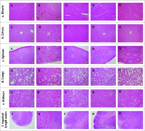
The samples of heart, liver, spleen, lungs, kidneys and lymphoid nodes from one normal animal and the animals infected with the Y. pestis Microtus 201 (9 × 10⁸ CFU), the Y. pestis Microtus 201 (1.28 × 10¹⁰ CFU), the EV vaccine (9.6 × 10⁸ CFU) or the EV vaccine (1.5 × 10¹⁰ CFU) were fixed in 10% neutral buffered formalin. The tissues were trimmed, paraffin embedded, sectioned and stained with haematoxylin and eosin (H and E). The slides prepared from these organs were observed under light microscope for pathological changes. Compared with normal tissues ( Panel E), no changes in histopathology were found in all examined tissues from 3 groups of animals infected with the Y. pestis Microtus 201 (9 × 10⁸ CFU), the EV vaccine (9.6 × 10⁸ CFU) and the EV vaccine (1.5 × 10¹⁰ CFU) (, Panel A, C and D), whereas the animals infected with the Y. pestis Microtus 201 (1.28 × 10¹⁰ CFU) showed evident alterations in these tissues (, Panel B). Vascular engorgement and slight liver cell degeneration were observed in heart tissues ( Panel B) and liver tissues (Fig. 4b, Panel B), respectively. Atrophic white pulp, splenic sinus congestion and reduced amounts of lymphocytes were found in the spleen tissues ( Panel B). Compared with lung tissues from the normal animal, lung tissues in panels A, C and D don't show evident changes. Alveolar walls are thickened in lungs of panels A, C and D, but the alveolar walls are also thickened in normal lungs. By contrast, disappearance of recognizable lung tissue architecture, edema, congestion and inflammatory cell infiltration were observed in the lung tissues of the animals infected with the Y. pestis Microtus 201 (1.28 × 10¹⁰ CFU) ( Panel B). Renal interstitium congestion and edema were observed in the kidney tissues ( Panel B). Lymphocyte hyperplasia, edema and neutrophil infiltration were found in the lymph node tissues ( Panel B).
Figure 4. Histopathologic observation of the tissues from the animals infected with the Y. pestis Microtus 201 and the vaccine EV. Tissue sections were stained with hematoxylin and eosin for pathological examination after infection with Y. pestis. (A) Tissue sections from the animals infected intravenously with 9 × 10⁸ CFU of the Y. pestis Microtus 201. (B) Tissue sections from the animals infected intravenously with 1.28 × 10¹⁰ CFU of the Y. pestis Microtus 201. (C) Tissue sections from the animals infected intravenously with 9.6 × 10⁸ CFU of the vaccine EV. (D) Tissue sections from the animals infected intravenously with 1.5 × 10¹⁰ CFU of the vaccine EV. (E) Tissue sections from one normal animal.

Discussion
Y. pestis biovar Microtus strains seem to have a low virulence to larger mammals,Citation18 whose major phenotypes are F1+ (able to produce fraction 1 capsule), LcrV+ (presence of V antigen), Pst+ (able to produce pesticin) and Pgm+ (pigmentation on Congo-red media). The Microtus strain 91001 has been demonstrated to be of low virulence for humans.Citation19 Our recent study showed that another Y. pestis Microtus 201 has a low virulence by the s.c. route in the Chinese-origin rhesus macaque model, and provides a protective efficacy similar to the EV vaccine against bubonic plague by generating strong Th1-type cellular and Th2-type humoral immune responses.Citation10 These results seem to signify that the Y. pestis Microtus 201 or 91001 may be a promising live attenuated plague vaccine candidate. However, although no plague symptom was observed in humans by superficial skin scarification with 5.1 × 10⁶ CFU of the Y. pestis strain 91001,Citation19 the dose and mode of inoculation were not enough to illustrate this strain to be completely safe for humans. In our previous study, we have demonstrated that the Y. pestis Microtus 201 strain has a low virulence by the s.c. route in the Chinese-origin rhesus macaque model,Citation10 but its virulence has not been evaluated by other routes of inoculation. Live attenuated vaccine EV is able to elicit both humoral and cellular immune responseCitation20-21 and is effective against bubonic and pneumonic plague in humans, but it can cause fatal plague in some non-human primates,Citation12 and retains virulence when administered by the intranasal (i.n.) and intravenous (i.v.) routes.Citation12-14 These side effects of varying severity made it not to be licensed for human use in Western countries.Citation15-17 Therefore, an ideal live attenuated vaccine for plague should at least have lower virulence than the vaccine strain EV. In the present study, the virulence of the Y. pestis Microtus 201 was assessed by the intravenous (i.v.) route, and compared with the vaccine EV.
To investigate whether the Y. pestis Microtus 201 has a lower virulence than the vaccine EV in higher animals when administered by intravenous infection, the virulence of the Y. pestis Microtus 201 and the vaccine EV were determined in the Chinese-origin rhesus macaque model. The results showed that the Chinese-origin rhesus macaques can tolerate about 109 CFU level of intravenous infection with the Y. pestis Microtus 201 or the EV vaccine. The animal infected with the vaccine EV eats less than normal animals from the second to third day, and the animal infected with the Y. pestis Microtus 201 eats less than normal animals from the sixth to twentieth day. Post-mortem examination did not show any clear gross changes in the organs from the animal infected with the Y. pestis Microtus 201 or the vaccine EV. These results indicated that the Y. pestis Microtus 201 and the vaccine EV might not cause serious damage to organs, and had similar virulence at a level of 109 CFU by intravenous infection. However, all the animals succumbed to 1010 CFU level of intravenous infection with the Y. pestis Microtus 201, whereas all the animals survive 1010 CFU level of intravenous infection with the vaccine EV. Multiple areas of severe abscess were only observed in the lungs of all the animals infected with the Y. pestis Microtus 201, indicating that the Microtus Y. pestis 201 is more virulent than the vaccine EV at a level of 1010 CFU by intravenous route. In addition, the result also indicates that bacteria attack mainly the lungs when administered by intravenous infection with up to 1010 CFU level of the Y. pestis Microtus 201. Compared with our previous study that all the animals survive subcutaneous infection with the Y. pestis Microtus 201 (1.4 × 10¹⁰ CFU), and no systemic symptoms were observed in the animals,Citation10 the Y. pestis Microtus 201 is more virulent to the Chinese-origin rhesus macaques by intravenous route than by subcutaneous route. To our surprise, all the animals survive 1010 CFU level of intravenous infection with the vaccine EV, and no plague symptom was observed in these animals. These results indicated that the Y. pestis Microtus 201 was low virulent in the Chinese-origin rhesus macaques, but it was more virulent than the vaccine strain EV when it was administered by intravenous route. The previous studies showed that the EV vaccine retained virulence in non-human primates when administered by the intranasal (i.n.) and intravenous (i.v.) routes,Citation12-14 but our current study showed that it was avirulent to the Chinese-origin rhesus macaques by the i.v. route, indicating that this species of non-human primate was less sensitive to the vaccine EV than other non-human primates. Although the Y. pestis Microtus 201 has higher virulence than the vaccine EV in the Chinese-origin rhesus macaques, but it might have a good potential to develop a live attenuated plague vaccine for humans, because the strain contains all the known protective antigens, can provide good protection against plague by generating strong Th1-type cellular and Th2-type humoral immune responses,Citation10 and have a lower virulence for non-human primates and humans. It is a promising strategy for the strain to develop live attenuated plague vaccines based on the virulence-associated determinants not essential for protection. In addition, its low virulence is not elicited by the deletion of pgm locus, which reduces the risk to reverse the virulence when used in hereditary hemochromatosis persons Citation11
In our previous study, it has been demonstrated that the Chinese-origin rhesus macaques can stand up to 1010 level of subcutaneous infection with the Y. pestis Microtus 201.Citation10 In this study, we found that approximately 109 level of intravenous infection with the Y. pestis Microtus 201 strain did not result in the death of a Chinese-origin rhesus macaque and induce any pathological changes in heart, liver, spleen, lungs, kidneys and lymph nodes, but the animals succumbed to 1010 CFU level of intravenous infection with the same strain. Post-mortem examination showed that multiple areas of severe abscess were observed in the lungs of the dead animals infected with 1010 level of Y. pestis Microtus 201, whereas other organs did not show any clear gross changes, indicating that the Y. pestis Microtus 201 attack mainly the lungs when administered by intravenous infection with up to 1010 CFU level of the Y. pestis Microtus 201. However, histopathology observation and immunohistochemistry staining showed that the Y. pestis Microtus 201 also invaded hearts, livers, spleens, kidneys and lymph nodes and caused different degree of pathological changes in these organs. These results revealed that the Y. pestis Microtus 201 was a low virulent strain for larger animals, but high dose of intravenous infection can cause the death of the Chinese-origin rhesus macaques, and that the animals died mainly from the lesions of lungs. Microbiological examination showed that bacteria can be eliminated from the animal infected with approximate 109 CFU of the Y. pestis Microtus 201 after the 21-day's post-infection observation, but when the infection dose reachs to 1010 CFU, bacteria can be isolated from the dead animals. By contrast, no bacterium was observed in the animals infected with 109 or 1010 CFU of the vaccine EV after the 21-day's post-infection observation. These results indicated that a good vaccine strain should have a limited ability to survive in animals. However, if a strain has a much lower ability to survive in animals, its immunogenicity is decreased. This may be due to the inability of the attenuated bacterium to invade and persist in the host long enough to initiate an immune response, because live vaccines must be able to reach, multiply in, and persist for a limited time in the lymphoid organs necessary to stimulate a protective immune response. Conversely, if a strain has a highly survival ability in animals, the bacterium will be highly virulent.Citation22
Taken together, we came to the conclusion that the Y. pestis Microtus 201 is a low virulent strain by s.c. or i.v. route, but it is more virulent to the Chinese-origin rhesus macaques than the vaccine strain EV. The Chinese-origin rhesus macaques can survive subcutaneous or intravenous infection with approximately 1010 or 109 CFU of the Y. pestis Microtus 201, but it cannot stand intravenous infection with approximately 1010 CFU of this strain, and mainly attacks the lungs of the animals, resulting in multiple areas of severe abscess, which may be the leading cause of animal death. In addition, the Y. pestis Microtus 201 also invaded hearts, livers, spleens, kidneys and lymph nodes and caused different degrees of pathological changes in these organs. By contrast, all the animals survive subcutaneous or intravenous infection with 1010 CFU of the vaccine EV, and do not show any pathological changes in hearts, livers, spleens, lungs, kidneys and lymph nodes. These results indicated that the vaccine EV is avirulent to the Chinese-origin rhesus macaques when administered by subcutaneous or intravenous route.
Y. pestis biovar Microtus strains are thought to be avirulent to larger mammals, such as guinea pigs, rabbits and humans.Citation18 Our previous study has demonstrated that the Y. pestis Microtus 201 is highly attenuated in Chinese-origin rhesus macaque model of infection by subcutaneous route.Citation10 In this study, we first demonstrated that the Y. pestis Microtus 201 is low virulent to Chinese-origin rhesus macaques by intravenous route, but all the animals infected with the Y. pestis Microtus 201 died when the infection dose increases up to 1010 CFU. The animal death is caused by the Y. pestis Microtus 201 invading hearts, livers, spleens, lungs, kidneys and lymph nodes, and resulting in different degrees of pathological changes in these organs after intravenous infection, but this strain of Y. pestis mainly attacks the lungs, which may be the leading cause of animal death. We also found that the vaccine EV is avirulent to the Chinese-origin rhesus macaques whether by the i.v. or by the s.c route, indicating that the Y. pestis Microtus 201 is more virulent than the vaccine EV. The Y. pestis Microtus 201 still needs to be further attenuated for developing a plague vaccine, and the vaccine EV is still the preferred live attenuated vaccine currently.
Materials and Methods
Bacterial strains and animals
The Y. pestis Microtus 201 was isolated from Microtus brandti in Inner Mongolia, China and has LD50 of 3 CFU for BALB ⁄ c mice by the subcutaneous route, while the LD50 in the i.v.-infected mice was 1.9 CFU.Citation23 The live attenuated strain EV was obtained from the Lanzhou Institute of Biological Products (LIBP), China.
Chinese-origin rhesus macaques (2 years old) were obtained from Laboratory Animal Research Center, Academy of Military Medical Science, China (licensed from the Ministry of Health in General Logistics Department of Chinese People's Liberation Army, Permit No. SCXK-2007-004). All protocols were approved by Committee of the Welfare and Ethics of Laboratory Animals, Beijing Institute of Microbiology and Epidemiology. All animals were raised in an air-conditioned laboratory with an ambient temperature of 21-25°C and a relative humidity of 40%–60%. All moribund animals after the infection with Y. pestis were euthanized by using pentobarbital sodium anesthesia. All animal experiments were conducted strictly in compliance with the Regulations of Good Laboratory Practice for nonclinical laboratory studies of drug issued by the National Scientific and Technologic Committee of People's Republic of China.
Animal infection and determination of virulence
Y. pestis was cultured in Luria broth at 28°C for 18 hours, quantified by Maxwell turbidimetry, and diluted in sterile phosphate-buffered saline (PBS). The number of Y. pestis in the dilution was verified by colony-forming unit (CFU) counts on Y. pestis selective agar medium. After the animals were infected with the Y. pestis Microtus 201 or the vaccine EV, they were closely observed for 21 d.
Firstly, 2 animals were selected to perform intravenous infection with the Y. pestis Microtus 201 (9 × 10⁸ CFU) and the EV vaccine (9.6 × 10⁸ CFU), respectively, in a preliminary experiment, and secondly, 10 Chinese-origin rhesus macaques were randomly divided into 2 experimental groups, each one of which contained 5 animals, and then were intravenously injected with the Y. pestis Microtus 201 (1.28 × 10¹⁰ CFU) and the EV vaccine (1.5 × 10¹⁰ CFU), respectively. All the animals were observed twice daily for signs of morbidity and, if moribund, animals were humanely euthanized by using pentobarbital sodium anesthesia.
Microbiological examination and immunohistochemistry (IHC)
All the moribund or survival animals were euthanized humanely by using pentobarbital sodium anesthesia for a post-mortem examination during the 21-day's post-infection observation period. Hearts, livers, spleens, lungs, kidneys and inguinal lymph nodes of the infected animals were harvested, homogenized, and then subjected to plate counts by using agar plates to confirm if Y. pestis was presented in these organs.
IHC staining was performed following the user's manual of the PV-9000 Kit (ZSGB-Bio).Citation24 Briefly, after the paraffin-embedded tissue sections were deparaffinized and rehydrated, the sections were subjected to antigen exposure in citrate buffer solution (0.1 M, pH 6.0) by microwaving at 95°C for 20 min, and incubated with 3% H2O2 in methanol for 10 min to block endogenous peroxidase activity. The sections were incubated for 12 h with the purified rabbit anti-F1 antigen of Y. pestis polyclonal antibody at 4°C. The sections were incubated with Polymer Helper for 20 min at 37°C, and then with polyperoxidase antirabbit IgG (ZSGB-Bio) for 10–20 min at 37°C. The slides were stained with 3, 3’-diaminobenzidine tetrahydrochloride (DAB). Finally, the sections were rinsed, counterstained, dehydrated, cleaned, mounted, and examined under light microscopy.Citation25
Histopathology observation
All the moribund animals after the infection and the surviving animals at Day 21 after the infection with the Y. pestis Microtus 201 and the vaccine strain EV were humanely euthanized by using pentobarbital sodium anesthesia. The tissues collected from these animals were placed into 10% neutral buffered formalin, dehydrated through a serial alcohol gradient (70%, 80%, 90%, 95%, and 100%), cleared with xylene, infiltrated with wax, and then embedded in paraffin.Citation26 Tissue sections were stained with hematoxylin and eosin (HE) for histopathological examination.
Statistical analysis
Statistical analyses were performed using the software GraphPad Prism version 5.0. The survival rates in the different infection groups were compared by a log-rank test. A probability value of < 0.05 was considered statistically significant.
Disclosure of Potential Conflicts of Interest
No potential conflicts of interest were disclosed.
Funding
Financial support for this study came from the National Natural Science Foundation of China (contract no. 81171529) and the National High Technology Research and Development Program of China (863 program) (contract no. 2012AA02A403).
References
- Perry RD, Fetherston JD. Yersinia pestis–etiologic agent of plague. Clin Microbiol Rev 1997; 10:35-66; PMID:8993858
- Welch TJ, Fricke WF, McDermott PF, White DG, Rosso ML, Rasko DA, Mammel MK, Eppinger M, Rosovitz MJ, Wagner D, et al. Multiple antimicrobial resistance in plague: an emerging public health risk. PLoS ONE 2007; 2:e309; PMID:17375195; http://dx.doi.org/10.1371/journal.pone.0000309
- WHO. Human plague in 1998 and 1999. Wkly Epidemiol Rec 2000; 75:338-43; PMID:11218330
- WHO. Human plague in 2002 and 2003. Wkly Epidemiol Rec 2004; 79:301-6; PMID:15369044
- Williamson ED. Plague vaccine research and development. J Appl Microbiol 2001; 91:606-8; PMID:11576295; http://dx.doi.org/10.1046/j.1365-2672.2001.01497.x
- Inglesby TV, Dennis DT, Henderson DA, Bartlett JG, Ascher MS, Eitzen E, Fine AD, Friedlander AM, Hauer J, Koerner JF, et al. Plague as a biological weapon: medical and public health management. Working Group on Civilian Biodefense. JAMA 2000; 283:2281-90; PMID:10807389; http://dx.doi.org/10.1001/jama.283.17.2281
- Wren BW. The yersiniae–a model genus to study the rapid evolution of bacterial pathogens. Nat Rev Microbiol 2003; 1:55-64; PMID:15040180; http://dx.doi.org/10.1038/nrmicro730
- Zhou D, Tong Z, Song Y, Han Y, Pei D, Pang X, Zhai J, Li M, Cui B, Qi Z, et al. Genetics of metabolic variations between yersinia pestis biovars and the proposal of a new biovar, microtus. J Bacteriol 2004; 186:5147-52; PMID:15262951; http://dx.doi.org/10.1128/JB.186.15.5147-5152.2004
- Song Y, Tong Z, Wang J, Wang L, Guo Z, Han Y, Zhang J, Pei D, Zhou D, Qin H, et al. Complete genome sequence of Yersinia pestis strain 91001, an isolate avirulent to humans. DNA Res 2004; 11:179-97; PMID:15368893; http://dx.doi.org/10.1093/dnares/11.3.179
- Zhang Q, Wang Q, Tian G, Qi Z, Zhang X, Wu X, Qiu Y, Bi Y, Yang X, Xin Y, et al. Yersinia pestis biovar Microtus strain 201, an avirulent strain to humans, provides protection against bubonic plague in rhesus macaques. Hum Vaccin Immunother 2014; 10:1-10; PMID:24832715; http://dx.doi.org/10.4161/hv.28050
- Frank KM, Schneewind O, Shieh W-J. Investigation of a researcher's death due to septicemic plague. N Engl J Med 2011; 364:2563-4; PMID:21714673; http://dx.doi.org/10.1056/NEJMc1010939
- Meyer KF, Smith G, Foster L, Brookman M, Sung M. Live, attenuated Yersinia pestis vaccine: virulent in nonhuman primates, harmless to guinea pigs. J Infect Dis 1974; 129:Suppl:S85-12; PMID:4207627; http://dx.doi.org/10.1093/infdis/129.Supplement_1.S85
- Smiley ST. Immune defense against pneumonic plague. Immunol Rev 2008; 225:256-71; PMID:18837787; http://dx.doi.org/10.1111/j.1600-065X.2008.00674.x
- Une T, Brubaker RR. In vivo comparison of avirulent Vwa- and Pgm- or Pstr phenotypes of yersiniae. Infect Immun 1984; 43:895-900; PMID:6365786
- Russell P, Eley SM, Hibbs SE, Manchee RJ, Stagg AJ, Titball RW. A comparison of Plague vaccine, USP and EV76 vaccine induced protection against Yersinia pestis in a murine model. Vaccine 1995; 13:1551-6; PMID:8578841; http://dx.doi.org/10.1016/0264-410X(95)00090-N
- Morton M, Garmory HS, Perkins SD, O'Dowd AM, Griffin KF, Turner AK, Bennett AM, Titball RW. A Salmonella enterica serovar Typhi vaccine expressing Yersinia pestis F1 antigen on its surface provides protection against plague in mice. Vaccine 2004; 22:2524-32; PMID:15193377; http://dx.doi.org/10.1016/j.vaccine.2004.01.007
- Williamson ED, Eley SM, Griffin KF, Green M, Russell P, Leary SE, Oyston PC, Easterbrook T, Reddin KM, Robinson A, et al. A new improved sub-unit vaccine for plague: the basis of protection. FEMS Immunol Med Microbiol 1995; 12:223-30; PMID:8745007; http://dx.doi.org/10.1111/j.1574-695X.1995.tb00196.x
- Fan Z, Ruo Y, Wang S, Jin L, Zhou X, Liu J, Zhang Y, Li F. Microtus brandti plague in the Xilin Gol Grassland was inoffensive to humans. Chin J Control Endem Dis 1995; 10:56-7
- Fan Z, Luo Y, Li F, Zhang C, Su X, Wang S. Pathogenicity test for detection of Xilinguole Plateau type of Y. pestis in humans. Chinese J Control of Endemic Dis 1994; 9:340-2
- Parent MA, Berggren KN, Kummer LW, Wilhelm LB, Szaba FM, Mullarky IK, Smiley ST. Cell-mediated protection against pulmonary Yersinia pestis infection. Infect Immun 2005; 73:7304-10; PMID:16239527; http://dx.doi.org/10.1128/IAI.73.11.7304-7310.2005
- Philipovskiy AV, Smiley ST. Vaccination with live yersinia pestis primes CD4 and CD8 T cells that synergistically protect against lethal pulmonary Y. pestis infection. Infect Immun 2007; 75:878-85; PMID:17118978; http://dx.doi.org/10.1128/IAI.01529-06
- Wang X, Zhang X, Zhou D, Yang R. Live-attenuated Yersinia pestis vaccines. Expert Rev Vaccines 2013; 12:677-86; PMID:23750796; http://dx.doi.org/10.1586/erv.13.42
- Yang F, Ke Y, Tan Y, Bi Y, Shi Q, Yang H, Qiu J, Wang X, Guo Z, Ling H, et al. Cell membrane is impaired, accompanied by enhanced type III secretion system expression in Yersinia pestis deficient in RovA regulator. PLoS One 2010; 5:e12840; PMID:20862262; http://dx.doi.org/10.1371/journal.pone.0012840
- Chen Z, Zhuo F-L, Zhang S-J, Tian Y, Tian S, Zhang J-Z. Modulation of tropoelastin and fibrillin-1 by infrared radiation in human skin in vivo. Photodermatol, Photoimmunol Photomed 2009; 25:310-6; PMID:19906166; http://dx.doi.org/10.1111/j.1600-0781.2009.00465.x
- Chen S, Cheng A, Wanga M, Zhu D, Luo Q, Liu F, Chen X. Immunohistochemical detection and localization of new type gosling viral enteritis virus in paraformaldehyde-fixed paraffin-embedded tissue. Vet Immunol Immunop 2009; 130:226-35; PMID:19304327; http://dx.doi.org/10.1016/j.vetimm.2009.02.011
- Rohr LR, Layfield LJ, Wallin D, Hardy D. A comparison of routine and rapid microwave tissue processing in a surgical pathology laboratory. Am J Clin Pathol 2001; 115:703-8; PMID:11345834; http://dx.doi.org/10.1309/15FB-FLD1-408X-JQA3

