
Abstract
This commentary highlights the effectiveness of optoelectronic properties of polymer semiconductors based on recent results emerging from our laboratory, where these materials are explored as artificial receptors for interfacing with the visual systems. Organic semiconductors based polymer layers in contact with physiological media exhibit interesting photophysical features, which mimic certain natural photoreceptors, including those in the retina. The availability of such optoelectronic materials opens up a gateway to utilize these structures as neuronal interfaces for stimulating retinal ganglion cells. In a recently reported work entitled “A polymer optoelectronic interface provides visual cues to a blind retina,” we utilized a specific configuration of a polymer semiconductor device structure to elicit neuronal activity in a blind retina upon photoexcitation. The elicited neuronal signals were found to have several features that followed the optoelectronic response of the polymer film. More importantly, the polymer-induced retinal response resembled the natural response of the retina to photoexcitation. These observations open up a promising material alternative for artificial retina applications.
Semiconducting polymers are an attractive choice as active materials for a variety of optoelectronic applications. These materials have a unique combination of optoelectronic and mechanical properties: namely for features related to mechanical conformity, large visible light absorption strength which results in sizable density of photogenerated carriers and solution processability.Citation1 The sizable optical absorption of these materials implies requirement in form of thin (sub-micron) films rather than thick layers as in the case of Silicon-type materials. These features offer the possibility of seamless integration of device structures based on these active materials with biological systems. In general, polymer surfaces with appropriate mechanical attributes such as adhesive strength, microtexture and nanotopography, surface wettability and stiffness are conducive for anchoring neuronal cells.Citation2,Citation3 The electronic characteristics of certain conjugated polymers have also been used for sensing and stimulating neuronal activity.Citation4-Citation7 The optoelectronic properties of these materials have recently been utilized as active triggers for neuronal stimulation.Citation8-Citation11 The present commentary highlights this growing usage of these novel optoelectronic features of organic semiconductor based polymer layers in contact with physiological media and the utilization of these structures as an interface to evoke neuronal signals in a blind retina.
The interfacing of organic semiconductors with physiological processes requires understanding and characterization of the interface between the semiconductor and physiological media, and surface potential across the interface. Earlier reports from our laboratory focused on the interface and transport characteristics across certain semiconducting polymer/electrolyte structures, which are reflected in the photocurrent, photovoltage and photocapacitance measurements across the interface.Citation12 The polymer layer utilized in our studies consisted of a binary blend-mixture, which is technically referred as bulkheterojunction (BHJ), with regio-regular poly-alkylthiophenes (rrP3AT) as the donor (p) component and a naphthalene derivative (N2200) as the acceptor (n) component.Citation12-Citation14 The BHJ aspect of the active layer is responsible for a wider absorption range and significant photoinduced carrier generation (higher quantum efficiency) compared with that in a pristine single component layer.
The photocurrent profiles across a BHJ/electrolyte interface are in form of transients, arising from a combination of bulk carrier concentration, diffusion length scales and the presence of different transport mechanisms prevalent in these structures. These optoelectronic features at the BHJ/electrolyte interfaces were highlighted in an earlier report from our laboratory ().Citation13 We demonstrated that the photocurrent profiles could be controlled by certain parameters, and that for an optimum combination of these parameters, the device structures could be used as single-pixel color sensing elements. These structures further revealed interesting similarities to natural photosensitive systems. Specifically, the antagonistic color detection property and the time scales, magnitudes and pulse profiles of the photocurrent profiles across the BHJ/electrolyte interface were similar to those found in retinal cone cells and certain photosensitive membrane proteins.Citation13 Such visual photoreceptor-type sensing layers operating in physiological environment are suitable stimulation elements for retinal neurons.
Figure 1. (A) Optoelectronic features across the BHJ/electrolyte interface have characteristic transient pulse profiles. (B) The photoexcitation of the BHJ/electrolyte interface results in stimulating a blind retina with dysfunctional photoreceptors.
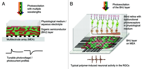
In our recently reported work,Citation14 we interfaced these optoelectronic polymer structures with a developing chick retina at a light-insensitive stage and showed their utility to elicit neuronal signals in the blind retina (). In another set of recent studies by Ghezzi et. al., a thiophene based pristine organic semiconductor was interfaced with degenerate rat retinas and was shown to partially restore light sensitivity in these tissues.Citation15 In our studies, the BHJ layer was coated on a multielectrode array (MEA) for stimulating the retina epiretinally and simultaneously recording its electrophysiological activity. The photoexcitation of the BHJ layer in contact with the retina in physiological conditions resulted in action potentials in the RGC layer of the otherwise blind retina. Such a response could be elicited with a wide range of wavelengths (400–600 nm). Utilizing a BHJ layer with a water-stable acceptor (n) component in our studies is advantageous over a pristine component for operation in physiological conditions.
It should be noted that in degeneration models like rd1 and light-induced damage in rats or mice, a reasonable fraction of residual photoreceptors could be present. This requires elaborate control experiments to distinguish the role of underlying polymer with that of residual receptors for retinal stimulation. The model of developing chick retina used in our experiments circumvents this issue. Though there are differences between the anatomy of the avian retina and the retina of higher mammals and a retinal degeneration model is absent in chicks, the usage at an embryonic stage where photoreceptors are insensitive serves as a suitable model to study the stimulation of a completely blind retina. Further, the animal handling protocol and retina preparation is relatively simple for the chick model.
Various groups have explored the stimulation of retina with μm-sized silicon photodiodes. However, it has been shown that that the current generated by a single microphotodiode is not sufficient to stimulate adjacent neurons with the ambient light and these detectors usually need to be supported by an external power source.Citation16,Citation17 Though the magnitude of photocurrents across a BHJ/electrolyte interface is lower than that of the silicon photodiodes, these structures are able to stimulate the retina under ambient light intensities. This points to the importance of better mechanical conformity of these soft structures, as well as to the similarity between the pulse-profile characteristics of BHJ-polymer/electrolyte structures with those of natural receptors.
The BHJ-induced neuronal response in the blind chick retina presented in our studies exhibited interesting features.Citation14 The temporal properties of the evoked retinal response correlated with the optoelectronic response of the polymer film. For instance, the response latency, the total number of spikes and the spike rate depended on light intensity, time duration and time interval of the incident light pulse used for photoexcitation of the BHJ layer. Further, at high intensities, the elicited neuronal response consisted of two distinct sets of spikes: a primary response with lower latency, followed by another set of sustained, longer latency spikes with a lower firing rate.Citation14
Such response of the light insensitive chick retina to the optical stimulation of the epiretinal BHJ interface, exhibiting a set of primary and secondary neuronal response, is similar to the response of a developed chick retina to opticalCitation18 and subretinal electricalCitation19 stimulation. The origin of such spike patterns has been interpreted to arise from the subretinal signaling in the inner retinal pathways between the bipolars and RGCs, or the horizontal/amacrine cells and the RGCs. The long latencies (>100 ms) and the dual-set spike pattern in the RGCs when the interface between the BHJ layer and the retina is epiretinal in nature, is intriguing because these features are usually absent in conventional epiretinal electrical stimulation of the retina. This points to the significance of non-pulsatile voltage/current profiles from the BHJ layer, which stimulate the presynaptic layers of the RGCs.Citation14 These observations are consistent with previous reports where the non-pulsatile profiles from an epiretinal electrical stimulation were shown to result in non-axonal response of the retina with longer latencies. These features demonstrate that the optoelectronic response profiles of the polymer film can be used for selective activation of the inner retinal layers, and have implications of a better control of neuronal activation over conventional profiles.
Successful operation and extended functioning of novel biointerfaces requires establishing the biocompatibility of the materials. The cytotoxicity and biocompatibilty of the interfaces are usually established by monitoring the growth of extended cell cultures, along with various cell viability assays like TUNEL and FDA/PI. Similar studies by our group have shown that the BHJ based polymer films are non-cytotoxic to retinal cells.Citation14 Implanting these structures in-vivo and monitoring the inflammatory response of the retina and surrounding tissues would further establish the biocompatibility of these materials.
In summary, organic, biocompatible optoelectronic polymer structures with biomimicking features are a potential alternative to existing silicon-based devices. The demonstration of active, photosensitive polymer-based structures to stimulate an embryonic-stage blind chick retinaCitation14 is encouraging. A wide range of polymer semiconductors available can be tapped in for wider range of spectral response and sensitivity, and the possibility of photoelectric signal initiated by these active materials to evoke neuronal activity opens up a promising route for vision restoration and augmentation. The approach presented in our recent studiesCitation14 utilized a simple, wiring free epiretinal interface, and has advantages to the conventional epi- and sub-retinal approaches which use inorganic elements and metal electrodes in contact with the tissue components. Initial studies on biocompatibility and cytotoxicity are also encouraging. The option of patterning the BHJ layer on flexible and conformable substrates having an array of transparent electrodes should then make it possible to utilize the polymer interface as an optoelectronic epiretinal interface for retinal prosthesis in in-vivo conditions.
Disclosure of Potential Conflicts of Interest
No potential conflicts of interest were disclosed.
Related Research Data
References
- Forrest SR. The path to ubiquitous and low-cost organic electronic appliances on plastic. Nature 2004; 428:911 - 8; http://dx.doi.org/10.1038/nature02498; PMID: 15118718
- Srivastava N, Venugopalan V, Divya MS, Rasheed VA, James J, Narayan KS. Neuronal differentiation of embryonic stem cell derived neuronal progenitors can be regulated by stretchable conducting polymers. Tissue Eng Part A 2013; 19:1984 - 93; http://dx.doi.org/10.1089/ten.tea.2012.0626; PMID: 23544950
- Srivastava N, James J, Narayan K. Morphology and electrostatics play active role in neuronal differentiation processes on flexible conducting substrates. Organogenesis 2013; 10:1; PMID: 24281142
- Khodagholy D, Doublet T, Quilichini P, Gurfinkel M, Leleux P, Ghestem A, Ismailova E, Hervé T, Sanaur S, Bernard C, et al. In vivo recordings of brain activity using organic transistors. Nat Commun 2013; 4:1575; http://dx.doi.org/10.1038/ncomms2573; PMID: 23481383
- Simon DT, Kurup S, Larsson KC, Hori R, Tybrandt K, Goiny M, Jager EWH, Berggren M, Canlon B, Richter-Dahlfors A. Organic electronics for precise delivery of neurotransmitters to modulate mammalian sensory function. Nat Mater 2009; 8:742 - 6; http://dx.doi.org/10.1038/nmat2494; PMID: 19578335
- Schmidt CE, Shastri VR, Vacanti JP, Langer R. Stimulation of neurite outgrowth using an electrically conducting polymer. Proc Natl Acad Sci U S A 1997; 94:8948 - 53; http://dx.doi.org/10.1073/pnas.94.17.8948; PMID: 9256415
- Moulton SE, Higgins MJ, Kapsa RMI, Wallace GG. Organic Bionics: A New Dimension in Neural Communications. Adv Funct Mater 2012; 22:2003 - 2014; http://dx.doi.org/10.1002/adfm.201102232
- Benfenati V, Toffanin S, Bonetti S, Turatti G, Pistone A, Chiappalone M, Sagnella A, Stefani A, Generali G, Ruani G, et al. A transparent organic transistor structure for bidirectional stimulation and recording of primary neurons. Nat Mater 2013; 12:672 - 80; http://dx.doi.org/10.1038/nmat3630; PMID: 23644524
- Ghezzi D, Antognazza MR, Dal Maschio M, Lanzarini E, Benfenati F, Lanzani G. A hybrid bioorganic interface for neuronal photoactivation. Nat Commun 2011; 2:166; http://dx.doi.org/10.1038/ncomms1164; PMID: 21266966
- Toffanin S, Benfenati V, Pistone A, Bonetti S, Koopman W, Posati T, Sagnella A, Natali M, Zamboni R, Ruani G, et al. N-type perylene-based organic semiconductors for functional neural interfacing. J Mater Chem B 2013; 1:3850 - 9; http://dx.doi.org/10.1039/c3tb20555j
- Cramer T, Chelli B, Murgia M, Barbalinardo M, Bystrenova E, de Leeuw DM, Biscarini F. Organic ultra-thin film transistors with a liquid gate for extracellular stimulation and recording of electric activity of stem cell-derived neuronal networks. Phys Chem Chem Phys 2013; 15:3897 - 905; http://dx.doi.org/10.1039/c3cp44251a; PMID: 23400105
- Gautam V, Bag M, Narayan KS. Dynamics of Bulk Polymer Heterostructure/Electrolyte Devices. J Phys Chem Lett 2010; 1:3277 - 3282; http://dx.doi.org/10.1021/jz101405v
- Gautam V, Bag M, Narayan KS. Single-pixel, single-layer polymer device as a tricolor sensor with signals mimicking natural photoreceptors. J Am Chem Soc 2011; 133:17942 - 9; http://dx.doi.org/10.1021/ja207853e; PMID: 21951019
- Gautam V, Rand D, Hanein Y, Narayan KS. A polymer optoelectronic interface provides visual cues to a blind retina. Adv Mater 2013; Forthcoming http://dx.doi.org/10.1002/adma.201304368
- Ghezzi D, Antognazza MR, Maccarone R, Bellani S, Lanzarini E, Martino N, Mete M, Pertile G, Bisti S, Lanzani G, et al. A polymer optoelectronic interface restores light sensitivity in blind rat retinas. Nat Photonics 2013; 7:400; http://dx.doi.org/10.1038/nphoton.2013.34
- Zrenner E. Will retinal implants restore vision?. Science 2002; 295:1022 - 5; http://dx.doi.org/10.1126/science.1067996; PMID: 11834821
- Loudin JD, Simanovskii DM, Vijayraghavan K, Sramek CK, Butterwick AF, Huie P, McLean GY, Palanker DV. Optoelectronic retinal prosthesis: system design and performance. J Neural Eng 2007; 4:S72 - 84; http://dx.doi.org/10.1088/1741-2560/4/1/S09; PMID: 17325419
- Zhou Y, Liu X, Liang PJ. The dual-peak light response of ganglion cells in chicken retina. Brain Res 2007; 1138:104 - 10; http://dx.doi.org/10.1016/j.brainres.2006.12.070; PMID: 17274958
- Stett A, Barth W, Weiss S, Haemmerle H, Zrenner E. Electrical multisite stimulation of the isolated chicken retina. Vision Res 2000; 40:1785 - 95; http://dx.doi.org/10.1016/S0042-6989(00)00005-5; PMID: 10814763