Abstract
The objective of the present study was to systematically explore the effects of 32K Da protein (32KP) on postmenopausal osteoporosis. Eighty 3-mo-old female Sprague-Dawley rats were employed and randomly divided into one sham-operated group (SHAM) and five ovariectomy (OVX) subgroups as OVX (control), OVX with 17-ethinylestradiol (E2, 25 g/kg/day), OVX with 32KP of graded doses (50, 50, or 150 mg/kg/day). 32KP or E2 diet was fed on week 4 after operation, for 16 weeks. Bone mass, bone turnover and strength were evaluated by dual-energy X-ray absorptiometry (DEXA), biochemical markers and three-point bending test, respectively. Femur marrow cavity was observed by light microscopy via hematoxylin-eosin staining. It is observed that different dosage treatment of 32KP increased the body weight and prevented the loss of bone mass induced by OVX. The prevention effect against bone loss was presumably due to the altering of the rate of bone remodeling. The bone mineral density and bone calcium content in OVX rats were lower than that in the control group, suggesting that 32KP was able to prevent significant bone loss. In addition, the data from three point bending test and femur sections showed that 32KP treatment enhanced bone strength and reduced the marrow cavity of the femur in OVX rats. In the serum and urine assay, 32KP decreased urinary deoxypyridinoline and calcium concentrations; however, serum alkaline phosphatase activities were not inhibited. It suggested that amelioration of bone loss was changed via inhibition of bone reabsorption. Our findings indicated that 32KP might be a potential alternative drug for the prevention and treatment of postmenopausal osteoporosis.
Introduction
Postmenopausal osteoporosis is a type of bone disease which often occurs in women after menopause. It is characterized by reduction in the bone mass and microarchitectural deterioration of bone tissue, resulting in skeletal fragility and susceptibility to fracturesCitation1. Anti-osteoporotic drugs mainly focus on preventing bone resorption include estrogen, calcitonin, bisphosphonates, calcium, vitamin D, and raloxifene, and on stimulating bone formation, include fluoride and anabolic steroids. Among these treatments, estrogen replacement therapy (ERT) used to be a popular regimen for prevention and treatment of postmenopausal osteoporosis.Citation2 However, previous studies showed that long-term ERT/HRT has been associated with increased risk of cancer in estrogen target tissues, including mammary gland and endometrium.Citation3 Therefore, it is urgent to develop “natural” products or synthetic substance with less undesirable side effects as new therapeutic choices.
Rumex crispus, popularly known as “tu-da huang” is well known for tonifying kidney in ancient medicine.Citation4 Based on theories of traditional Chinese medicine (TCM), the kidney is responsible for the nourishment of bone and supports gonadal functions. In this respect, Rumex crispus, known to have kidney-tonifying activities, is one of the most commonly used herbs in formulas prescribed for the treatment of osteoporosis in China.Citation5 In our previous study, we have successfully harvested several types of proteins from Rumex crispus (related results were submitted to Chinese Journal of Natural Medicines for consideration). We’ve become especially curious about the preventive effect of 32k Da against osteoporosis induced by estrogen deficiency.
In the present study, we’ve investigated the prevention or ameliorative activity of 32k Da protein (32KP) against bone loss by different dosages in ovariectomized rats.
Results
Body and uterine weights
Six groups of rats had a similar initial mean body weight. In spite of pair feeding in the animals, the final body weight of OVX group were significantly higher than SHAM group (p < 0.01) (). 32KP was not able to prevent this Ovx-induced weight gain. Another finding of this study is that Ovx induced atrophy of uterine tissue, as is shown in previous studies.Citation6,Citation7 It indicated the success of the surgical procedure, and yet demonstrated that different dosage 32KP treatment did not have any uterotropic effects (). The final body weight of OVX group were significantly higher than that in SHAM group (p < 0.01), however, the body weight of OVX group remained significantly lower than SHAM group throughout the study (p < 0.01 for all). E2 completely prevented the increase of body weight associated with E2 deficiency and the body weight was equivalent to that of the SHAM group after treatment (p < 0.01 for all). All three doses of 32KP were able to significantly reduce the OVX-induced body weight gain at different scales (p < 0.05 or 0.01 vs. OVX group) .OVX lead to significant atrophy of uterine tissue compared with SHAM group (p < 0.01), indicating the success of the surgical procedure, and the administration of E2 significantly increased the uterine weight compared with OVX group (p < 0.01). As is expected, 32KP at all dose levels failed to elicit any uterotrophic effect.
Table 1. Effect of 32Kda protein on the weight of body and uterus
Biochemical parameters in serum and urine
In the present study, the measured values of Ca and P in serum had no significant differences among all groups (p > 0.05 for all), while the measured values of Ca and P from the urine in OVX group were significantly higher compared with that of the SHAM group (p < 0.01 for both). All three doses of 32KP (p < 0.05 or 0.01) had significantly preventive effect on the increase of Ca level in Urine in OVX group ().
Table 2. Effects of 16 weeks treatment with 32KP or E2 on biochemical parameters in serum and urine of ovariectomized (OVX) rats
The bone resorptin marker (Urinary DPD) and bone formation markers (plasma OC and ALP activity) were significantly increased in OVX group compared with SHAM group (p < 0.01 for all). Compared with OVX group, 16 weeks of treatment with 100 or 150 mg/kg/day 32KP significantly suppressed the increase in serum ALP level (p < 0.01 for both); 32KP at higher doses (100 or 150 mg/kg/day) significantly reduced serum OC level (p < 0.01 for both). In the analysis of urinary DPD/Cr ratio, treatment with 100 or 150 mg/kg/day 32KP significantly decreased the ratio compared with OVX group (p < 0.05 or 0.01, respectively). In , we found that E2 treatment had a similar effect as the highest dose of 32KP in reducing bone turnover (p < 0.01 for all)
Total bone mineral content and density of the femur
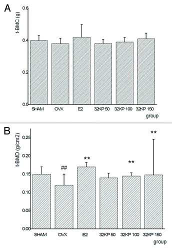
We surprisingly found that there were no significant difference of the right femur t-BMC among any of the treatment groups (). However, OVX significantly lowered the right femur t-BMD compared with the SHAM group (p < 0.01). 32KP at higher doses (100 or 150 mg/kg/day) significantly increased the right femur t-BMD compared with the OVX group (p < 0.05 or 0.01, respectively). E2 also increased the right femur t-BMD, significantly higher than OVX group (p < 0.01) ().
Biomechanical quality of the femur
The maximum stress and Young’s modulus of the right femur was significantly decreased in the OVX group, as compared with SHAM group (p < 0.01 for both). Compared with the OVX group, both E2 and 32KP at higher doses (100 or 150 mg/kg/day) significantly increased the Young’s modulus and maximum stress (p < 0.05 or 0.01). OVX also induced a decrease in maximum load, energy and stiffness, but the treatment with 32KP and E2 could not reverse the process (p > 0.05). ().
Table 3. Effects of 16 weeks treatment with 32KP or E2 on bone biomechanical paremeters in the femoral diaphysis of ovariectomized (OVX) rats
Hematoxylin-eosin staining results
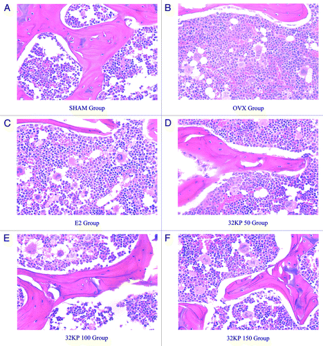
The OVX group has the smallest right femur marrow cavity () andthe SHAM group has the largest (). The femur marrow cavity was larger in the 50, 100, 150 mg/kg of 32KP, E2 groups than in the SHAM group ().
Discussion
All developing or even the developed countries face an increasing number of elderly people, and consequently, a growing prevalence of chronic age-related conditions. As a result, osteoporosis has become a major public health concern.Citation6 Although traditional Chinese medicines (TCM) for tonifying kidney has been widely used in clinical practice to treat bone disease for thousands of years and will undoubtedly continue to be used as a cost-effective alternative to commercial pharmaceutical products by traditional users, their international acceptance as an alternative therapeutic regimen for the prevention and treatment of osteoporosis still require extensive research using modern science. The present study was designed to systematically evaluate the protection effect of 32KP against ovariectomy-induced bone loss in mature rats.
Normally, ovariectomized rats have significantly higher body weights compared with sham-operated rats due to fat deposition caused by estrogen deficiency.Citation7,Citation8 These have noted the increase in body weight as a mechanism to provide an additional stimulus for bone neoformation, serving as a partial protection against the osteopenia which occurs in long bones due to supporting the body weight.Citation9 Our data are consistent with these findings that all three doses of 32KP were able to significantly reduce the OVX-induced body weight gain at different scales. This suggested that estrogen deficiency induced by OVX could alter the energy metabolism and 32KP could partly reverse the effect. A marked atrophy of the uterus has been used as evidence of the success of ovariectomy for estrogen directly affects uterine weight.Citation10 Indeed, ovariectomy resulted in a significant decrease in uterine weight. Ironically, 32KP did not exhibit any uterotrophic activity; this lack of uterotrophic activity could be beneficial in reducing the risk of diseases associated with E2 treatment. Furthermore, long-term oral administration of 32KP had no effect on other organs.
Biochemical markers of bone turnover have been widely used as research tools to measure the effects of various drugs on bone remodeling.Citation11 Bone tissue consists of mineral and organic compounds, which play a role in supporting the body. The mineral compounds can make bone to keep strength and stiffness. Phosphorus is an important component in bone tissue. Evidence exists that many American women who are at high risk of developing osteoporosis is typically high in serum level of phosphorus and low in calcium.Citation6 It indicates that osteoporosis can be prevented and cured by decreasing the level of phosphorus and increasing the level of calcium in serum. So, it is necessary to maintain normal or sub-normal blood phosphorus level. From current studying, 32KP could significantly prevent the increase Ca level and decrease P level in Urine in OVX group. The loss of bone mass was accompanied by a significant increase in bone remodeling, as was evidenced by the enhanced levels of the bone turnover markers DPD, OC and the urinary DPD/Cr ratio. All three doses of 32KP could significantly increase in urinary Ca excretion, and 32KP at the highest dose (150 mg/kg/day) significantly decreased urinary P excretion. It showed only higher doses of 32KP consistent with the maintenance of bone mass by inhibiting bone remodeling. The reason is possible that lower dosage 32KP was destroyed by stomach via oral administration.
BMD is the gold standard for the evaluation of individuals at risk for osteoporosis, as it best predicts the fracture risk in people without previous fractures.Citation12 In our experiment, the results showed that 32KP at higher doses (100 or 150 mg/kg/day) significantly increased the right femur t-BMD compared with the OVX group. It indicated that high dose 32k Da protein could be benefit for not only inhibiting bone resorption, but also increasing the mineral concentration in the bones of OVX rats. As for how much concentration of 32k Da protein would be the best dosage will be evaluated by further experiments.
In the current study, three-point bending tests of bone sp.ecimens were performed to study the effects of 32KP on the biomechanical properties of femoral diaphysis in OVX rats. As expected, Ovariectomy has been shown to decrease mechanical properties. Treatment with 32KP at higher doses (100 or 150 mg/kg/day) significantly prevented the maximum stress and Young’s modulus decreases in OVX rats. Our data indicated that different 32k Da dose groups exhibited superior mechanical bone properties compared with the OVX groups, which is consistent with the findings that ovariectomy results in decreased bone density as well as a reduced biomechanical strength of bones.Citation13,Citation14
Bone histomprphometry studies demonstrated that the normal control group had the smallest femur marrow cavity stained by Hematoxylin-eosin and the SHAM group had the largest. These results indicate that the bone structures of osteoporosis rats are very loose, and after 16 weeks treatment of 32KP at different dosage, the marrow cavities of the femur of rats with osteoporosis have been obviously diminished, suggesting that 32KP can prevent the development of bone loss induced by ovariectomy in rats from the histomprphometry level.
In summary, the results obtained in the current study provide evidence that daily oral administration of 32KP contributes significantly to the prevention of bone loss induced by ovariectomy in rats. The evidence was evaluated by dual-energy X-ray absorptiometry, biochemical markers three-point bending test, and hematoxylin-eosin staining. However, 32KP did not have a stimulatory effect on the uterus similar to E2. We believe that 32KP has the potential for further development as a natural alternative drug for postmenopausal osteoporosis.
Patients, Materials and Methods
Preparation of 32KP
32k Da protein was extracted from Rumex crispus by ultrasonic assistant technology in alkali. Afterwards, the protein was further purified on Sephadex G-25 and G-50 columns, respectively and determined by Electrophoresis. The collected 32k Da containing solution was dehydrated by freeze-drying to obtain purified powder of 32k Da.
Animals and treatments
All rats were pair-fed and allowed free access to distilled water and fed with standard rat chow (The acclimatized rats underwent either bilateral laparotomy (SHAM, n = 10) or bilateral ovariectomy (OVX, n = 70). Four weeks after recovering from surgery, the ovariectomized (OVX) rats were randomly divided into five groups: OVX with vehicle (OVX, n = 50); OVX with E2, n = 15, 25 g/kg body weight/day); OVX with 32KP of graded doses (32KP50, n = 15, 50 mg/kg body weight/day), (32KP50, n = 15, 50 mg/kg body weight/day) and (32KP150, n = 15, 150 mg/kg body weight/day). Six weeks later, osteoporosis was successfully induced. Rats in each group were interfered according to above-mentioned medications. After 16-week treatment, all rats were sacrificed, and the blood samples were collected. The serum levels of calcium and phosphorus were detected by biochemistry methods. At the same time, the femurs were also harvested to determine BMD by dual energy X-ray absorptiometry, and for histomorphometric analysis by hematoxylin-eosin staining, as well as bone biomechanical quality measuring by a three-point bending test.
Assay for serum and urine chemistry
Urine and blood samples were collected at week 0, 4, 8, 12, 16, and 20. Urine was collected directly from the bladder. Blood samples were taken from the tail vein and then centrifuged to collect the serum. Both urine and blood samples were immediately sent to test in the department of examination at Jiujiang Hospital.
DEXA analysis
According to previous method,Citation15 femur BMD (g/cm2) was detected by dual-energy X-ray absorptiometry (DEXA) (GE Healthcare). BMD was calculated by BMC of the measured area.
Histomorphometric analysis
The right femurs of rats were fixed with 10% formalin for 48 h, and decalcified with 8% nitric acid overnight. Thereafter, the femur samples were washed with distilled water for 5 times, and dehydrated with a series of ethanol (70, 80 and 90%,) for 12 h, successively. Then, all samples were immersed in dimethylbenzene, embedded with paraffin, sliced into 5 μm sections, and stored at −60 °C overnight for staining. The glass slides with the sections were immersed in dimethylbenzene for 3 min, in 95% ethanol for 3 min, and in 80% ethanol for 3 min. Subsequently, sections were stained with hematoxylin for 8 min, immersed in 1% chlorhydric acid-ethanol for 30 sections, and placed in Li3CO3 for 1 s. There were tap water washes between each step. Thereafter, sections were stained with eosin for 25 min, immersed in a series of ethanol (80% for 3 min, 95% for 3 min, and 50% for 3 min), and treated with dimethylbenzene for 3 min. Finally, the marrow cavity of the femurs was observed by light microscopy with X40 (Olympus).
Three-point bending test
According to previously method,Citation16 Prior to mechanical testing, the right femurs were collected and stored at 37 °C until the day of test. Briefly, the length of the femurs was first measured with a micrometer and the middle part of the diaphysis.Citation17 Then the femur was placed in the 858 Mini Bionix II material testing machine (MTS, Eden Prairie) on two supports separated by a distance of 20 mm. Afterwards the load was applied to the middle part of the diaphysis, thus a three-point bending test was created. The right femoral diaphysis was determined at the speed of 4 mm/min by material testing machine. The biomechanical quality parameters including load–deformation curve, maximum load, stiffness, and energy absorption, maximum stress and and Young’s modulus were obtained from the right femoral diaphysis via keeping record of the load and displacement until the specimen was broken.
Statistical evaluation
Results were expressed as mean ± SD. All experimental data were analyzed using one-way analysis of variance with Dunn.qqett’s test. Values of p < 0.05 were considered statistically significant.
Disclosure of Potential Conflicts of Interest
No potential conflicts of interest were disclosed.
Acknowledgments
This work was supported by grants from National Natural Science Foundation of China (81000075).
References
- Zhang R, Hu SJ, Li C, Zhang F, Gan HQ, Mei QB. Achyranthes bidentata root extract prevent OVX-induced osteoporosis in rats. J Ethnopharmacol 2012; 139:12 - 8; http://dx.doi.org/10.1016/j.jep.2011.05.034; PMID: 21669273
- Zhao X, Wu ZX, Zhang Y, Yan YB, He Q, Cao PC, Lei W. Anti-osteoporosis activity of Cibotium barometz extract on ovariectomy-induced bone loss in rats. J Ethnopharmacol 2011; 137:1083 - 8; http://dx.doi.org/10.1016/j.jep.2011.07.017; PMID: 21782010
- Davison S, Davis SR. Hormone replacement therapy: current controversies. Clin Endocrinol (Oxf) 2003; 58:249 - 61; http://dx.doi.org/10.1046/j.1365-2265.2003.01774.x; PMID: 12608928
- Suh HJ, Lee KS, Kim SR, Shin MH, Park S, Park S. Determination of singlet oxygen quenching and protection of biological systems by various extracts from seed of Rumex crispus L. J Photochem Photobiol B 2011; 102:102 - 7; http://dx.doi.org/10.1016/j.jphotobiol.2010.09.008; PMID: 21185197
- Shiwani S, Singh NK, Wang MH. Carbohydrase inhibition and anti-cancerous and free radical scavenging properties along with DNA and protein protection ability of methanolic root extracts of Rumex crispus. Nutr Res Pract 2012; 6:389 - 95; http://dx.doi.org/10.4162/nrp.2012.6.5.389; PMID: 23198017
- Jakubowitz E, Seeger JB, Kretzer JP, Heisel C, Kleinhans JA, Thomsen M. The influence of age, bone quality and body mass index on periprosthetic femoral fractures: a biomechanical laboratory study. Med Sci Monit 2009; 15:BR307 - 12; PMID: 19865047
- Breitman PL, Fonseca D, Cheung AM, Ward WE. Isoflavones with supplemental calcium provide greater protection against the loss of bone mass and strength after ovariectomy compared to isoflavones alone. Bone 2003; 33:597 - 605; http://dx.doi.org/10.1016/S8756-3282(03)00243-6; PMID: 14555264
- Iwamoto J, Seki A, Sato Y, Matsumoto H. Vitamin K(2) improves renal function and increases femoral bone strength in rats with renal insufficiency. Calcif Tissue Int 2012; 90:50 - 9; http://dx.doi.org/10.1007/s00223-011-9548-3; PMID: 22080166
- Lau BY, Fajardo VA, McMeekin L, Sacco SM, Ward WE, Roy BD, Peters SJ, Leblanc PJ. Influence of high-fat diet from differential dietary sources on bone mineral density, bone strength, and bone fatty acid composition in rats. Appl Physiol Nutr Metab 2010; 35:598 - 606; http://dx.doi.org/10.1139/H10-052; PMID: 20962915
- Kandziora F, Bail H, Schmidmaier G, Schollmeier G, Scholz M, Knispel C, Hiller T, Pflugmacher R, Mittlmeier T, Raschke M, et al. Bone morphogenetic protein-2 application by a poly(D,L-lactide)-coated interbody cage: in vivo results of a new carrier for growth factors. J Neurosurg 2002; 97:Suppl 40 - 8; PMID: 12120650
- Nian H, Ma MH, Nian SS, Xu LL. Antiosteoporotic activity of icariin in ovariectomized rats. Phytomedicine 2009; 16:320 - 6; http://dx.doi.org/10.1016/j.phymed.2008.12.006; PMID: 19269147
- Ettinger B, San Martin J, Crans G, Pavo I. Differential effects of teriparatide on BMD after treatment with raloxifene or alendronate. J Bone Miner Res 2004; 19:745 - 51; http://dx.doi.org/10.1359/jbmr.040117; PMID: 15068497
- Jiang Y, Zhao J, Genant HK, Dequeker J, Geusens P. Bone mineral density and biomechanical properties of spine and femur of ovariectomized rats treated with naproxen. Bone 1998; 22:509 - 14; http://dx.doi.org/10.1016/S8756-3282(98)00027-1; PMID: 9600785
- Tong HY, Hu SM, Zhou P, Fu Q, Li J, Gao XM, Zhang JJ. [Effects on rats’ bone mineral density and bone biomechanics by suspensory simulated weightlessness and removing suspension]. Zhongguo Gu Shang 2008; 21:276 - 9; PMID: 19102188
- Pierce MC, Valdevit A, Anderson L, Inoue N, Hauser DL. Biomechanical evaluation of dual-energy X-ray absorptiometry for predicting fracture loads of the infant femur for injury investigation: an in vitro porcine model. J Orthop Trauma 2000; 14:571 - 6; http://dx.doi.org/10.1097/00005131-200011000-00010; PMID: 11149504
- Ko YJ, Wu JB, Ho HY, Lin WC. Antiosteoporotic activity of Davallia formosana. J Ethnopharmacol 2012; 139:558 - 65; http://dx.doi.org/10.1016/j.jep.2011.11.050; PMID: 22155390
- Marcelli C. [Osteoporosis in children and adolescents]. Presse Med 2007; 36:1078 - 83; http://dx.doi.org/10.1016/j.lpm.2007.03.002; PMID: 17395422

