Abstract
The co-existence of certain peptides influenced the kinetic rate of aggregation and the lag-time of fibril formation of rbPrP. Using recently developed structural conversion assay system, peptides have been screened from bovine brain peptide library. Peptide sequences of positive components have been elucidated by mass spectrometry and chemically synthesized to confirm actions.
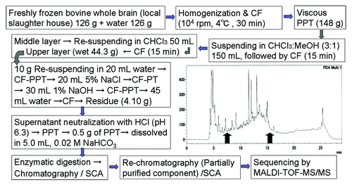
A novel bio-detection system using structured peptides as protein mimetic has been developed. The protein (as analyte) and peptide (as capture molecules) interaction cause structural changes and this is reflected in fluorescent-intensity changes of the labeled peptides in a dose dependent manner reviewed in.Citation1 Human prion protein related overlapping peptides have been synthesized to construct libraries. From these libraries peptides had been found that discriminated between infected/normal brain homogenate and prion strains and/or animal species.Citation2 Proteins designated as molecular chaperones have been known to assist structural conversion or folding. We hypothesize that certain peptides may induce or enhance the conformational change of proteins as reverse phenomenon of β-sheet breaker peptides.Citation3 That is, the self-assembly or aggregation of proteins is influenced by peptides derived from proteins, as BSE is believed to be caused by animal proteins in the feed. In order to investigate the validity of this hypothesis an assay system is necessary to define the peptide(s) that are involved or are causative factors associated with prion diseases. The conversion is a phenomenon in which helix structured segments convert to sheet structures. In addition to the above libraries the peptide HPPSH had been designed by the concept with following two modules connected to give a conjugate. One was the α-helical region of human prion protein, hPrP180–195,Citation4 as a conversion module which converts to β-sheet, and another was hPrP170–175, which has been reported to contain a β-sheet structure,Citation5 on which Tyr residue was added (at position 169) to allow UV quantization expected as an inducer (). The secondary structures of the present peptides were characterized by circular dichroism spectra, which were described in,6 taking into account the influence of a hydrophobic environment as described in.Citation7 We have found that the influence on aggregation has been evaluated from determination of the lag-time when the Thioflavin T (ThT) fluorescence change starts to increase and the kinetics from the incremental change in the fluorescent intensities until a plateau is reached.Citation6 Peptides P2-P5 () and HPPSH decrease the lag-time, but the other peptides have no or very little influence. Each peptide alone using ThT no significant effects on aggregation. P8 shows the largest rate constant increase (ca. 10x that of rbPrP alone) indicating a significant acceleration of aggregation. HPPSH shows the lowest rate constant (ca. 50% of rbPrP alone) and hence indicates a deceleration of aggregation. All the other peptides have little effect on the rate. HPPSH changes the kinetic rate of aggregation, lag-time of fibril formation and morphologies of nanostructures of rbPrP. We have shown that the co-existence of certain peptides influenced the kinetic rate of aggregation and the lag-time of fibril formation of rbPrP.6 There are peptides which influence, more precisely accelerate, the structural change of recombinant prion protein and this model may mimic the formation of the pathogenic isoform of prion protein (PrPSc). By using the above assay method to monitor structural changes in rbPrP the responsible peptides for the structural conversion have seen searched from partial digests of bovine brain. For this purpose the peptide library has been constructed from natural bovine brain obtained from a local slaughter house (). Fatty acids have been removed by extraction and the resulting material was partially hydrolyzed and chromatographed to give the component libraries. Using the above conversion assay method, we have screened peptides responsible for structural conversion. The structural elucidation of active components in mixtures can be directly analyzed using high resolution mass spectrometer to give multiple precursor ions in MALDI-TOF-MS which can be further analyzed by MS/MS to reveal the sequence of each component. All the peptides found have been characterized by using the MS BLAST algorithm and chemically synthesized to confirm their effects on both the lag time and rate of structural conversion of rbPrP. Only one peptide, of which the sequence is “MDVVNQLVAGGQFR” and originates from synaptophysin,Citation8 showed significant effects. Fitting a Gompertz curve indicates the effect starts at a peptide concentration of 3.3 μM. According to the cellular component in Gene Onthology, synaptophysin localizes in synaptic vesicle membrane and presynaptic membrane. Since PrPC is highly expressed at synapses,Citation9 this suggests that PrPC and synaptophysin may colocalize and may interact each other. The interaction of synaptophysin with PrPC plays a role in synaptic function,Citation10 although the structural conversion by the peptides derived from synaptophysin has not been described. The present approach allows minimization of the time required to discovery active compounds. A dose-dependency was established using the synthetic peptide, as unknown amounts of target were assayed at the stage of screening of the natural library.
Figure 1. Prion related synthetic peptide fragments and the de novo designed peptide for the structural conversion, designated HPPSH. Each Cys-residue was replaced by a Ser-residue to avoid disulfide bond formation. Hence hPrP170–175 (SNQNNF) formed parallel β-sheet [PDB ID: 2OL9] and hPrP180–195 (VNITIKQHTVTTTTKG) formed α-helix [PDB ID: 2IV4].
![Figure 1. Prion related synthetic peptide fragments and the de novo designed peptide for the structural conversion, designated HPPSH. Each Cys-residue was replaced by a Ser-residue to avoid disulfide bond formation. Hence hPrP170–175 (SNQNNF) formed parallel β-sheet [PDB ID: 2OL9] and hPrP180–195 (VNITIKQHTVTTTTKG) formed α-helix [PDB ID: 2IV4].](/cms/asset/9fefd712-4ed6-4a82-8b76-1064fd32919e/kprn_a_10927961_f0001.gif)
Figure 2. Scheme for the preparation of peptide library derived bovine brain. Abbreviations used are CF, centrifugation (10 000 rpm, 4 °C); PPT, precipitate; SCA, structural conversion assay. Chromatogram is a tryptic digests and fractions eluted at 7–8 and 15–16 min (arrow) exhibited significance in SCA.

Disclosure of Potential Conflicts of Interest
No potential conflicts of interest were disclosed.
Acknowledgments
Authors thanks to Drs K Kasai, T Yokoyama and S Mohri, National Institute for Animal Health for variable discussions and Dr V Wray, Helmholtz Centre for Infection Research, Braunschweig, Germany, for linguistic advice. A part of this work was funded by the Research and Development Program for New Bio-industry Initiatives, National Agriculture and Food Research Organization (2007–2011).
References
- Nokihara K, Ohyama T, Usui K, Yonemura K, Takahashi M, Mihara H. Development of peptide-chips focusing on a high throughput protein-detection system. Solid-Phase Synthesis and Combinatorial Chemical Libraries. Epton, R. ed.; Mayflower Scientific, UK 2004; pp83-8.
- Kasai K, Hirata A, Ohyama T, Nokihara K, Yokoyama T, Mohri S. Novel assay with fluorescence-labelled PrP peptides for differentiating L-type atypical and classical BSEs, and scrapie. FEBS Lett 2012; 586:325 - 9; http://dx.doi.org/10.1016/j.febslet.2012.01.012; PMID: 22285492
- Soto C, Kascsak RJ, Saborío GP, Aucouturier P, Wisniewski T, Prelli F, Kascsak R, Mendez E, Harris DA, Ironside J, et al. Reversion of prion protein conformational changes by synthetic beta-sheet breaker peptides. Lancet 2000; 355:192 - 7; http://dx.doi.org/10.1016/S0140-6736(99)11419-3; PMID: 10675119
- Ronga L, Palladino P, Ragone R, Benedetti E, Rossi F. A thermodynamic approach to the conformational preferences of the 180-195 segment derived from the human prion protein α2-helix. J Pept Sci 2009; 15:30 - 5; http://dx.doi.org/10.1002/psc.1086; PMID: 19035579
- Sawaya MR, Sambashivan S, Nelson R, Ivanova MI, Sievers SA, Apostol MI, Thompson MJ, Balbirnie M, Wiltzius JJ, McFarlane HT, et al. Atomic structures of amyloid cross-beta spines reveal varied steric zippers. Nature 2007; 447:453 - 7; http://dx.doi.org/10.1038/nature05695; PMID: 17468747
- Hirata A, Yajima S, Yasuhara T, Nokihara K. Structural conversion rate changes of recombinant bovine prion by designed synthetic peptides. Int J Pept Res Ther 2012; 18:217 - 25; http://dx.doi.org/10.1007/s10989-012-9294-z
- Wray V, Kakoschke C, Nokihara K, Naruse S. Solution structure of pituitary adenylate cyclase activating polypeptide by nuclear magnetic resonance spectroscopy. Biochemistry 1993; 32:5832 - 41; http://dx.doi.org/10.1021/bi00073a016; PMID: 8504103
- Nokihara K, Yajima S, Hirata A, Sogon T, Yasuhara T. Characterization of peptides obtained from digests of bovine brain which accelerate structural conversions of the recombinant bovine prion protein. FEBS Lett 2013; 587:673 - 6; http://dx.doi.org/10.1016/j.febslet.2013.01.033; PMID: 23376025
- Herms J, Tings T, Gall S, Madlung A, Giese A, Siebert H, Schürmann P, Windl O, Brose N, Kretzschmar H. Evidence of presynaptic location and function of the prion protein. J Neurosci 1999; 19:8866 - 75; PMID: 10516306
- Russelakis-Carneiro M, Hetz C, Maundrell K, Soto C. Prion replication alters the distribution of synaptophysin and caveolin 1 in neuronal lipid rafts. Am J Pathol 2004; 165:1839 - 48; http://dx.doi.org/10.1016/S0002-9440(10)63439-6; PMID: 15509552
