Abstract
Prions, the causative agent of chronic wasting disease (CWD) enter the environment through shedding of bodily fluids and carcass decay, posing a disease risk as a result of their environmental persistence. Plants have the ability to take up large organic particles, including whole proteins, and microbes. This study used wheat (Triticum aestivum L.) to investigate the uptake of infectious CWD prions into roots and their transport into aerial tissues. The roots of intact wheat plants were exposed to infectious prions (PrPTSE) for 24 h in three replicate studies with PrPTSE in protein extracts being detected by western blot, IDEXX and Bio-Rad diagnostic tests. Recombinant prion protein (PrPC) bound to roots, but was not detected in the stem or leaves. Protease-digested CWD prions (PrPTSE) in elk brain homogenate interacted with root tissue, but were not detected in the stem. This suggests wheat was unable to transport sufficient PrPTSE from the roots to the stem to be detectable by the methods employed. Undigested PrPTSE did not associate with roots. The present study suggests that if prions are transported from the roots to the stems it is at levels that are below those that are detectable by western blot, IDEXX or Bio-Rad diagnostic kits.
Introduction
Chronic Wasting Disease (CWD) is a fatal neurodegenerative prion disease affecting wild and farmed cervids. Specifically, CWD has been found in deer (mule and white-tail), elk and moose.Citation1 Experimental transmission has also been demonstrated in reindeer.Citation2 Prion diseases, also termed Transmissible Spongiform Encephalopathies (TSE), are caused by misfolding of the cellular prion protein, PrPC, into the disease-causing conformation designated PrPTSE. Misfolded PrPTSE is broadly accepted as the disease causing agent responsible for misfolding of PrPC into PrPTSE.Citation3,Citation4
Prevalence of CWD is high in endemic areas like Wyoming (USA) where, in a number of herds, 50% of wild mule deer are CWD positive.Citation1 Indirect transmission of CWD has been confirmed in studies where deer became infected following exposure to areas which previously held CWD-positive deer.Citation5,Citation6 CWD PrPTSE remains infectious for more than two years ex vivo in the environment representing a long-term hazard for transmission.Citation5 Infected cervids have been reported to shed PrPTSE in bodily fluids, disseminating infectious prions in urine, feces, blood and saliva.Citation7-Citation10
PrPTSE binds to whole soil and interaction with some soil minerals has been shown to increase the infectivity of PrPTSE.Citation11,Citation12 Although PrPTSE binds to soil, it requires 60 d for CWD PrPTSE to become fully bound in sandy soil as compared with 30 d for prions associated with transmissible mink encephalopathy (TME).Citation13 In seawater, scrapie PrPTSE remains stable with a one log reduction only observed after 30 d, suggesting unbound PrPTSE may persist and flow with surface or ground water.Citation14 Taken together, these factors suggest PrPTSE may remain unbound within the soil environment for substantial periods.
Plants acquire nutrients from the surrounding soil environment. Nitrogen (N) is one of the main macronutrients vital for plant growth and reproduction. There are multiple forms of N in the soil environment and the importance of organic N, including amino acids and peptides, to plant health is well documented.Citation15,Citation16 The nutritional value of protein N to plants is contentious but, the potential for uptake has been demonstrated.Citation17-Citation19 Uptake of green fluorescent protein into roots has been shown for Arabidopsis thaliana and Hakea actites.Citation19 Earlier work suggested ovalbumin is not only taken up by roots, but is subsequently transported to upper regions of tomato plants when roots are damaged.Citation18
The ability of plants to take up larger organic particles raises the possibility that PrPTSE may enter plants through roots. This study used wheat as a model to investigate the potential uptake of CWD PrPTSE into roots and its subsequent transport to the stem. If such a phenomenon was confirmed it would suggest the potential for plants to act as a vector for the transmission of CWD in cervids.
Results
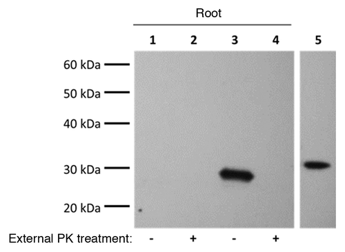
PrPC exposures. Wheat plants with roots exposed to recombinant mouse PrPC contained a prion band in the root protein extract as identified by western blotting (, lane 3). The prion band in the root extract had a slightly smaller molecular weight than PrPC in the solution roots were exposed to (, lane 5). When the outer surface of roots was exposed to 10 µg/mL proteinase K (PK) for 5 min at room temperature, no prion signal in the root protein extract was observed (, lane 4). No prion signal was seen in stem or leaf protein extracts (data not shown).
Figure 1. Recombinant PrPC binds to the outside of wheat roots. Wheat plant roots were exposed to 50 µg/mL PrPC for 24 h then roots were either digested with (10 µg/mL) proteinase K (PK) for 5 min or left undigested. Control plants were exposed to the same solution lacking PrPC. Western Blotting on root protein extracts used the 8H4 mAb (1:10000). The last lane confirming antibody specificity is from the same blot with irrelevant lanes omitted. Results are representative of three independent replicates (n = 3). Lanes 1 and 2: control plants; Lanes 3 and 4: PrPC exposed plants; Lane 5: positive control (50 ng PrPC).

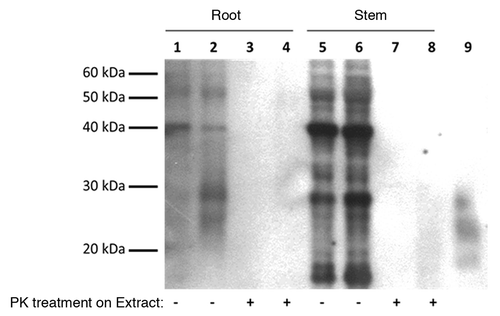
PrPTSE exposures. In wheat plants exposed to PrPTSE, the root extract had three prion reactive bands <30 kDa that were not present in control extracts (, lanes 1 and 2). Plant proteins cross-reacted with P4 mAb in both the root and stem extracts with prominent bands at ~40 kDa and ~29 kDa (). When root extracts were digested with PK (10 µg/mL, 37 °C, 30 min) there were no PK-resistant proteins present in either the control or CWD exposed samples (, lanes 3 and 4). Only PK-sensitive cross reaction bands were present in the lower stem extract (, lanes 5–8).
Figure 2. Prion signal found in wheat roots exposed to CWD PrPTSE was protease sensitive but no prion signal in lower stem (Stem) extract was visible. CWD PrPTSE was purified from brain homogenate (BH) with Bio Rad-TeSeE® purification and re-suspended in phosphate buffered saline to a 1% solution (w/v) based on the initial BH solution. Wheat plant roots were exposed to the purified solution for 24 h. Normal BH processed with TeSeE® served as a negative control. Plant protein extracts were digested with proteinase K (PK) (10 µg/mL, 30 min, 37 °C) to determine PK-resistance of any proteins. Western blotting of plant protein extracts (plant total protein extraction kit) was done using P4 mAb (1:5000) and Prionics®-Check Western kit. Results are representative of three independent replicates (n = 3). Lanes 1, 3, 5, 7: plants exposed to normal BH processed with Bio Rad kit; Lanes 2, 4, 6, 8: plants exposed to CWD infected BH processed with Bio Rad kit; Lane 9: CWD infected BH (0.1%) processed with Bio Rad kit.

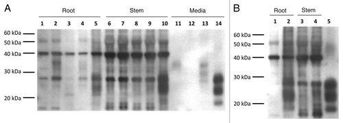
Wheat plants were also exposed to either infectious or non-infectious elk brain homogenates (BH), which were either digested with PK to leave only PK-resistant PrPTSE protein, or not digested, to represent all brain proteins (). The root extract from plants exposed to PK-digested CWD BH had prion reactivity with bands <30 kDa not present in the control extract (, lanes 1 and 5). Similarly, the lower stem extract from the same PK-digested CWD BH treatment had three prion reactive bands which were not present in the control lower stem extract when the outside of the stem was not rinsed with dH2O (, lanes 6 and 10). When the outside of the stem was rinsed with dH2O following exposure, the suspected PrPTSE bands were no longer visible in the lower stem extract, leaving only cross-reaction bands (, lanes 3 and 4). There were no definitive bands in the root or stem extracts of the other treatments (normal BH-digested and undigested; and CWD BH-undigested) which differed from the control protein extracts (, lanes 2–4 and 7–9).
Figure 3. Proteinase K (PK) digested CWD PrPTSE in a brain homogenate (BH) solution interacts with wheat roots but signal from lower stem (Stem) disappears when the stem is rinsed. (A) Wheat plant roots were exposed for 24 h to BH solutions either CWD positive (CWD BH) or CWD negative (Normal BH) and both digested with PK (50 µg/mL, 30 min, 37 °C) or not. Control plants were exposed to dH2O for 24 h. Only the roots were rinsed for 1 min in dH2O, with protein extracted from both the root and lower stem. Western blotting of plant extracts was done using P4 mAb (1:5000) and Prionics®-Check Western kit. Lanes 1 and 6: plants exposed to H2O; Lanes 2 and 7: plants exposed to Normal BH; Lanes 3 and 8: plants exposed to Normal BH digested with PK; Lanes 4 and 9: plants exposed to CWD BH; Lanes 5 and 10: plants exposed to CWD BH digested with PK; Lane 11: 0.1% Normal BH; Lane 12: 0.1% Normal BH digested with PK; Lane 13: 0.1% CWD BH; Lane 14: 0.1% CWD BH digested with PK. (B) Wheat plant roots were exposed to PK-digested (50 µg/mL, 30 min, 37 °C) CWD BH for 24 h then both the roots and stem were rinsed in dH2O for 1 min. Western blotting of extracted protein used P4 mAb (1:5000) and Prionics®-Check Western kit. Results are representative of three independent replicates (n = 3). Lanes 1 and 3: plants exposed to H2O; Lanes 2 and 4: plants exposed to CWD BH digested with PK; Lane 5: 0.1% CWD BH digested with PK.

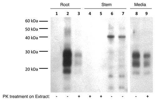
The plant total protein extraction kit (Sigma Aldrich) resulted in PrPTSE becoming sensitive to a mild PK digestion (10 µg/mL, 30min, 37 °C) (data not shown). An alternate extraction with 1% sodium dodecyl sulfate (SDS) was used in order to preserve PK-resistance of any PrPTSE present in plant tissue associated with the PK-digested CWD BH treatment (). With the 1% SDS extraction, no cross-reaction bands were observed in the control root extract (, lane 1). Prion protein in the root extract was resistant to mild PK treatment (10 µg/mL, 30min, 37 °C) (, lanes 2–3). There were no PK-resistant proteins in the lower stem and only cross-reaction bands were visible when extracts were not digested (, lanes 4–7).
Figure 4. Proteinase K (PK) digested CWD PrPTSE interacts with wheat roots and remains slightly PK resistant after extraction while no CWD PrPTSE was detected in the lower stem (Stem). CWD positive brain homogenates (BH) were digested with PK (50 µg/mL, 30 min, 37 °C) prior to being exposed to wheat roots for 24 h. Wheat roots and stems were rinsed with dH2O for 1 min then protein was extracted with 1% SDS. Extracts were digested with PK (10µg/mL, 30 min, 37 °C) to determine any PK-resistant bands prior to blotting. Western blotting on protein extracts was done using P4 mAb (1:5000) and Prionics®-Check Western kit (Prionics). Images are from the same blot with irrelevant lanes omitted. Results are representative of three independent replicates (n = 3). Lanes 1, 4, 6: plants exposed to H2O; Lanes 2, 3, 5, 7: plants exposed to CWD BH digested with PK; Lanes 8 and 9: 0.1% CWD BH in presence of 1% SDS digested with PK.

Protein extracted with 1% SDS from plants exposed to PK-digested CWD BH was analyzed using Bio-Rad TeSeE® and IDEXX HerdChek® diagnostic tests (). TeSeE® analysis revealed all root and stem extracts were negative (). When PK digestion was omitted from TeSeE® protocol, the root extract from the PK-digested CWD BH treatment was positive and control root extract was negative (). Stem extracts were negative when PK treatment was omitted from the TeSeE® protocol (). All samples were negative when tested with IDEXX HerdChek® ().
Table 1. Analysis of plant protein extracts (1% SDS) from proteinase K-digested (PK) CWD BH treatment with Bio-Rad and IDEXX diagnostic tests
Discussion
This study shows that protease-digested CWD PrPTSE interacts with wheat roots, both when exposed in a brain homogenate (BH) and purified solution. Similarly, when in a purified solution, recombinant PrPC interacted with the outside of wheat roots. Transport of PrPTSE or PrPC from roots to stems was not detectable using the methods employed. This suggests that plants with damaged root hairs do not uptake PrPTSE or transport it to stems at high concentrations, an outcome that may change if more extensive root damage occurs.
One interesting observation was that only PK-digested PrPTSE associated with roots, while undigested PrPTSE did not (). Proteinase K digestion is known to alter the structure of PrPTSE by removing the N-terminus.Citation20 With this altered structure, PrPTSE may have exhibited an enhanced affinity for wheat roots. When considering the strong interaction between recombinant PrPC and the roots, it is worth noting that this prion protein is also N-terminally truncated as compared with mammalian PrPC (). The fact that PrPTSE and PrPC lacking the N-terminus interact with roots is supported by studies showing the N-terminus plays a role in the binding of PrPTSE to soil components.Citation11,Citation13 Alternatively, in an undigested BH it is possible that other brain proteins may occupy binding sites, thereby blocking any interaction of PrPTSE with the root surface. Degradation of PrPTSE by microbes has been suggested to occur during composting, suggesting that partial degradation of PrPTSE in the soil environment is possible,Citation21 an outcome that may increase its interaction with plant roots.
Initially, total plant protein extractions were undertaken using a kit to maximize the amount of protein obtained ( and ). The high concentration of detergents in the extraction buffer affected the PK-resistance of prions, as other studies have confirmed.Citation22 A 1% SDS solution was used as an alternate extraction procedure to preserve PK-resistance of PrPTSE based on other studies that extracted PrPTSE from compost ().Citation21 It should be noted that 1% SDS extracted protein less efficiently than the extraction kit and likely lowered our detection limit in plant tissue as demonstrated by the decreased intensity of cross-reaction bands in the root and stem (). Efforts to specifically identify the proteins responsible for the cross-reaction bands in the root and stem were not undertaken owing to the lack of identifiable targets for the P4 antibody epitope as determined by BLAST analysis of the wheat proteome. The closest match for the cross reaction bands at ~40 kDa and ~29 kDa were the Cysteine Proteinase protein (71% coverage, 80% similarity) and R2R3-MYB protein (57% coverage, 100% similarity), respectively. Western blotting without the P4 antibody failed to produce the > 20 kDa cross reaction bands in the plant stem, confirming that the cross-reaction was primarily attributable to P4 (data not shown).
When extracted with 1% SDS, the PrPTSE signal from the roots was reduced with a mild PK digestion (10 µg/mL, 30 min, 37 °C) (). When digested under the same conditions, the PrPTSE signal from the solution roots were exposed to also decreased in the presence of SDS (). There was a larger decrease in PrPTSE signal as a result of digestion of the root extract as compared with PrPTSE in solution (). Together, these results suggest PrPTSE was more PK-sensitive when associated with wheat roots than when in solution. The interaction with roots could have altered PrPTSE conformation making it more PK-sensitive. This hypothesis is supported by the negative reading obtained from the IDEXX HerdChek® test, which identifies PrPTSE based on misfolded conformation by Seprion-capture (). The lack of any positive samples from IDEXX HerdChek® suggests even though there are PK-resistant prions in the roots, the conformation of the prion species is no longer identifiable as CWD PrPTSE. This finding is also supported by Bio-Rad TeSeE® which also did not identify any of the plant tissues as positive for PrPTSE. Conversely, when Bio-Rad TeSeE® protocol was altered by removing PK-treatment of samples, roots from the PK-digested CWD BH treatment were positive for prions, while control roots were negative (). The Bio-Rad TeSeE® manufacturer’s protocol was altered by removing PK-digestion in order to determine if more PK-sensitive prion species could be identified. The observation that western blotting of the PK-digested CWD BH treatment solution exhibited the only reactivity to P4 mAb with PK-resistant PrPTSE present, along with TeSeE® results confirms these roots contained PrPTSE. These results suggest the PrPTSE in the root extract underwent significant changes with the impact of these conformational changes on infectivity being unknown.
The loss of the possible PrPTSE signal in the unwashed stem of plants exposed to PK-digested CWD BH after a simple dH2O wash was surprising (). It is unlikely that this observation is due to external contamination from the media solution as care was taken to have only the roots submerged in media. The fact that other treatments with prion reactive protein in the media (undigested CWD and normal BH) did not have a prion signal in unwashed stems that were handled in the same manner, supports that these findings are not due to contamination from the treatment solution ().
The interaction of protease-digested PrPTSE with wheat roots may have implications for the behavior of prions in the environment. Previous studies have shown CWD PrPTSE remains infectious for over two years outside of the host.Citation5 Other work has also outlined that CWD PrPTSE are relatively resistant to degradation in a brain homogenate, but that the N-terminus is rapidly cleaved.Citation20 In subsequent work, the role of the N-terminus in soil interactions was investigated with truncated hamster adapted transmissible mink encephalopathy (TME) HY PrPTSE exhibiting a greater affinity for sand and sandy loam soil as compared with undigested TME HY PrPTSE.Citation13 This suggests that removal of the N-terminus of PrPTSE as a result of microbial activity could affect the mobility of prions within soil. Our work shows PK-digested CWD PrPTSE binds to wheat roots when soil is not present, but the effect of soil particles on this interaction are unknown. Based on the documented affinity of PrPTSE to different soils and minerals, like montmorillonite, it is likely there would be competition between wheat roots and soil for binding of PrPTSE.Citation11,Citation12 We predict N-terminally truncated CWD PrPTSE would have the best possibility of binding to wheat roots in a silty clay loam soil, as PK-digested PrPTSE has a lower affinity for this soil type.Citation13
The lack of detectable PrPTSE inside the stem of wheat plants suggests that if it is transported to the stem, it is at levels that were below the level of detection for the methods employed in the current study. It is possible that trace amounts of PrPTSE could be transported to the stem. We also observed cross-reactivity of plant proteins with the P4 mAb antibody during western blotting, suggesting that plants do contain proteins with linear epitopes similar to prions. The Prionics®-Check WESTERN kit had a detection limit on the elk PrPTSE source used in this study of approximately 102 mg-/mg+ brain tissue (based on mono-glycosylated band) similar to other work on BSE with the same kit (Fig. S1).Citation23 Given the bioassay titration was 107.2 i.c. ID50/g in transgenic mice, infectious levels of prions could be contained in plant tissue that would not have been identified by western blotting. Cervids grazing on plants with trace PrPTSE at this level could potentially develop CWD.
Early studies have suggested transport of lysozyme and ovalbumin proteins to aerial portions of tomato plants occurs and is aided by root damage.Citation18 Recently, uptake of whole bacteria into plants and transport distal from the roots was shown as pathogenic Salmonella enterica entered the roots of sweet basil plants and moved internally to the stem and leaves.Citation24 Similar to protein uptake, root damage has also been identified as a factor enhancing the uptake of viruses and bacteria into plants.Citation25,Citation26 In soil, the activity of nematodes or other parasites could form lesions in roots that could serve as a port of entry for prions into plants.Citation27 In our study, plants were grown in solid agarose media and subsequently exposed to PrPTSE solution, an approach that deliberately inflicted root damage to fine root hairs but avoided major damage to the main branches. Previous work in our lab, has shown that ovalbumin can enter wheat roots and be transported to the stem using the same growth and exposure system employed in this study.Citation28 This difference may be explained by the aggregating properties of prions. It has been shown in previous studies that the size of an infectious aggregate for PrPTSE is ~26 protein monomers.Citation29 This behavior could be responsible for differences in the uptake of ovalbumin monomers (~45kDa) as compared with prion aggregates.
In conclusion, our data shows that PrPTSE is not transported to aerial tissues at concentrations detectable by western blot, however, it still remains possible for infectious levels to be achieved, especially if significant root damage occurs. The findings suggest that PK digestion may facilitate the interaction of PrPTSE with plant roots which is interesting based on the affects this also has on affinity of these molecules for soil. This study used wheat as a model but it would be valuable to test native grasses such as crested wheat grass and fescue which make up a significant portion of the diet of elk and deer. It is probable that both biotic and abiotic factors affect uptake of PrPTSE into plants and the introduction of other factors such as soil organisms and particles into the system is something to consider in future investigations. Regardless of system, if uptake occurs, animal infectivity assays are needed to confirm if the prion protein remains in an infective state after plant uptake. These experiments provide the first known investigation of the involvement of plants in prion transmission and provide a solid base for future studies.
Materials and Methods
Plant growth
Triticum aestivum L. (cv AC Andrew) seeds were germinated for five d after surface sterilization with 70% ethanol and 3% hypochlorite. Germinated seeds were transferred to Magenta® boxes containing sterile Murashige and Skoog (MS) agarose media adjusted to pH 5.8.Citation30 Filter-sterilized media (0.2 µm) was combined with autoclaved phytagel (P8169, Sigma Aldrich) to a final concentration of 0.3%. All plants were grown axenically for 30–45 d prior to uptake experiments. Growth boxes were placed in a growth room with light conditions of 16 h d/8 h night (150 µmol m−2 s−1) at 20 °C ± 2 °C.
PrPC exposures
Recombinant mouse PrPC (amino acids 23–230) was expressed in BL21 Escherichia coli (Stratagene) and purified as previously described.Citation31 The PrPC gene contained a His6 affinity tag at the C-terminus that was removed by a thrombin cleavage site after purification on a Ni-NTA agarose column (Qiagen). After re-folding, PrPC was dialyzed against dH2O and concentrated using Amicon Ultra centrifugal filters (3kDa, Millipore). Purity was confirmed by SDS-PAGE and concentration was determined by Bradford assay.Citation32 Wheat plants were removed from growth boxes, roots were rinsed with dH2O and patted dry with paper towel. Roots were exposed to 4 mL of 50 µg/mL PrPC for 24 h in nitrogen-free MS media: 29.21 mM sucrose, 2.99 mM CaCl2·2H2O, 1.50 mM MgSO4·7H2O, 1.25 mM KH2PO4, 0.11 mM EDTA ferric sodium salt, 0.10 mM H3BO3, 0.10 mM MnSO4·H2O, 29.91 nM ZnSO4·7H2O, 5.00 nM KI, 1.03 nM Na2MoO4·2H2O, 0.11 nM CoCl2·6H2O, and 0.10 nM CuSO4·5H2O.Citation30 Plants were removed from the PrPC solution and roots were rinsed for 1 min in dH2O. Roots from one half of the plants were exposed to 10 µg/mL proteinase K (0.7 U/s L, PK, Life Technologies) for 5 min followed by another rinse with dH2O to remove loosely adherent protein on the outer surface. Plants were separated into roots, stems and leaves and each fraction was flash frozen in liquid nitrogen. Separated fractions were pulverized with a mortar and pestle and protein was extracted using a plant total protein extraction kit (Sigma Aldrich). Protein samples were heated at 96 °C for 5 min in sample buffer (2.2% SDS, 48 µM Tris, 10% glycerol, 1% 2-mercaptoethanol, pH 6.8) prior to loading on a polyacrylamide gel. Samples were electrophoresed using a 4–12% NuPAGE® Bis-Tris polyacrylamide gels at 200 V for 50 min in 1X MOPS buffer (Life Technologies). Gels were transferred (wet) to nitrocellulose membranes (0.2 µm pore, Genscript, Piscataway, USA) at 100V for 60 min in 1X NuPAGE® transfer buffer (Life Technologies) with 10% methanol. Western blotting was done with 8H4 mAb (1:10000, Sigma Aldrich) and an Advanced One-Hour Western Kit (Genscript).
Prion source
All experiments using infectious material were completed in enhanced BSL 2 containment laboratories at the National Centre for Animal Disease Lethbridge Laboratory, Canadian Food Inspection Agency and material was also obtained through this collaboration. Elk (Cervus canadensis) brain stems positive for CWD were homogenized in PBS (CWD positive) or dH2O (CWD negative) with a Dounce tissue grinder to make 20% solutions. The titer of CWD positive elk homogenate used in this study was 107.2 i.c. ID50/g as determined by bioassay in Tg(CerPrP)1536+/− transgenic mice expressing elk PrP. For experiments with purified PrPTSE, both CWD positive and negative brain homogenates (BH) were processed using a TeSeE® purification kit (Bio-Rad, Mississauga, Canada) following manufactures specifications based on its use in other publications for quick removal of other brain homogenate components.Citation33,Citation34 Pelleted PrPTSE was re-suspended in Dulbecco’s phosphate-buffered saline (PBS, Sigma Aldrich), vortexed, and subject to 8, 10 s rounds of sonication at maximum intensity using a Misonix Sonicator 3000 Ultrasonic Cell Disruptor (Misonix). Solutions containing purified PrPTSE were diluted to be equivalent to 1% (w/v) of the BH input material. For BH experiments, digestion of homogenates before plant exposure were done using the homogenized 20% brain solutions at 37 °C for 30 min with 50 µg/mL PK (Life Technologies). The BH were then diluted to 1% (w/v) with dH2O for plant exposure.
PrPTSE exposure and extraction
Wheat plants were removed from growth boxes, roots were rinsed in dH2O and patted dry with a paper towel. Plants with roots were placed in conical tubes containing 4 mL of either CWD positive or negative 1% BH or 1% purified solution so as to ensure the roots were immersed in the solution for 24 h. Exposures were independently replicated three times. After exposure, roots and stems were immediately separated with a razor blade and handled separately to avoid cross-contamination. Roots and the lower half of stems were then rinsed in dH2O for 1 min and flash frozen in liquid nitrogen. Frozen tissue was pulverized using a tissue TUBE™ Impactor (Covaris Inc.). Protein was extracted either using a plant total protein extraction kit (Sigma Aldrich) following manufactures specifications or by incubation of tissue with 4 µL 1% sodium dodecyl sulfate (SDS)/mg tissue for 15 min at room temperature. For both the kit and 1% SDS extraction, the protein concentration was kept consistent for roots and stems by using the same volume of extraction buffer per mg of tissue.
Detection
Protease digestion of protein extracts was done with 10 µg/mL PK at 37 °C for 30 min. Samples were heated at 96 °C for 5 min in sample buffer (2.2% SDS, 48 µM Tris, 10% glycerol, 1% 2-mercaptoethanol, pH 6.8) prior to loading onto a polyacrylamide gel. Samples were electrophoresed using a 12% NuPAGE® Bis-Tris polyacrylamide gels at 150 V for 60 min in 1X MOPS buffer with NuPAGE® antioxidant (Life Technologies). Gels were transferred (wet) to polyvinyl difluoride membranes (PVDF, 0.45 µm pore, Millipore) at 150V for 60 min in 1X NuPAGE® Transfer buffer (Life Technologies) with 10% methanol. Western blotting was done using P4 mAb (1:5000, R-biopharm), Prionics®-Check WESTERN kit (Prionics) and were developed with CDP-Star (Roche).
Samples extracted with 1% SDS were subjected to diagnostic testing with Bio-Rad TeSeE® and IDEXX HerdChek® (Westbrook). Sample analyses using Bio-Rad TeSeE® were done in duplicate, with one replicate following manufacture’s specifications and the other without the addition of PK (undigested). Samples were also analyzed using Seprion-capture technology with the IDEXX HerdChek® Ag Test and were performed according to manufacturer’s specifications.
| Abbreviations: | ||
| CWD | = | chronic wasting disease |
| PrPTSE | = | infectious prion protein |
| PrPC | = | cellular prion protein |
| TSE | = | transmissible spongiform encephalopathy |
| TME | = | transmissible mink encephalopathy |
| N | = | nitrogen |
| MS | = | murashige and skoog media |
| PK | = | proteinase K |
| BH | = | brain homogenate |
| PBS | = | phosphate-buffered saline |
| SDS | = | sodium dodecyl sulphate |
| TME HY | = | hyper strain TME |
Additional material
Download Zip (142.4 KB)Disclosure of Potential Conflicts of Interest
No potential conflicts of interest were disclosed.
Acknowledgments
This work was supported by funding from the Alberta Prion Research Institute and the Peer-Review Project plan of Agriculture and Agri-Food Canada. NSERC and Alberta Innovates GSS scholarships awarded to J. R. are also gratefully acknowledged. Technical assistance by Renee Clark is greatly appreciated.
Supplemental Materials
Supplemental materials may be found here: http://www.landesbioscience.com/journals/prion/article/27963/
References
- Saunders SE, Bartelt-Hunt SL, Bartz JC. Occurrence, transmission, and zoonotic potential of chronic wasting disease. Emerg Infect Dis 2012; 18:369 - 76; http://dx.doi.org/10.3201/eid1803.110685; PMID: 22377159
- Mitchell GB, Sigurdson CJ, O’Rourke KI, Algire J, Harrington NP, Walther I, Spraker TR, Balachandran A. Experimental oral transmission of chronic wasting disease to reindeer (Rangifer tarandus tarandus). PLoS One 2012; 7:e39055; http://dx.doi.org/10.1371/journal.pone.0039055; PMID: 22723928
- Deleault NR, Harris BT, Rees JR, Supattapone S. Formation of native prions from minimal components in vitro. Proc Natl Acad Sci U S A 2007; 104:9741 - 6; http://dx.doi.org/10.1073/pnas.0702662104; PMID: 17535913
- Wang F, Wang X, Yuan CG, Ma J. Generating a prion with bacterially expressed recombinant prion protein. Science 2010; 327:1132 - 5; http://dx.doi.org/10.1126/science.1183748; PMID: 20110469
- Miller MW, Williams ES, Hobbs NT, Wolfe LL. Environmental sources of prion transmission in mule deer. Emerg Infect Dis 2004; 10:1003 - 6; http://dx.doi.org/10.3201/eid1006.040010; PMID: 15207049
- Mathiason CK, Hays SA, Powers J, Hayes-Klug J, Langenberg J, Dahmes SJ, Osborn DA, Miller KV, Warren RJ, Mason GL, et al. Infectious prions in pre-clinical deer and transmission of chronic wasting disease solely by environmental exposure. PLoS One 2009; 4:e5916; http://dx.doi.org/10.1371/journal.pone.0005916; PMID: 19529769
- Haley NJ, Mathiason CK, Carver S, Zabel M, Telling GC, Hoover EA. Detection of chronic wasting disease prions in salivary, urinary, and intestinal tissues of deer: potential mechanisms of prion shedding and transmission. J Virol 2011; 85:6309 - 18; http://dx.doi.org/10.1128/JVI.00425-11; PMID: 21525361
- Mathiason CK, Powers JG, Dahmes SJ, Osborn DA, Miller KV, Warren RJ, Mason GL, Hays SA, Hayes-Klug J, Seelig DM, et al. Infectious prions in the saliva and blood of deer with chronic wasting disease. Science 2006; 314:133 - 6; http://dx.doi.org/10.1126/science.1132661; PMID: 17023660
- Pulford B, Spraker TR, Wyckoff AC, Meyerett C, Bender H, Ferguson A, Wyatt B, Lockwood K, Powers J, Telling GC, et al. Detection of PrPCWD in feces from naturally exposed Rocky Mountain elk (Cervus elaphus nelsoni) using protein misfolding cyclic amplification. J Wildl Dis 2012; 48:425 - 34; http://dx.doi.org/10.7589/0090-3558-48.2.425; PMID: 22493117
- Tamgüney G, Miller MW, Wolfe LL, Sirochman TM, Glidden DV, Palmer C, Lemus A, DeArmond SJ, Prusiner SB. Asymptomatic deer excrete infectious prions in faeces. Nature 2009; 461:529 - 32; http://dx.doi.org/10.1038/nature08289; PMID: 19741608
- Johnson CJ, Phillips KE, Schramm PT, McKenzie D, Aiken JM, Pedersen JA. Prions adhere to soil minerals and remain infectious. PLoS Pathog 2006; 2:e32; http://dx.doi.org/10.1371/journal.ppat.0020032; PMID: 16617377
- Johnson CJ, Pedersen JA, Chappell RJ, McKenzie D, Aiken JM. Oral transmissibility of prion disease is enhanced by binding to soil particles. PLoS Pathog 2007; 3:e93; http://dx.doi.org/10.1371/journal.ppat.0030093; PMID: 17616973
- Saunders SE, Bartz JC, Bartelt-Hunt SL. Influence of prion strain on prion protein adsorption to soil in a competitive matrix. Environ Sci Technol 2009; 43:5242 - 8; http://dx.doi.org/10.1021/es900502f; PMID: 19708348
- Maluquer de Motes C, Cano MJ, Torres JM, Pumarola M, Girones R. Detection and survival of prion agents in aquatic environments. Water Res 2008; 42:2465 - 72; http://dx.doi.org/10.1016/j.watres.2008.01.031; PMID: 18321558
- Hill PW, Quilliam RS, DeLuca TH, Farrar J, Farrell M, Roberts P, Newsham KK, Hopkins DW, Bardgett RD, Jones DL. Acquisition and assimilation of nitrogen as peptide-bound and D-enantiomers of amino acids by wheat. PLoS One 2011; 6:e19220; http://dx.doi.org/10.1371/journal.pone.0019220; PMID: 21541281
- Jamtgard S, Nasholm T, Huss-Danell K. Characteristics of amino acid uptake in barley. Plant Soil 2008; 302:221 - 31; http://dx.doi.org/10.1007/s11104-007-9473-4
- Adamczyk B, Godlewski M, Zimny J, Zimny A. Wheat (Triticum aestivum) seedlings secrete proteases from the roots and, after protein addition, grow well on medium without inorganic nitrogen. Plant Biol (Stuttg) 2008; 10:718 - 24; http://dx.doi.org/10.1111/j.1438-8677.2008.00079.x; PMID: 18950429
- Ulrich JM, McLaren AD. The absorption and translocation of C14-labeled proteins in young tomato plants. Am J Bot 1965; 52:120 - 6; http://dx.doi.org/10.2307/2440226; PMID: 14268296
- Paungfoo-Lonhienne C, Lonhienne TGA, Rentsch D, Robinson N, Christie M, Webb RI, Gamage HK, Carroll BJ, Schenk PM, Schmidt S. Plants can use protein as a nitrogen source without assistance from other organisms. Proc Natl Acad Sci U S A 2008; 105:4524 - 9; http://dx.doi.org/10.1073/pnas.0712078105; PMID: 18334638
- Saunders SE, Bartz JC, Telling GC, Bartelt-Hunt SL. Environmentally-relevant forms of the prion protein. Environ Sci Technol 2008; 42:6573 - 9; http://dx.doi.org/10.1021/es800590k; PMID: 18800532
- Xu S, Reuter T, Gilroyed BH, Dudas S, Graham C, Neumann NF, Balachandran A, Czub S, Belosevic M, Leonard JJ, et al. Biodegradation of specified risk material and fate of scrapie prions in compost. J. Environ. Sci. Health, Part A: Toxic/Hazard. Subst Environ Eng 2013; 48:26 - 36; http://dx.doi.org/10.1080/10934529.2012.707599
- Breyer J, Wemheuer WM, Wrede A, Graham C, Benestad SL, Brenig B, Richt JA, Schulz-Schaeffer WJ. Detergents modify proteinase K resistance of PrP Sc in different transmissible spongiform encephalopathies (TSEs). Vet Microbiol 2012; 157:23 - 31; http://dx.doi.org/10.1016/j.vetmic.2011.12.008; PMID: 22226540
- Gray JG, Dudas S, Czub S. A study on the analytical sensitivity of 6 BSE tests used by the Canadian BSE reference laboratory. PLoS One 2011; 6:e17633; http://dx.doi.org/10.1371/journal.pone.0017633; PMID: 21412419
- Gorbatsevich E, Sela Saldinger S, Pinto R, Bernstein N. Root internalization, transport and in-planta survival of Salmonella enterica serovar Newport in sweet basil. Environ Microbiol Rep 2013; 5:151 - 9; http://dx.doi.org/10.1111/1758-2229.12008; PMID: 23757144
- Urbanucci A, Myrmel M, Berg I, von Bonsdorff CH, Maunula L. Potential internalisation of caliciviruses in lettuce. Int J Food Microbiol 2009; 135:175 - 8; http://dx.doi.org/10.1016/j.ijfoodmicro.2009.07.036; PMID: 19720414
- Bernstein N, Sela S, Pinto R, Ioffe M. Evidence for internalization of Escherichia coli into the aerial parts of maize via the root system. J Food Prot 2007; 70:471 - 5; PMID: 17340885
- van Dam NM. Belowground Herbivory and Plant Defenses. Annu Rev Ecol Evol Syst 2009; 40:373 - 91; http://dx.doi.org/10.1146/annurev.ecolsys.110308.120314
- Rasmussen J, Gilroyed BH, Reuter T, Badea A, Eudes F, Graf R, Laroche A, Kav NNV, McAllister TA. Ovalbumin protein can be taken up by wheat roots and transported to the stem. Submitted
- Silveira JR, Raymond GJ, Hughson AG, Race RE, Sim VL, Hayes SF, Caughey B. The most infectious prion protein particles. Nature 2005; 437:257 - 61; http://dx.doi.org/10.1038/nature03989; PMID: 16148934
- Murashige T, Skoog F. A revised medium for rapid growth and bio assays with tobacco tissue cultures. Physiol Plant 1962; 15:473 - 97; http://dx.doi.org/10.1111/j.1399-3054.1962.tb08052.x
- Baral PK, Wieland B, Swayampakula M, Polymenidou M, Rahman MH, Kav NN, Aguzzi A, James MN. Structural studies on the folded domain of the human prion protein bound to the Fab fragment of the antibody POM1. Acta Crystallogr D Biol Crystallogr 2012; 68:1501 - 12; http://dx.doi.org/10.1107/S0907444912037328; PMID: 23090399
- Bradford MM. A rapid and sensitive method for the quantitation of microgram quantities of protein utilizing the principle of protein-dye binding. Anal Biochem 1976; 72:248 - 54; http://dx.doi.org/10.1016/0003-2697(76)90527-3; PMID: 942051
- Arsac JN, Andreoletti O, Bilheude JM, Lacroux C, Benestad SL, Baron T. Similar biochemical signatures and prion protein genotypes in atypical scrapie and Nor98 cases, France and Norway. Emerg Infect Dis 2007; 13:58 - 65; http://dx.doi.org/10.3201/eid1301.060393; PMID: 17370516
- Simon S, Nugier J, Morel N, Boutal H, Créminon C, Benestad SL, Andréoletti O, Lantier F, Bilheude JM, Feyssaguet M, et al. Rapid typing of transmissible spongiform encephalopathy strains with differential ELISA. Emerg Infect Dis 2008; 14:608 - 16; http://dx.doi.org/10.3201/eid1404.071134; PMID: 18394279
