

Abstract
A thorough understanding of interactions occurring at the interface between nanocarriers and biological systems is crucial to predict and interpret their biodistribution, targeting, and efficacy, and thus design more effective drug delivery systems. Upon intravenous injection, nanoparticles are coated by a protein corona (PC). This confers a new biological identity on the particles that largely determines their biological fate. Liposomes have great pharmaceutical versatility, so, as proof of concept, their PC has recently been implicated in the mechanism and efficiency of their internalization into the cell. In an attempt to better understand the interactions between nanocarriers and biological systems, we analyzed the plasma proteins adsorbed on the surface of multicomponent liposomes. Specifically, we analyzed the physical properties and ultrastructure of liposome/PC complexes and the aggregation process that occurs when liposomes are dispersed in plasma. The results of combined confocal microscopy and flow cytometry experiments demonstrated that the PC favors liposome internalization by both macrophages and tumor cells. This work provides insights into the effects of the PC on liposomes’ physical properties and, consequently, liposome–liposome and liposome–cell interactions.
Supplementary materials
Figure S1 AFM elastic map of a single liposome/PC complex.
Note: The single liposome/PC vesicles are stiffer than bare liposomes, but softer than liposome/PC aggregates.
Abbreviations: AFM, atomic force microscopy; PC, protein corona.
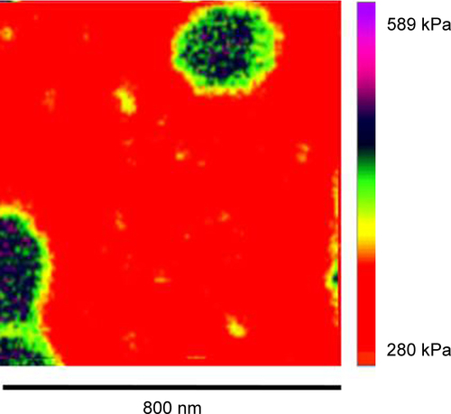
Figure S2 AFM analysis of the height of a liposome/PC complex.
Notes: Offline image processing of an AFM image of a representative liposome/PC complex. The original scan of this single spherical liposome was obtained in height mode. Using offline Nanoscope software, the liposome was magnified further, and then a section analysis was performed. Along the whole particle profile, there are regions at different heights (differences =5 nm).
Abbreviations: AFM, atomic force microscopy; PC, protein corona.
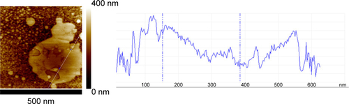
Figure S3 AFM elastic map of another area of the liposome/PC samples.
Notes: The image reveals a more complete picture of the mechanical properties of the particle aggregates. In detail, the image shows three different regions imputed to the presence of a cluster of three particles (yellow dotted line).
Abbreviations: AFM, atomic force microscopy; PC, protein corona.
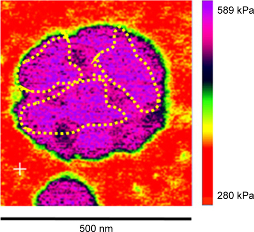
Figure S4 Confocal images of macrophages and cancer cells treated with liposomes in the absence and presence of a PC.
Note: The increased uptake and the presence of particles’ cluster (white arrows) in both J774 and 4T1 cells in the liposomes after incubation in plasma.
Abbreviation: PC, protein corona.
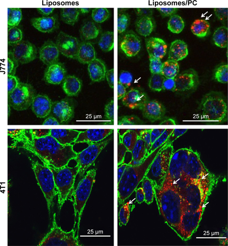
Table S1 Mass spectrometry details of the proteins identified in the corona
Acknowledgments
The authors would like to thank Kemi Cui and HMRI Advanced Cellular and Tissue Microscope Core Facility for traditional confocal scanning services, David Haviland and the HMRI Flow Cytometry Core Facility for flow cytometry setup and acquisition, and DA Engler, RK Matsunami, and the HMRI Proteomics Programmatic Core Laboratory for mass spectrometry analyses. The authors acknowledge the Sealy Center for Structural Biology and Molecular Biophysics at the University of Texas Medical Branch at Galveston for providing research resources. We thank Jean Ann Gilder (Scientific Communication srl. Naples, Italy) and Megan Livingston for editing the text. We thank Associazione Bianca Garavaglia, Via C Cattaneo 8, 21052 Busto Arsizio, Varese, Italy. This work was supported by grants RF-2010-2318372 and RF-2010-2305526 from Italian Ministry of Health, and by grants 1R21CA173579-01A1 from NIH/NCI, 5U54CA143837 PSOC Pilot project from NIH/NCI, W81XWH-12-10414 BCRP Innovator Expansion from Department of Defense, William Randolph Hearst Foundation, and The Regenerative Medicine Program Cullen Trust for Health Care to ET and by POR Campania FSE 2007–2013 Project DIAINTECH, Italy (to FS).
Disclosure
The authors report no conflicts of interest in this work.