

Abstract
Micro/nanoparticles could cause adverse effects on cardiovascular system and increase the risk for cardiovascular disease-related events. Nanoparticles prepared from poly(ethylene glycol) (PEG)-b-poly(ε-caprolactone) (PCL), namely PEG-b-PCL, a widely studied biodegradable copolymer, are promising carriers for the drug delivery systems. However, it is unknown whether polymeric PEG-b-PCL nano-micelles give rise to potential complications of the cardiovascular system. Zebrafish were used as an in vivo model to evaluate the effects of PEG-b-PCL nano-micelle on cardiovascular development. The results showed that PEG-b-PCL nano-micelle caused embryo mortality as well as embryonic and larval malformations in a dose-dependent manner. To determine PEG-b-PCL nano-micelle effects on embryonic angiogenesis, a critical process in zebrafish cardiovascular development, growth of intersegmental vessels (ISVs) and caudal vessels (CVs) in flk1-GFP transgenic zebrafish embryos using fluorescent stereomicroscopy were examined. The expression of fetal liver kinase 1 (flk1), an angiogenic factor, by real-time quantitative polymerase chain reaction (qPCR) and in situ whole-mount hybridization were also analyzed. PEG-b-PCL nano-micelle decreased growth of ISVs and CVs, as well as reduced flk1 expression in a concentration-dependent manner. Parallel to the inhibitory effects on angiogenesis, PEG-b-PCL nano-micelle exposure upregulated p53 pro-apoptotic pathway and induced cellular apoptosis in angiogenic regions by qPCR and terminal deoxynucleotidyl transferase dUTP nick end labeling (TUNEL) apoptosis assay. This study further showed that inhibiting p53 activity, either by pharmacological inhibitor or RNA interference, could abrogate the apoptosis and angiogenic defects caused by PEG-b-PCL nano-micelles, indicating that PEG-b-PCL nano-micelle inhibits angiogenesis by activating p53-mediated apoptosis. This study indicates that polymeric PEG-b-PCL nano-micelle could pose potential hazards to cardiovascular development.
Supplementary materials
Figure S1 Effect of PEG-b-PCL nano-micelle on embryo hatch.
Notes: Embryos were exposed to 0, 60, 120, and 240 μg/mL PEG-b-PCL nano-micelles. At 72 hpf, hatched embryos were counted. Data are expressed as mean ± SEM from three independent experiments (n=60), *P<0.05.
Abbreviations: hpf, hours postfertilization; PEG-b-PCL, poly(ethylene glycol)-b-poly(ε-caprolactone); SEM, standard error of mean.
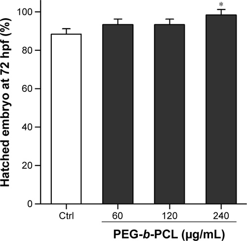
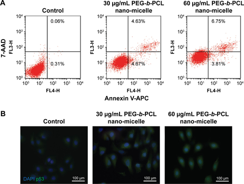
Figure S2 PEG-b-PCL nano-micelle induces apoptosis in endothelial cells in vitro.
Notes: (A) Apoptosis analysis of HUVECs by flow cytometry. HUVECs with or without PEG-b-PCL nano-micelles treatment were co-stained with APC-conjugated Annexin V and 7-AAD. Cells positive for Annexin V are in early apoptotic stage (lower right quadrant), and cells positive for Annexin V/7-AAD are in late apoptotic stage (upper right quadrant). (B) p53 immunostaining in HUVECs with or without PEG-b-PCL nano-micelles treatment. HUVECs were fixed with 4% PFA and co-stained with antibody against p53 (green) and 4′,6-diamidino-2-phenylindole for DNA (blue).
Abbreviations: HUVECs, human umbilical vein endothelial cell; PEG-b-PCL, poly(ethylene glycol)-b-poly(ε-caprolactone); PFA, paraformaldehyde; AAD, aminoactinomycin D; APC, allophycocyanin.
Acknowledgments
We would like to thank Yiming Zheng from the Department of Biomedical Sciences in Florida State University for help with manuscript preparation. We thank Dr Xiang Xie from the College of Bioengineering in Chongqing University for technical assistance. This study was supported by grants from the National Natural Science Foundation of China (111572064, 11332003), by the cooperative project of Chongqing Key Laboratory of Nano/Micro Composite Materials and Devices (CQKL-1502) and the National Key Technology R & D Program of China (2012BAI18B02). The authors also thank the support of experimental instruments from the National “111 Plan” Base (B06023) and the Public Experiment Center of State Bioindustrial Base, Chongqing, People’s Republic of China.
Disclosure
The authors report no conflicts of interest in this work.