

Abstract
There is a need for novel nanomaterials with properties not yet exploited in regenerative nanomedicine. Based on lessons learned from the oldest metazoan phylum, sponges, it has been recognized that two previously ignored or insufficiently recognized principles play an essential role in tissue regeneration, including biomineral formation/repair and wound healing. Firstly, the dependence on enzymes as a driving force and secondly, the availability of metabolic energy. The discovery of enzymatic synthesis and regenerative activity of amorphous biosilica that builds the mineral skeleton of siliceous sponges formed the basis for the development of successful strategies for the treatment of osteochondral impairments in humans. In addition, the elucidation of the functional significance of a second regeneratively active inorganic material, namely inorganic polyphosphate (polyP) and its amorphous nanoparticles, present from sponges to humans, has pushed forward the development of innovative materials for both soft (skin, cartilage) and hard tissue (bone) repair. This energy-rich molecule exhibits a property not shown by any other biopolymer: the delivery of metabolic energy, even extracellularly, necessary for the ATP-dependent tissue regeneration. This review summarizes the latest developments in nanobiomaterials based on these two evolutionarily old, regeneratively active materials, amorphous silica and amorphous polyP, highlighting their specific, partly unique properties and mode of action, and discussing their possible applications in human therapy. The results of initial proof-of-concept studies on patients demonstrating complete healing of chronic wounds are outlined.
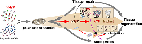
Graphical Abstract
Acknowledgments
We thank our colleagues Prof. Dr W. Müller-Klieser and Prof. Dr S. Walenta (Institute of Pathophysiology; University Medical Center Mainz; Germany) for providing us with the cryosections through the sponge for the visualization of the ATP accumulation. W.E.G. M. is the holder of an ERC Advanced Investigator Grant (Grant No.: 268476). In addition, W.E.G. M. has obtained three ERC-PoC Grants (Si-Bone-PoC, Grant No.: 324564; MorphoVES-PoC, Grant No.: 662486; and ArthroDUR, Grant No.: 767234). In addition, this work was supported by Grants from the European Commission (Grant Nos.: 604036 and 311848), the International Human Frontier Science Program and the BiomaTiCS research initiative of the University Medical Center, Mainz. Further support came from the Grant of the BMBF (SKIN-ENERGY, Grant No.: 13GW0403A/B) and the Grant of the BMWi (Grant No.: ZF4294002AP9).
Author Contributions
All authors made a significant contribution to the work reported, whether that is in the conception, study design, execution, acquisition of data, analysis and interpretation, or in all these areas; took part in drafting, revising or critically reviewing the article; gave final approval of the version to be published; have agreed on the journal to which the article has been submitted; and agree to be accountable for all aspects of the work.
Disclosure
The authors report no conflicts of interest in this work.