

Abstract
Background
In the current scenario, the synthesis of nanoparticles (NPs) using environmentally benign methods has gained significant attention due to their facile processes, cost-effectiveness, and eco-friendly nature.
Methods
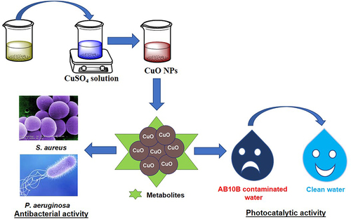
In the present study, copper oxide nanoparticles (CuO NPs) were synthesized using aqueous extract of Coelastrella terrestris algae as a reducing, stabilizing, and capping agent. The synthesized CuO NPs were characterized by X-ray diffraction (XRD), UV-visible spectroscopy (UV-Vis), Fourier transform infrared spectroscopy (FTIR), dynamic light scattering (DLS), and field emission scanning electron microscopy (FE-SEM) coupled with energy-dispersive X-ray spectroscopy (EDS).
Results
XRD investigation revealed that the biosynthesized CuO NPs were nanocrystalline with high-phase purity and size in the range of 4.26 nm to 28.51 nm. FTIR spectra confirmed the existence of secondary metabolites on the surface of the synthesized CuO NPs, with characteristic Cu–O vibrations being identified around 600 cm−1, 496 cm−1, and 440 cm−1. The FE-SEM images predicted that the enhancement of the algal extract amount converted the flattened rice-like structures of CuO NPs into flower petal-like structures. Furthermore, the degradation ability of biosynthesized CuO NPs was investigated against Amido black 10B (AB10B) dye. The results displayed that the optimal degradation efficacy of AB10B dye was 94.19%, obtained at 6 pH, 50 ppm concentration of dye, and 0.05 g dosage of CuO NPs in 90 min with a pseudo-first-order rate constant of 0.0296 min−1. The CuO-1 NPs synthesized through algae exhibited notable antibacterial efficacy against S. aureus with a zone of inhibition (ZOI) of 22 mm and against P. aeruginosa with a ZOI of 17 mm.
Conclusion
Based on the findings of this study, it can be concluded that utilizing Coelastrella terrestris algae for the synthesis of CuO NPs presents a promising solution for addressing environmental contamination.
Graphical Abstract
Associated Content
All experimental details are explained in the Supplementary Material 1.
Data Sharing Statement
All data generated or analyzed during this study are included in this published article. Additional data can be provided upon request from the corresponding author.
Acknowledgments
The authors thank the Department of Chemistry, Mohanlal Sukhadia University, Udaipur (Raj.), India, for providing the necessary laboratory facilities. The authors are grateful to the Department of Physics, Department of Botany, and Department of Environmental Science, Mohanlal Sukhadia University, Udaipur, for providing the XRD facility, algal culture, and water sample analysis, respectively. The authors acknowledge the facilities of Manipal University Jaipur (MUJ), India, for UV-Vis, FE-SEM coupled with EDS, and antibacterial analysis facilities.
Author Contributions
All authors made a significant contribution to the work reported, whether that is in the conception, study design, execution, acquisition of data, analysis and interpretation, or in all these areas; took part in drafting, revising or critically reviewing the article; gave final approval of the version to be published; have agreed on the journal to which the article has been submitted; and agree to be accountable for all aspects of the work.
Disclosure
The authors have no competing interests to declare that are relevant to the content of this article.