

Abstract
Nanotechnology-derived platforms, such as dendrimers, are very attractive in several biological applications. In the case of human immunodeficiency virus (HIV) infection, polyanionic carbosilane dendrimers have shown great potential as antiviral agents in the development of novel microbicides to prevent the sexual transmission of HIV-1. In this work, we studied the mechanism of two sulfated and naphthylsulfonated functionalized carbosilane dendrimers, G3-S16 and G2-NF16. They are able to inhibit viral infection at fusion and thus at the entry step. Both compounds impede the binding of viral particles to target cell surface and membrane fusion through the blockage of gp120–CD4 interaction. In addition, and for the first time, we demonstrate that dendrimers can inhibit cell-to-cell HIV transmission and difficult infectious synapse formation. Thus, carbosilane dendrimers’ mode of action is a multifactorial process targeting several proteins from viral envelope and from host cells that could block HIV infection at different stages during the first step of infection.
Supplementary materials
Figure S1 Structure of polyanionic carbosilane dendrimers.
Notes: (A) (i) Third-generation G3-S16, with 16 sulfated end groups. C256H508N48Na16O64S16Si29; MW: 6,978.41 g/mol. (ii) Computer model of G3-S16 equilibrated in salt water. (B) (iii) Second-generation G2-NF16, with 16 naphthylsulfonated end groups. C184H244N24Na16O56S16Si13; MW: 4,934.02 g/mol. (iv) Model of G2-NF16 dendrimer equilibrated in salt water. The dendrimer Si core atom is colored in magenta. The color coding for all remaining atoms is C – gray, O – red, Si – beige, and S – yellow.
Abbreviation: MW, molecular weight.
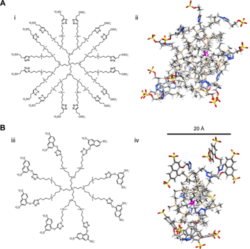
Figure S2 Specificity of antiviral activity.
Notes: Activated PBMCs were treated for 1 hour with nontoxic concentration of dendrimers and then infected overnight with env-X4-luc and env-VSVG-luc HIV-derived lentivirus. After 2 days of infection, viral infection was quantified measuring luciferase expression levels. AZT was used as positive control. (**P<0.01, ***P<0.001 vs control). Data represent the mean ± SD of three independent experiments.
Abbreviations: PBMCs, peripheral blood mononuclear cells; HIV, human immunodeficiency virus; SD, standard deviation; NT, nontreated; AZT, azidothymidine.
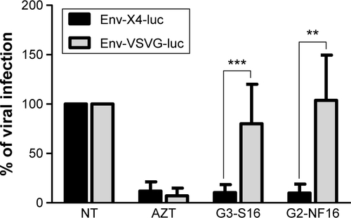
Figure S3 Equilibrated structure of gp120 after 100 ns of simulation (left) and superposition of this “unbound like” conformation with the original (bound) gp120 conformation (right).
Notes: Color coding: bridging sheet (terminal parts of V1/V2 loops) in green (“unbound like” configuration) and cyan (bound configuration) and V3 loop in magenta (“unbound like” configuration) and red (bound configuration).
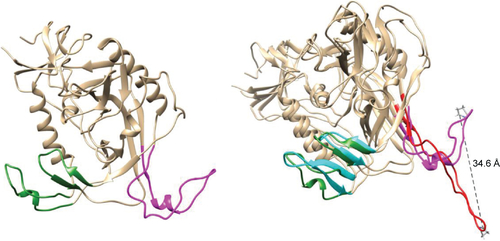
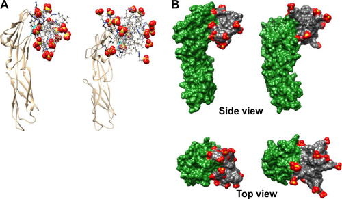
Figure S4 Molecular modeling of CD4–dendrimer complexes.
Notes: (A) CD4–G2-S16 (left) and CD4–G3-S16 (right). Just the closest (≤3 Å) amino acids to the dendrimer are shown (in ball and stick) using black color for carbons to distinguish them from dendrimer carbons (in gray, stick representation). Colors of the other elements are O – red, S – yellow, H – white, and Si – tan. Part of the small alpha helix on CD4 colored in cyan localizes two neighboring arginines that contribute to cationic character of the surrounding area. This small helix is one of the important parts of the CD4–gp120 or CD4–anionic dendrimer binding interface. For the terminal SO3 groups, the sphere representation is used. (B) CD4–G2-NF16 (left) and CD4–G3-S16 (right) complexes with visualized molecular surfaces to better see mutual dendrimer–protein integration. The CD4 molecular surface is in green and dendrimer molecular surface is in gray, except the terminal SO3 groups where red is used for oxygen and yellow for sulfur.
Acknowledgments
We acknowledge the Center of Transfusion of Madrid for the buffy coats and the Spanish HIV HGM BioBank for their process. We thank Rafael Samaniego and Laura Díaz from confocal microscopy and cytometry analysis units of HGUGM, respectively. This work has been (partially) funded by the RD12/0017/0037 project as part of Acción Estratégica en Salud, Plan Nacional de Investigación Científica, Desarrollo e Innovación Tecnológica 2008–2011 and cofinanced by Instituto de Salud Carlos III (ISCIII) (Subdirección General de Evaluación) and Fondo Europeo de Desarrollo Regional. RETIC PT13/0010/0028, El Fondo de Investigación Sanitaria (grant number PI13/02016), CTQ2011-23245 and CTQ2014-54004-P (MIMECO), Comunidad de Madrid (grant numbers S-2010/BMD-2351 and S-2010/BMD-2332), CYTED 214RT0482. CIBER-BBN is an initiative funded by the VI National R&D&i Plan 2008–2011, Iniciativa Ingenio 2010, the Consolider Program, and CIBER Actions and financed by the ISCIII with assistance from the European Regional Development Fund. This work was supported partially by a Marie Curie International Research Staff Exchange Scheme Fellowship within the 7th European Community Framework Programme, project number PIRSES-GA-2012-316730 NANOGENE, cofinanced by the Polish Ministry of Science and Higher Education (grant number W21/7PR/2013). MP is supported by Spanish MICINN through the Ramón y Cajal (RYC-2009-05486) and by Fondos de Investigación Sanitaria ISCIII (PI12_01763).
Author contributions
EV-C, MP, and M ÁM-F designed, performed experiments, and composed the figures. MM performed the computational work for molecular modeling. FJM and RG synthetized the dendrimers. All authors contributed toward data analysis, drafting, and critically revising the paper and agree to be accountable for all aspects of the work.
Disclosure
The authors report no conflicts of interest in this work.