1,927
Views
27
CrossRef citations to date
0
Altmetric
Articles

From molecular evolution to biobricks and synthetic modules: a lesson by the bacterial flagellum

, , &
Pages 49-64 | Received 03 Jan 2014, Accepted 21 Mar 2014, Published online: 14 Jul 2014

Figures & data

Figure 1. The bacterial flagellum. (a) Flagellar architecture and protein composition of Gram-negative (left) and Gram-positive (right) bacteria. (b) Domain architecture of Flagellin. The variable domains that are inserted into the conserved D1 domain (i.e. D1-N and D1-C in yellow and cyan, respectively) are shown in green. The conserved D0 that comprises N- and C-terminus of Flagellin is shown in red and blue, respectively. (c) Models of the Flagellin structures from S. typhimurium (pdb: 1UCU), Sphingomonas (based on pdb: 2ZBI), and Bacillus subtilis (this work). The color code is as in Figure (b). (d) Cartoon of the flagellar filament with the different Flagellins from Figure (c). The color code is as in Figure (b).
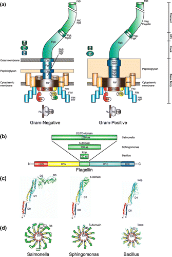
Figure 1. The bacterial flagellum. (a) Flagellar architecture and protein composition of Gram-negative (left) and Gram-positive (right) bacteria. (b) Domain architecture of Flagellin. The variable domains that are inserted into the conserved D1 domain (i.e. D1-N and D1-C in yellow and cyan, respectively) are shown in green. The conserved D0 that comprises N- and C-terminus of Flagellin is shown in red and blue, respectively. (c) Models of the Flagellin structures from S. typhimurium (pdb: 1UCU), Sphingomonas (based on pdb: 2ZBI), and Bacillus subtilis (this work). The color code is as in Figure 1(b). (d) Cartoon of the flagellar filament with the different Flagellins from Figure 2(c). The color code is as in Figure 1(b).

Figure 2. Bacterial flagellation patterns. (a) Polar flagellation patterns are colored in blue, and lateral patterns are marked in green. Sheathed flagella are shadowed in red. The listed organisms are grouped according to the occurrence and absence of FlhF and FlhG. (b) Domain architecture of SRP-GTPases. The conserved NG-domain is in green. The B-domain of FlhF is in blue. M- and A- domains of Ffh and FtsY, respectively, are in grey. (c) Domain architecture of MinD and FlhG. The N-terminus of FlhG contains the conserved ‘DQAxxLR’ motif. (d) Molecular evolution and diversification of the regulatory circuit of FlhF and FlhG. ‘T’ and ‘D’ stand for ATP/GTP and ADP/GDP, respectively. For further description see text.
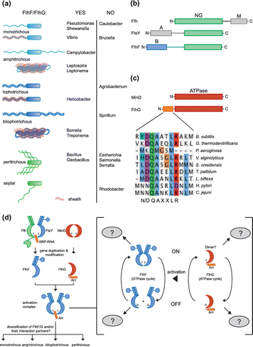
Figure 2. Bacterial flagellation patterns. (a) Polar flagellation patterns are colored in blue, and lateral patterns are marked in green. Sheathed flagella are shadowed in red. The listed organisms are grouped according to the occurrence and absence of FlhF and FlhG. (b) Domain architecture of SRP-GTPases. The conserved NG-domain is in green. The B-domain of FlhF is in blue. M- and A- domains of Ffh and FtsY, respectively, are in grey. (c) Domain architecture of MinD and FlhG. The N-terminus of FlhG contains the conserved ‘DQAxxLR’ motif. (d) Molecular evolution and diversification of the regulatory circuit of FlhF and FlhG. ‘T’ and ‘D’ stand for ATP/GTP and ADP/GDP, respectively. For further description see text.

Table 1. Roles of FlhF and FlhG in different bacterial species.

Reprints and Corporate Permissions

Please note: Selecting permissions does not provide access to the full text of the article, please see our help page How do I view content?

To request a reprint or corporate permissions for this article, please click on the relevant link below:

Academic Permissions

Please note: Selecting permissions does not provide access to the full text of the article, please see our help page How do I view content?

Obtain permissions instantly via Rightslink by clicking on the button below:

If you are unable to obtain permissions via Rightslink, please complete and submit this Permissions form. For more information, please visit our Permissions help page.