Figures & data
Table I. Clinical findings in patients with PCD.
Figure 1 Photographs of a histology specimen of a murine trachea. Lower(a) and higher (b) magnification photographs depict the respiratory epithelium, which covers the cartilage layer. The ciliated respiratory epithelial cells appear to be brighter and carry hair‐like projections (cilia). Intercalated mucous cells do not carry cilia and are darker in colour.
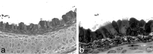
Figure 2 Clinical findings in PCD subjects.a. Radiograph of chest shows dextrocardia in a patient with situs inversus. Note also pulmonary dystelectasis and infiltrates. b. Chest radiograph of an adult PCD patient with chronic airway disease and bronchiectasis.
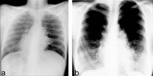
Figure 3 a. Schematic diagram of a cilium cross‐section (left). b. Electron micrograph of a cilium cross‐section from a healthy proband analyzed by transmission electron microscopy. Please note outer dynein arms are visibly attached at almost all nine peripheral doublets. In contrast, inner dynein arms are visible only on a few peripheral doublets (arrow). c. Transmission electron micrograph of a cross‐section from a respiratory cilium originating from a patient carrying DNAH5 mutations shows absence of outer dynein arms.
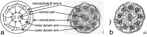
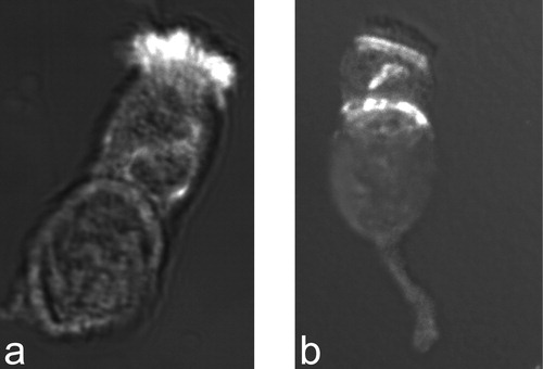
Figure 4 Staining of respiratory epithelial cells with an antibody directed against the axonemal dynein heavy chain DNAH5.a. Respiratory epithelial cell of a healthy proband shows positive staining (white, original red) for the entire length of all cilia. b. In a PCD patient with outer dynein arm defect documented by electron microscopy, no staining of the ciliary compartment is observed. However, there is DNAH5 accumulation in the cytoplasm and at the ciliary base (microtubules organizing centers).