
Figures & data

Figure 1. Identification of A2780 cell-released exosomes. (A) Morphological characteristics of exosomes under TEM. (B) NTA of isolated exosomes shows the distribution of particle size. (C) Western blotting of CD63 and CD81 levels on cell lysates and extracted exosomes. TEM: transmission electron microscope; NTA: nanoparticle tracking analysis.
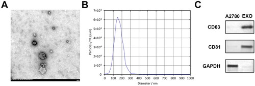
Figure 2. Hypoxic A2780 cell derived exosomes promote macrophage M2 polarisation. (A, B) RT-qPCR measured the iNOS, IL-1β, IL-6, CD206, Arg-1, and IL-10 expression ratio of macrophages co-incubated with hypo-exos and nor-exos. Hypo-exo: hypoxia A2780-derived exosome; nor-exo: normoxic A2780-derived exosome. **p < 0.01; ***p < 0.001.
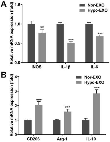
Figure 3. Exosomal miR-1225-5p contributed to the differentiation of macrophages. (A) Volcano plot of aberrant expressed miRNAs in hypoxia and normoxia groups. (B) miR-1225-5p and miR-1207-5p expression in A2780 cells treated with hypoxia and normoxia. (C) miR-1225-5p levels in A2780 cells after its knockout. (D, E) RT-qPCR detected the iNOS, IL-1β, IL-6, CD206, Arg-1, and IL-10 expression of macrophages co-incubated with hypo-exos and nor-exos. *p < 0.05; **p < 0.01; ***p < 0.001 (vs. Nor, scramble, and nor-EXO). #p < 0.05; ##p < 0.01; ###p < 0.001 (vs. Hypo-EXO + scramble).
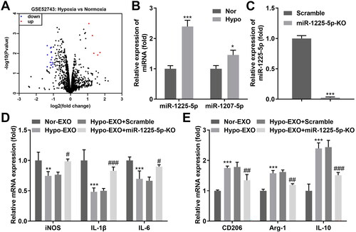
Figure 4. Exosomal miR-1225-5p facilitates polarization of macrophages. mRNA levels of exosomal miR-1225-5p isolated from (A) A2780 cells and (B) exosomoes derived from A2780 cells after transfection. (C) FAM-labeled miR-122-5p were transfected into A2780 cells, the released exosomes were incubated with macrophages. The trail of miR-1225-5p was observed under a confocal microscope. (D, E) RT-qPCR was used to detect the iNOS, IL-1β, IL-6, CD206, Arg-1, and IL-10 expression of macrophages co-incubated with hypo-exos and nor-exos. **p < 0.01; ***p < 0.001 (vs. mimic nc, and nor-EXO). ##p < 0.01; ###p < 0.001 (vs. Hypo-EXO + mimic nc).
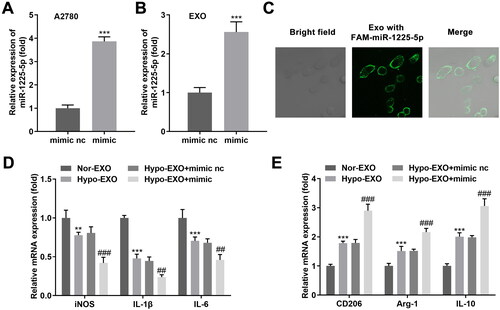
Figure 5. TLR2 was a miR-1225-5p downstream gene. (A) The binding domain between miR-1225-5p and TLR2 was predicted by bioinformatics. (B) Dual-luciferase reporter analysis confirmed the association between TLR2 and miR-1225-5p. (C) qRT-PCR as well as (D) western blot analyses for the expression of TLR2. ***p < 0.001 (vs. mimic nc).
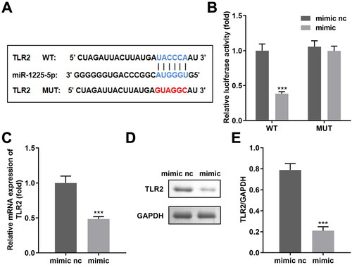
Figure 6. Exosomal miR-1225-5p promotes polarisation of macrophages into M2-like macrophages by modulating TLR2. mRNA levels of exosomal TLR2 isolated from (A) A2780 cells and (B) exosomes derived from A2780 cells after transfection. (C) Exosomes released from GFP-labeled TLR2 transfected A2780 cells were incubated with macrophages. The trail of TLR2 was observed under a confocal microscope. (D, E) RT-qPCR was used to detect the iNOS, IL-1β, IL-6, CD206, Arg-1 and IL-10 expression of macrophages co-incubated with hypo-exos overexpressing miR-122-5-5p and TLR2. **p < 0.01; ***p < 0.001 (vs. vector, mimic + vector).
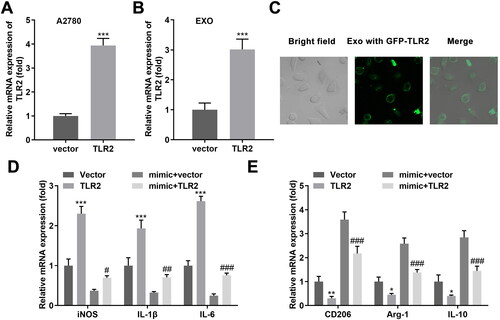
Figure 7. Hypoxia promoted the migration-promoting function of M2-like macrophages on ovarian cancer cells by the activation of the wnt/β-catenin pathway. (A, B) Cell viability, (C, D) migration, (E, F) and invasion of ovarian cancer cells cultured in CM hypo-exo treated macrophages. (G–S) Proteins of the wnt/β-catenin, PI3K, Notch, p65, and STAT3 pathways were examined using Western blotting. ***p < 0.001 [vs. CM(nor-exo)].
![Figure 7. Hypoxia promoted the migration-promoting function of M2-like macrophages on ovarian cancer cells by the activation of the wnt/β-catenin pathway. (A, B) Cell viability, (C, D) migration, (E, F) and invasion of ovarian cancer cells cultured in CM hypo-exo treated macrophages. (G–S) Proteins of the wnt/β-catenin, PI3K, Notch, p65, and STAT3 pathways were examined using Western blotting. ***p < 0.001 [vs. CM(nor-exo)].](/cms/asset/9ecf8328-9b8f-45e0-8171-59c7ba252ac6/iaut_a_2281226_f0007_c.jpg)
Data availability statement
The datasets used and/or analyzed during the current study are available from the corresponding author on reasonable request.