
Figures & data
Table 1. Assessment of surface reactivity of test dusts by measured in vitro generation of hydroxyl free radicals.
Figure 1. Changes in acellular biomarkers of toxicity in BALFs from rats 1 and 4 wk after instillation with three lunar dusts, TiO2, and quartz (SiO2). Rats were instilled with 0 (Control), 1.0, 2.5, or 7.5 mg/rat of test dust. Figs. (A) Albumin, (B) LDH, and (C) Macrophage chemoattractant protein-1. Graph labels: LD-jm: jet-mill-ground lunar dust; LD-ug: unground lunar dust; LD-bm: ball-mill-ground lunar dust; lighter color bars: 1 wk; darker color bars: 4 wk. Each bar represents mean ± SD from 6 rats. Statistical analyses of differences between treatment groups are shown in and .
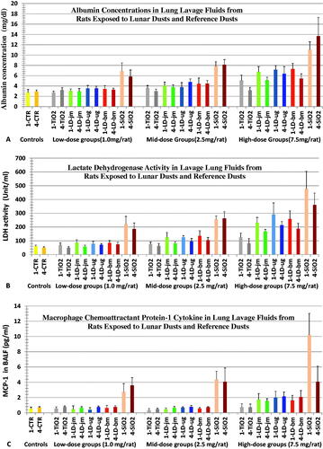
Figure 2. Changes in cell counts in BALFs from rats 1 and 4 wk after instillation with three lunar dusts, TiO2, and quartz (SiO2). Rats were instilled with 0 (Control), 1.0, 2.5, or 7.5 mg/rat of test dust. Figs., (A) total BAL cell counts, (B) macrophage counts, (C) neutrophil counts. Graph labels: LD-jm: jet-mill-ground LD; LD-ug: unground LD; LD-bm: ball-mill-ground LD; lighter color bars: 1 wk; darker color bars: 4 wk. Each bar represents mean ± SD from 6 rats. Statistic differences between treatments are shown in and .
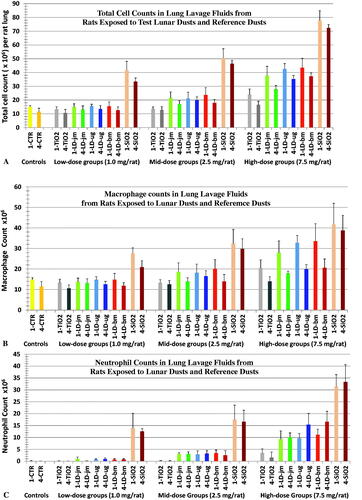
Figure 3. Lungs and lymph nodes (left tracheobronchial and parathymic lymph nodes). These tissues were isolated from rats of the high-dosed groups 13 wk after the dust instillation. Groups of 6 rats were each instilled with vehicle containing no dust (Controls, (A)), or 7.5 mg of TiO2 (B), quartz (C), LD-jm (Moon dust A), LD-ug (Moon Dust C) or LD-bm (Moon dust E). Lungs of TiO2-treated rats were unremarkable (B). The lungs of rats instilled with three LDs were similar (D–F), some superficial whitish nodules were visible. Nodules in the lungs of quartz-treated rats were larger; the lungs were lumpy and rough (C). The lymph nodes (LNs) of quartz-treated rats were much bigger and firm, the left tracheobronchial LN is visible as a translucent lump of the size a peanut front of the trachea and at the center above the lung in (C). LNs isolated from LD-exposed rats were smaller (grayish LD particles inside the LNs made the LNs appeared darker) (D–F); LNs of control animals were too small and could not be found, except from one animal (A).
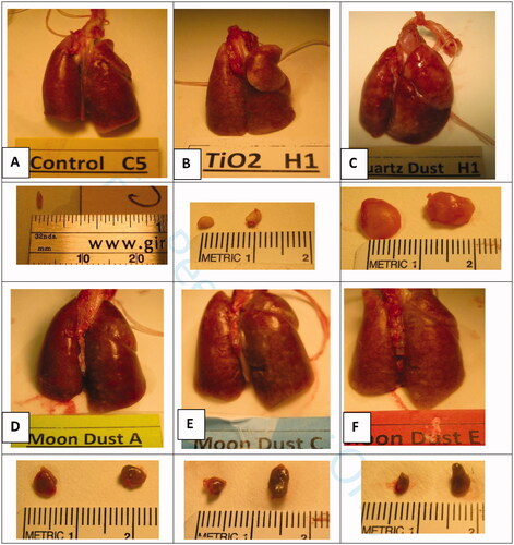
Figure 4. Lung and lymph node tissues 13 weeks after intratracheal instillation with unground lunar dust (LD-ug) and reference dusts (TiO2, and quartz). (A) Control rat: normal lung structure; (B) LD-ug, low dose: some particles in macrophages, no lesions; (C) LD-ug, mid dose: a gathering of dust particles, macrophages, and inflammatory cells in alveoli, some alveolar walls were thicker than those of more normal adjacent alveoli; (D) LD-ug, high dose (hd): a low-grade granuloma (microscopic nodule) consisting of PLMs, inflammatory cells, and thickened alveolar septa. (E) LD-ug, hd: an accumulation predominately composed of lymphocytes and particle-laden macrophages with some neutrophils visible around a blood vessel, characterized as vasculitis; (F) LD-ug, hd: low-grade granulomas (pink areas) containing fine LD grains mostly inside macrophages among lymphoid cells (in blue) in a mediastinal lymph node with no/minimal fibrosis, (G) TiO2, hd: showed TiO2 particles, minimal or no lesions; (H) Quartz, hd: Many large granulomas with moderate fibrosis, severe vasculitis; (I) Quartz, hd: A large structureless granuloma with diffuse and moderate fibrosis in a mediastinal lymph node (Magnifications: 10× to 50×.).
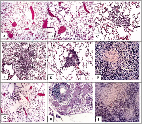
Figure 5. Histopathological responses of different tissues in the lungs of dust-instilled rats 13 weeks after instilled with 1.0 (L), 2.5 (M), or 7.5 (H) mg/rat of LD-jm (jet-mill ground), LD-ug (unground), LD-bm (ball-mill ground), TiO2, or quartz. Lesion scoring scale: 0 = none/normal, 1 = minimal, 2 = mild, 3= moderate, 4= marked, 5 = severe (two major effects in each pulmonary tissue area are shown in the figure). Each bar represents mean ± SD from 6 rats.
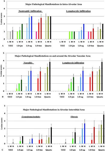
Figure 6. Histopathological responses in the lungs and lymph nodes of dust-instilled rats 13 weeks after instilled with 1.0 (L), 2.5 (M), or 7.5 (H) mg/rat of LD-jm (jet-mill ground), LD-ug (unground), LD-bm (ball-mill ground), TiO2, or quartz. Lesion scoring scale: 0 = none/normal, 1 = minimal, 2 = mild, 3= moderate, 4= marked, 5 = severe (two major effects in each pulmonary tissue area are shown in the figure). Each bar represents mean ± SD from 6 rats.
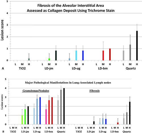
Table 2. Statistical comparison of BALF toxicity biomarker induction by lunar dusts, quartz and TiO2.
Table 3. Statistical comparison of BALF inflammatory biomarker induction by three lunar dusts.