8,588
Views
26
CrossRef citations to date
0
Altmetric
Review Articles

Simulated biological fluids – a systematic review of their biological relevance and use in relation to inhalation toxicology of particles and fibres

, , , &
Pages 217-248 | Received 29 Sep 2020, Accepted 11 Mar 2021, Published online: 27 Apr 2021

Figures & data

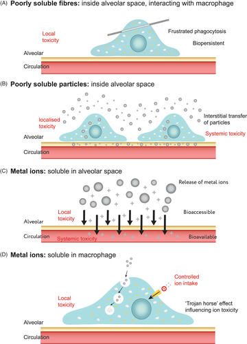
Figure 1. Schematic highlighting the pathogenicity pathways of (a) fibres inside the alveolar space interacting with macrophages; (b) poorly soluble metals remaining in the alveolar space, alongside transfer through cellular mechanisms; and (c, d) soluble metals in intra- and extra-cellular regions.

Figure 1. Schematic highlighting the pathogenicity pathways of (a) fibres inside the alveolar space interacting with macrophages; (b) poorly soluble metals remaining in the alveolar space, alongside transfer through cellular mechanisms; and (c, d) soluble metals in intra- and extra-cellular regions.

Figure 2. Schematic demonstrating the diversity of simulated biological fluids and the differences in their composition and physiochemical properties. In particular, the alveoli and lysosome have been highlighted indicating the locations and characteristics of the lysosomal fluid, lung lining fluid and interstitial fluid. [adapted with permission from (Plumlee et al. Citation2006)].

Figure 2. Schematic demonstrating the diversity of simulated biological fluids and the differences in their composition and physiochemical properties. In particular, the alveoli and lysosome have been highlighted indicating the locations and characteristics of the lysosomal fluid, lung lining fluid and interstitial fluid. [adapted with permission from (Plumlee et al. Citation2006)].

Table 1. Composition (g/L) of frequently used biofluids used to simulate the neutral conditions of the lung lining fluid.

Table 2. Composition (g/L) of frequently used biofluids for simulation of the acidic conditions of the lysosome.

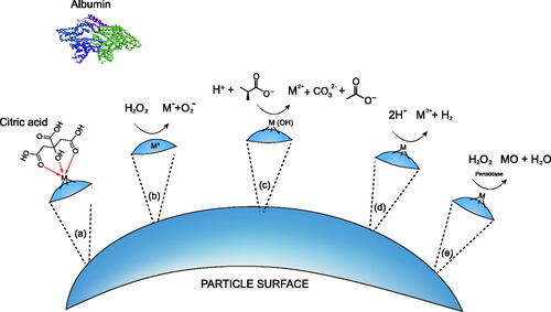
Figure 3. Selected mechanisms that effect dissolution on the surface of a particle including: (a) complexation, (b,c) redox activities, (d) proton-promoted dissolution and (e) enzymatic action. The prevalence of each dissolution mechanism will be material specific, driven by the chemistry of the materials surface.

Figure 3. Selected mechanisms that effect dissolution on the surface of a particle including: (a) complexation, (b,c) redox activities, (d) proton-promoted dissolution and (e) enzymatic action. The prevalence of each dissolution mechanism will be material specific, driven by the chemistry of the materials surface.

Table 3. Correlation of results from in vitro acellular assays with measurements taken in vivo or within cells grown in vitro.

Table 4. Studies comparing “simple” and complex fluids, including an indication of whether the more complex fluid caused less (−), equal (0), or greater (+) dissolution.