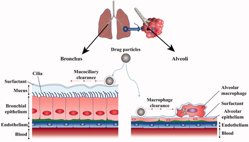
4,711
Views
25
CrossRef citations to date
0
Altmetric
Review Article

Nanocrystals based pulmonary inhalation delivery system: advance and challenge

, , , , , , & show all
Pages 637-651 | Received 03 Jan 2022, Accepted 01 Feb 2022, Published online: 21 Feb 2022

Figures & data

Figure 1. The structure of lung and pulmonary mucus barrier.

Figure 1. The structure of lung and pulmonary mucus barrier.

Figure 2. Preparation methods of nanocrystals based pulmonary inhalation delivery system.

Figure 2. Preparation methods of nanocrystals based pulmonary inhalation delivery system.

Table 1. Examples of nanocrystal preparation.

Table 2. Examples of nanocrystals pulmonary inhalation system.

Figure 3. (A) Schematic diagram of BUD-INAM in vitro and in vivo release of BUD, (B) in vitro release profile, (C) retention on the porcine tracheal mucosa surface as a function of time, and (D) plasma concentration–time profiles. Adapted with permission from Liu et al. (Citation2018).

Figure 3. (A) Schematic diagram of BUD-INAM in vitro and in vivo release of BUD, (B) in vitro release profile, (C) retention on the porcine tracheal mucosa surface as a function of time, and (D) plasma concentration–time profiles. Adapted with permission from Liu et al. (Citation2018).

Figure 4. (A) Schematic diagram of PEGylated mucus-penetrating nanocrystals and lung treatment in vivo, (B) in vitro release profile, (C) inhibition ability B. cenocepacia J2315 biofilm, cytotoxicity of C109 formulations to wild type 16HBE (D) and CF (CFBE41o−) bronchial epithelial cells (E). Adapted with permission from Costabile et al. (Citation2020).

Figure 4. (A) Schematic diagram of PEGylated mucus-penetrating nanocrystals and lung treatment in vivo, (B) in vitro release profile, (C) inhibition ability B. cenocepacia J2315 biofilm, cytotoxicity of C109 formulations to wild type 16HBE (D) and CF (CFBE41o−) bronchial epithelial cells (E). Adapted with permission from Costabile et al. (Citation2020).

Figure 5. (A) Schematic illustration of curcumin nanocrystals on dissolution, airway mucosa penetration, lung tissue distribution, and absorption by pulmonary delivery. (B) Confocal microscopy images of calu-3 cells after incubation with CUR-NCs for 2 h (the scale bar is 50 μm); (C) calu-3 cell viability upon exposure to NC formulations at different drug concentrations; (D) mean fluorescence intensity of cells determined by flow cytometry (n = 3); (E) z-stack confocal images of the calu-3 cell layer with the polyester membrane after transport of NCs formulations for 1 h (the scale bar in xy plane was 50 μm). Adapted with permission from He et al. (Citation2020).

Figure 5. (A) Schematic illustration of curcumin nanocrystals on dissolution, airway mucosa penetration, lung tissue distribution, and absorption by pulmonary delivery. (B) Confocal microscopy images of calu-3 cells after incubation with CUR-NCs for 2 h (the scale bar is 50 μm); (C) calu-3 cell viability upon exposure to NC formulations at different drug concentrations; (D) mean fluorescence intensity of cells determined by flow cytometry (n = 3); (E) z-stack confocal images of the calu-3 cell layer with the polyester membrane after transport of NCs formulations for 1 h (the scale bar in xy plane was 50 μm). Adapted with permission from He et al. (Citation2020).