
Figures & data
Figure 1. 1a and 1b Patient with homocystinuria, 60 years of age, with bilateral ectopia lentis (subluxated lenses) and severe myopia.
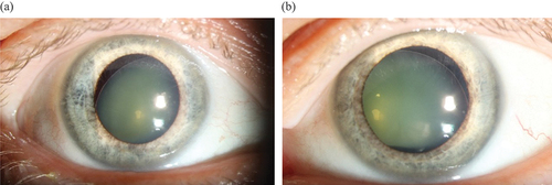
Table 1. Summary of visual and ocular findings in patients with homocystinuria, including symptoms, ectopia lentis and myopia prior to diagnosis, best-corrected visual acuity, strabismus, refraction, surgery due to ectopia lentis, retinal pathology, and other typical homocystinuria manifestations.
Figure 2. Exterior image from 30-year-old patient displaying dense corneal opacities with growth of vessels in the right eye which was blind after repeated surgery for retinal detachment after ectopia lentis.
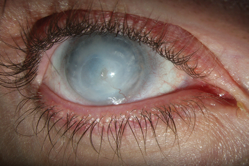
Figure 3. Patient with homocystinuria, 30 years of age, who underwent ocular surgery at 25 years of age because of ectopia lentis, where the crystalline luxated lens was removed and replaced with an intraocular lens (IOL). Several surgical re-operations were needed as the IOL also luxated. Diagnosis of HCU was established three years after the first surgery.
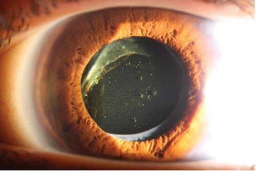
Figure 4. a, b and c OCT- and UWFI-images from a 70-year-old patient with homocystinuria. OCT showing bilateral retinal atrophy. UWFI-images showing bilateral peripheral pigmentary changes and retinal atrophy.
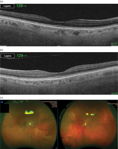
Figure 5. Duration from onset of first ocular manifestation to diagnosis in patients presenting with ocular manifestations, such as ectopia lentis or severe myopia, prior to a diagnosis of homocystinuria. Several patients had both before being diagnosed. Duration from the onset of the first ocular manifestation of homocystinuria to diagnosis is shown in blue. The eye symbol indicates ocular manifestation, and the black box indicates a thrombotic event. “D” indicates time of diagnosis, “EL” indicates ectopia lentis, and “SM” indicates severe myopia.
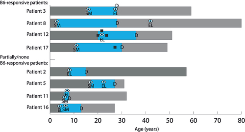
Table 2. Demographic data, visual and ocular findings, and results of visual quality of life in a group of 18 patients with homocystinuria.
Data availability statement
The data that support the findings of this study are available on request from the corresponding author, KTF. The data are not publicly available due to containing information that could compromise the privacy of research participants.