724
Views
17
CrossRef citations to date
0
Altmetric
Research Article

β-Lactam type molecular scaffolds for antiproliferative activity: Synthesis and cytotoxic effects in breast cancer cells

, , , &
Pages 668-685 | Received 02 Jan 2008, Accepted 21 Mar 2008, Published online: 20 Oct 2008

Figures & data

Figure 1 Structure of tamoxifen, raloxifene and related SERMs.

Figure 1 Structure of tamoxifen, raloxifene and related SERMs.

Scheme 1 Synthesis of β-lactams 12au. Scheme Reagents: (a) CH3CH2OCOCH2Br, Zn, TMCS, Benzene, reflux, 6h. (b) aldehyde/ketone, LDA, THF, 78°C, 30min. (c) R3R4N-CH2CH2Cl, Acetone, reflux, 2 h.
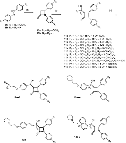
Scheme 1 Synthesis of β-lactams 12a–u. Scheme Reagents: (a) CH3CH2OCOCH2Br, Zn, TMCS, Benzene, reflux, 6h. (b) aldehyde/ketone, LDA, THF, 78°C, 30min. (c) R3R4N-CH2CH2Cl, Acetone, reflux, 2 h.

Scheme 2 Synthesis of β-lactams 13ae. Scheme Reagents: (a) pyridinium chlorochromate, CH2Cl2, 25°C

Scheme 2 Synthesis of β-lactams 13a–e. Scheme Reagents: (a) pyridinium chlorochromate, CH2Cl2, 25°C

Table I.  Antiproliferative and cytotoxic effects of compounds 12al in MCF-7 cells..

Table II.  Antiproliferative and cytotoxic effects of compounds 13ae containing carbonyl at C-5 in MCF-7 cells.

Table III.  Antiproliferative and cytotoxic effects of compounds 12mu with modifications at the C-5 position in MCF-7 cells.

Figure 2 Compound 12u inhibited proliferation and induced cytotoxicity of MCF-7 cells. Antiproliferative and cytotoxic activity of compounds 12u on oestrogen sensitive MCF-7 breast cancer cells, IC50 = 5.51 μM. The optical density values are given as a ratio of the treated cells and control cells × 100% and are means of at least 9 replicates. The absence of error bars indicates that the error was smaller than the size of the symbol.
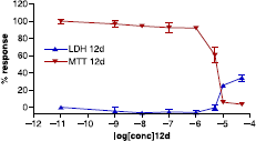
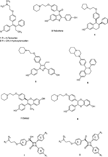
Figure 2 Compound 12u inhibited proliferation and induced cytotoxicity of MCF-7 cells. Antiproliferative and cytotoxic activity of compounds 12u on oestrogen sensitive MCF-7 breast cancer cells, IC50 = 5.51 μM. The optical density values are given as a ratio of the treated cells and control cells × 100% and are means of at least 9 replicates. The absence of error bars indicates that the error was smaller than the size of the symbol.
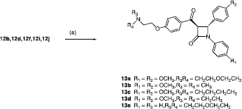
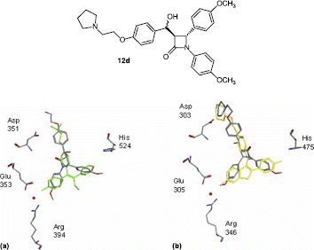
Figure 3 Docked structure for compound 12d in (a) ERα superposed with 4-hydroxytamoxifen (green) and (b) ERβ superposed with raloxifene (yellow).

Figure 3 Docked structure for compound 12d in (a) ERα superposed with 4-hydroxytamoxifen (green) and (b) ERβ superposed with raloxifene (yellow).

Reprints and Corporate Permissions

Please note: Selecting permissions does not provide access to the full text of the article, please see our help page How do I view content?

To request a reprint or corporate permissions for this article, please click on the relevant link below:

Academic Permissions

Please note: Selecting permissions does not provide access to the full text of the article, please see our help page How do I view content?

Obtain permissions instantly via Rightslink by clicking on the button below:

If you are unable to obtain permissions via Rightslink, please complete and submit this Permissions form. For more information, please visit our Permissions help page.