Figures & data
Figure 1. Enhanced expression of autophagy markers in pulmonary endothelial cells on combined treatment with morphine and Tat compared to monotreatments. Confluent Human pulmonary microvascular endothelial cells (HPMEC) were treated with 1 µM morphine (M) and/or 25 ng/ml Tat (T) in 0.5% serum-containing medium from 3 h to 9 d followed by western blot for (A) ULK1, (B) BECN1, (C) ATG5 and (D) ATG7. The graphs are the densitometry analysis of protein expression at various time points from at least 3 independent experiments. The western blots above each graph represent analysis of the 6-h treatment. The blot probed for BECN1 was stripped and reprobed for ATG5 and ATG7. (E) Cells were pretreated with naloxone (1 µM) and/or Su5416 (0.5 µM) 20 min before 6 h morphine and/or Tat treatment followed by western blot of ULK1 and BECN1 expression. The representative blots are shown with densitometry graphs representing mean +SEM from 3 independent experiments. *P < 0.05, **P < 0.01, ***P < 0.001 vs. control, #P < 0.05, ##P < 0.01, ###P < 0.001 vs. morphine, $P < 0.05, $$P < 0.01, $$$P < 0.001 vs. Tat, @P < 0.001 vs. M+T.
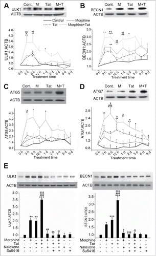
Figure 2. Increased number of autophagosomes or autolysosomes in HPMEC on combined treatment with morphine and Tat. (A) Confluent HPMEC were treated with morphine (1 μM) and/or HIV-Tat (25 ng/ml) for 24 h. Live cells were stained with LysoTracker Red dye for 30 min followed by immunofluorescence staining for LC3B (green). Magnification 60X. Scale bar: 50 μm. (B) HPMECs were transfected with 300 ng of pBABE-puro mCherry-EGFP-LC3B plasmid followed by morphine and/or Tat treatment at 48 h post-transfection. After 24 h of treatment, cells were fixed with 4% paraformaldehyde and viewed using a Nikon Eclipse E2000-U inverted confocal microscope. Green puncta (GFP) represent autophagosomes and red puncta (mCherry) represent autolysosomes. Magnification 100X. Scale bar: 50 μm. (C) Transmission electron microscopy (TEM) analysis of HPMEC treated with morphine and/or Tat. Cells treated for 24 h were trypsinized followed by fixation in glutaraldehyde and processed for TEM (3000X magnification). The column on the right shows magnified images of the insets. Scale bars: 500 nm.
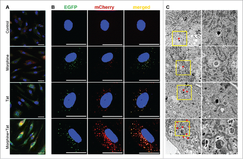
Figure 3. Enhanced autophagic flux on combined treatment with morphine and Tat. HPMECs untransfected (A, C, D) or transfected with pBABE-puro mCherry-EGFP-LC3B plasmid (B) were treated with morphine and/or HIV-Tat in the absence or presence of BAF. ((A)and B) For MAP1LC3B immunostaining (A) and visualization of GFP or mCherry LC3B puncta (B) cells were fixed with 4% paraformaldehyde at 24 h post-treatment and viewed using a confocal microscope. ((C)and D) Western blot analysis of MAP1LC3B-II (C) and SQSTM1 (D) at 24 h and 48 h post treatment, respectively. The graphs represent the densitometry analysis of western blots from 2 or 3 independent experiments. Mean +SEM, *P < 0.05, **P < 0.01, ***P < 0.001 vs. control, ##P < 0.01, ###P < 0.001 vs. morphine, $P < 0.05 vs. Tat, @P < 0.05 vs. combined morphine and Tat (M+T), θP < 0.05, θθP < 0.01, θθθP < 0.001 vs BAF. Note: In order to visualize autophagosomes (yellow puncta) more clearly in M+T+BAF, we lowered the green and increased the red fluorescence intensities compared to the images captured for .
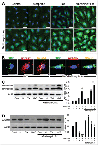
Figure 4. Transcription-independent increase in the expression of autophagy proteins on morphine and Tat treatment. (A) Confluent HPMEC were treated with morphine and/or Tat in 0.5% FBS containing endothelial cell medium from 30 min to 6 h followed by RNA extraction, cDNA preparation and qPCR for ULK1, BECN1, ATG5 and ATG7. (B and C) HPMEC at 80% confluence were treated with either (B) actinomycin D (3 μg/ml) or (C) cycloheximide (10 μg/ml)15 min before morphine and Tat treatment for 6 h followed by protein extraction for western blot analyses of ULK1, BECN1, ATG5 and ATG7. The graphs represent the densitometry of 4 independent experiments. Mean +SEM, *P < 0.01, **P < 0.001 vs. control, @P < 0.001 vs. combined morphine and Tat (M+T).
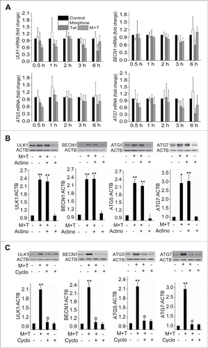
Figure 5. Autophagy protects HPMEC from morphine and Tat-mediated apoptosis. HPMEC (1.25 × 10⁴ cells / well) were plated on 96-well plates followed by pretreatment with either 3-methyladinine (3-MA) (A) or rapamycin (C) before morphine and/or Tat treatment for 3 d followed by Cell Death Detection ELISA. Alternatively, cells were transfected with ULK1 siRNA or scrambled (scrm) siRNA (B) or with ULK1 expression plasmid or empty vector (EV) (D) using HiPerfect or GeneJuice reagent respectively as per the manufacturer's instructions before treatment with morphine and/or Tat. The data are average of 3 independent experiments performed in triplicates. Mean + SD, *P < 0.05, **P < 0.01, ***P < 0.001 vs. control, #P < 0.05, ##P < 0.01, ###P < 0.001 vs. morphine, $$$P < 0.001 vs. Tat, @P < 0.05, @@@P < 0.001 vs. morphine+Tat treatment.
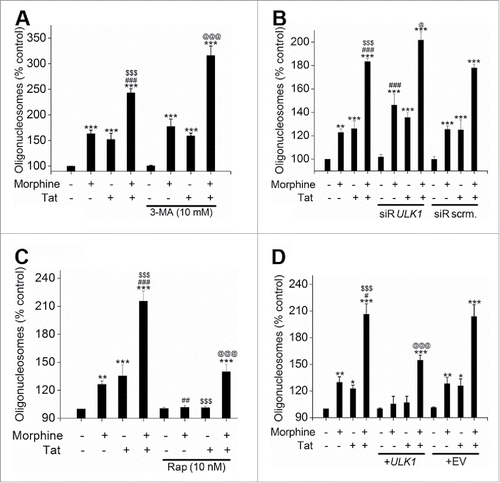
Figure 6. Autophagy promotes the morphine and Tat-treated HMPEC toward hyperproliferation. Autophagy was inhibited by treatment with 3-MA (A) or ULK1 knockdown (B) and was stimulated by rapamycin (C) or ULK1 overexpression (D) followed by morphine and/or Tat treatment. MTS Cell Proliferation Assay was later done at 6 d post-treatment. Cells (3 × 10³ cells/well) were plated on 96-well plates followed by pretreatment with either 3-MA (A) or rapamycin (C) before morphine and/or Tat treatment. Cells were transfected with ULK1 siRNA or scrambled (scrm) siRNA using HiPerfect (B) or transfected with ULK1 expression plasmid or empty vector (EV) using GeneJuice reagent (D) as per the manufacturer's instructions followed by morphine and/or Tat treatment. The data shown are mean + SD of 3 independent experiments done in triplicates. *P < 0.05, ** P <0.01, ***P < 0.001 vs. control, #P < 0.05, ###P < 0.001 vs. morphine, $P < 0.05, $$$P < 0.001 vs. Tat, @@@P < 0.001 vs. morphine+Tat treatment.
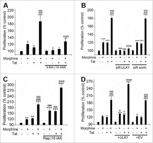
Figure 7. Morphine and Tat-mediated enhanced oxidative stress activates autophagy in endothelial cells. (A) Analysis of H2O2 or (B) superoxide levels in morphine (1 μM) and/or HIV-Tat (25 ng/ml)-treated HPMEC for the indicated time points by amplex red assay or SOD-inhibitable cytochrome C reductase assay, respectively. C) HPMEC (2 × 10⁴ cells/well) plated on a 96-well plate were pretreated with naloxone (1 μM) and/or Su5416 (0.5 µM) followed by morphine and/or Tat for 3 h and measurement of total reactive oxygen species using the DCF assay kit. (D and E) Confluent HPMEC were pretreated with antioxidants for 30 min followed by 6 h Tat and morphine treatment for western blot analysis for ULK1 and BECN1 expression (D) and for MAP1LC3B immunofluorescence staining (100X magnification) (E). (F) HPMEC were pretreated with antioxidant cocktail (Aox) and/or BAF followed by combined morphine-Tat treatment for 24 h for western blot analyses of MAP1LC3B-II and SQSTM1 expression. The graphs represent densitometry from 3 independent experiments, mean+ SEM. (G and H) Modulation of morphine and Tat induced oxidative stress with autophagy inhibition and stimulation. Confluent HPMEC pretreated with either 3MA (10 mM) or rapamycin (10 nM) before morphine and Tat were assayed for (G) H2O2 and (H) O2− generation using amplex red and SOD-inhibitable cytochrome c reductase assays, respectively. Mean +SEM of at least 3 independent experiments. A, D, G) *P < 0.05, **P < 0.01, ***P < 0.001 vs. control, #P < 0.05, ##P < 0.01, ###P < 0.001 vs. morphine, $P < 0.05, $$P < 0.01, $$$0.001 vs. Tat, @P < 0.05, @@P < 0.01, @@@P < 0.001 vs. combined morphine and Tat (M+T). (B, C, F, H) *P < 0.001 vs. control, #P < 0.001 vs. morphine, $0.001 vs. Tat, @P < 0.001 vs. M+T treatment. Scale bars: 50 μm .
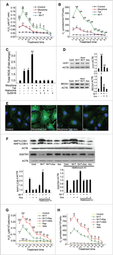
Figure 8. Enhanced autophagy in SIV-infected macaques exposed to morphine. (A) Total cellular extract from frozen macaque lung tissues from SIV (V group, n = 4), morphine (M group, n = 4) or SIV+morphine-injected macaques (VM group, n = 6) were probed or reprobed for the expression of autophagic markers BECN1, ATG5 and ATG7 by western blot. Graphs represent the average densitometry of the protein expression from each group. (B) The paraffin-embedded macaque lung sections immunostained for LC3B and endothelial marker (VWF). Magnification: 60X. Scale Bar 50 μm. (C) Representative TEM images of macaque lung sections. The upper panel represents 4000X magnification of endothelial cells lining the blood vessels from the macaques in the respective groups with red triangles pointing at the autophagic bodies (autophagosomes or autolysosomes). Lower panel images are of C088 macaque from the VM group. The first image (20X) is a crystal violet stained section from the cut block used for processing the tissue for TEM imaging, representing one of the areas with proliferating endothelial cells. The red box represents the approximate area of the TEM image shown in the middle, lower panel (250X). Further higher magnification image (2000X) of the inset shows a remarkably higher number of autophagic bodies in one of the cells. The graph is the average number of autophagic bodies in at least 30 endothelial cells per group (n = 3 for V or M group and n = 4 for VM group). #P < 0.05, ###P < 0.001 vs. M, $P < 0.05, $$$P < 0.001 vs V. Scale bars: 500 nm.
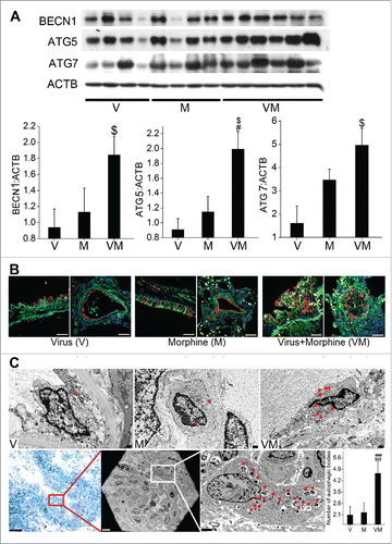
Figure 9. Significant increase in the expression of autophagy related proteins and number of autophagosomes or autolysosomes in the human lungs from HIV-infected IVDUs. (A) Western blot analysis of total lung extracts from normal, HIV-infected and /or IVDUs for BECN1, ATG5 and ATG7 expression. Graphs represent the densitometry analysis of the blots. (B) TEM analysis showing autophagic bodies (autophagosomes or autolysosomes) in the endothelial lining of the pulmonary blood vessels (3000X magnification). Lower panel shows the 800X magnification image with an area (red box) of the blood vessel from where representative 3000X image of HIV+ IVDU group was captured. The graph represents the average number of autophagic bodies counted in at least 24 cells per group (n = 2 per group). *P < 0.05, **P < 0.01 vs. normal, #P < 0.05 vs. IVDU, $P < 0.05 vs HIV. Scale bars: 500 nm.
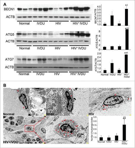
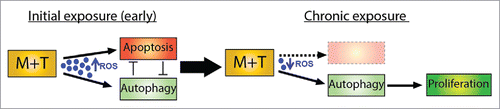
Figure 10. Schematic representation of autophagy-mediated hyperproliferation of pulmonary endothelial cells on chronic exposure to morphine and Tat.