Figures & data
Table 1. Summary of (eco)toxicological methods used in this study.
Table 2. Physico-chemical properties of the studied nanomaterials (NMs were ≥99% pure).
Figure 1. Surface charge (ζ-potential) of five nanomaterials in five different test media analyzed at concentration 100 mg metal/l using Malvern Zetasizer.
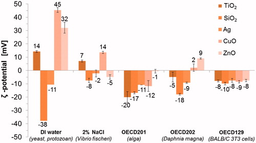
Table 3. Dissolution (share of dissolved metal to total metal) of Ag, CuO and ZnO nanomaterials was determined by atomic absorption spectroscopy in supernatants of ultracentrifuged 10 mg metal/l NM suspensions after 30-min, 24-h, 48-h and 72-h incubation in deionized water (DI water) or test media.
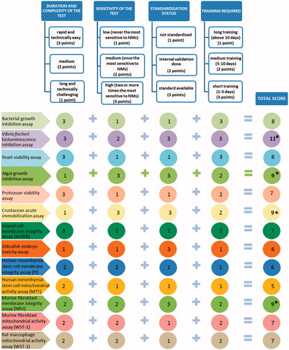
Table 4. Toxicity (EC50, MBC or LOEC) values of nanomaterials expressed in a heat map.
Figure 2. Shematic representation of embryonic development (0.2 h and 72 h are shown) of zebrafish (Kimmel et al., Citation1995, reprinted with the permission of John Wiley and Sons) (a). Representative images of development of unexposed zebrafish embryo (control) or exposed to 100 mg/l TiO2 NMs or to 10 mg/l Ag NMs after 72 h (b). Red arrow indicate undeveloped embryo exposed to Ag NMs. Scale bar = 1 mm.
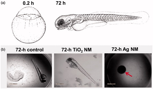
Figure 3. Algae Raphidocelis subcapitata after 72-h exposure in OECD medium (a), with 100 mg/l of nano-SiO2 (b) and 100 mg/l of TiO2, photographed in parallel using phase contrast (c) and fluorescence microscopy (d).
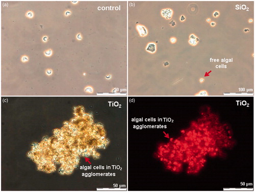
Figure 4. Images of Daphnia magna: not exposed (a, c) and after 24-h incubation with MWCNTs (b, d). Red arrows indicate the gut of Daphnia. Upper panels depict the gut of Daphnia after 24-h incubation and lower panels after transfer of daphnids to artificial freshwater. Note that even after the 24-h recovery in the clean artificial freshwater the gut remained filled with MWCNTs (d). Images were taken with Olympus BX61. Scale bar = 50 μm.
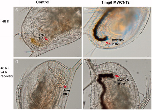
Figure 5. Decision tree for the selection of optimal toxicity assays for screening and hazard ranking of NMs. MTT, 3-(4,5-dimethylthiazol-2-yl)-2,5-diphenyltetrazolium bromide; NRU, neutral red uptake; PI, propidium iodide; WST-1, 2-(4-Iodophenyl)-3-(4-nitrophenyl)-5-(2,4-disulfophenyl)-2H-tetrazolium.