14,854
Views
303
CrossRef citations to date
0
Altmetric
Review Article

Inhibiting extracellular vesicles formation and release: a review of EV inhibitors

& ORCID Icon
Article: 1703244 | Received 08 Mar 2019, Accepted 05 Dec 2019, Published online: 19 Dec 2019

Figures & data

Figure 1. Exosomes are released from intracellular compartments known as multi-vesicular bodies (MVBs). MVBs biogenesis is associated with two different mechanisms: ESCRT-dependent and ESCRT-independent pathways.

ESCRT is a multi-molecular machinery composed of different proteins able to interact with ubiquitylated cargos and promote the formation of Intra-Luminal Vesicles (ILVs). The ESCRT pathways seems to be associated to RAS, a small GTPases able to orchestrate different cellular processes including exosomes production and cell proliferation, differentiation, adhesion and migration. In order to function properly, RAS must be linked to a lipid chain, achieved by the activity of specific enzymes called farnesyltransferases. Manumycin A is proposed to be able to inhibit these latter enzymes; thus, in turn, it is able to prevent RAS activation and function, preventing the formation of exosomes through the ESCRT pathway. The ESCRT-independent pathway is orchestrated by neutral sphingomyelinases, a family of enzymes able to convert sphingomyelin, present inside lipid raft, in ceramide. The resulting ceramides, interacting with each other, form large microdomains inducing the budding and the formation of ILVs into MVBs. GW4869 is a potent neutral sphingomyelinases inhibitor that preventing the formation of ILVs is able to block exosomes production.
Figure 1. Exosomes are released from intracellular compartments known as multi-vesicular bodies (MVBs). MVBs biogenesis is associated with two different mechanisms: ESCRT-dependent and ESCRT-independent pathways.

Table 1. Most commonly used compounds reported to prevent EVs (MVs or exosomes) release and their biological properties.

Figure 2. Calpains, once activated through calcium binding, can activate different cellular processes including cell migration (through their interaction with Talin and FAK), cell invasion (thorough the promotion of MMP release and activation), and MVs formation and release (through their activity on cytoskeletal proteins including cortactin). Calpain inhibitors, of which calpeptin is one of the most studied, are under investigation for clinical application.

Figure 2. Calpains, once activated through calcium binding, can activate different cellular processes including cell migration (through their interaction with Talin and FAK), cell invasion (thorough the promotion of MMP release and activation), and MVs formation and release (through their activity on cytoskeletal proteins including cortactin). Calpain inhibitors, of which calpeptin is one of the most studied, are under investigation for clinical application.

Table 2. EV separation and characterisation methods used in the studies discussed in this review.
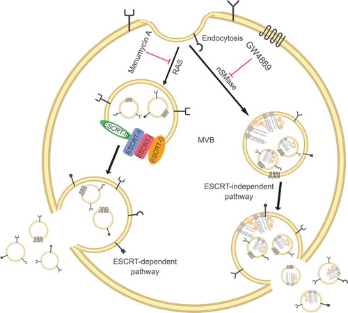
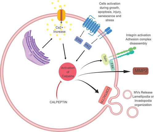
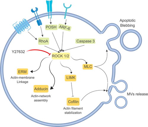
Figure 3. Rho-associated protein kinases (ROCK) are serine-threonine kinases involved in cytoskeleton re-organisation. Once activated by multiple stimuli, ROCK regulate the shape and the movement of the cells, through the activation of Adducin or ERM (ezrin, radixin and moesin), but they can also interact with MLC (myosin light chain) and LIMK (LIM kinases, able to inactivate cofilin, fundamental for actin filament stabilisation) both involved in MVs release. Y27632 is a competitive inhibitor of both ROCK1 and ROCK2 and by blocking these proteins it can inhibit MVs release.

Figure 3. Rho-associated protein kinases (ROCK) are serine-threonine kinases involved in cytoskeleton re-organisation. Once activated by multiple stimuli, ROCK regulate the shape and the movement of the cells, through the activation of Adducin or ERM (ezrin, radixin and moesin), but they can also interact with MLC (myosin light chain) and LIMK (LIM kinases, able to inactivate cofilin, fundamental for actin filament stabilisation) both involved in MVs release. Y27632 is a competitive inhibitor of both ROCK1 and ROCK2 and by blocking these proteins it can inhibit MVs release.

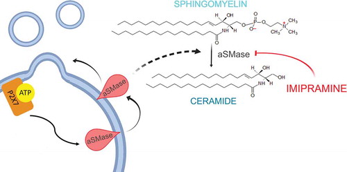
Figure 4. MVs present unique lipid characteristics, including an enrichment of sphingomyelin and ceramide, thus every enzyme able to interfere with membrane composition, i.e. calpains, scramblases and acid sphingomyelinases, plays a key role in MVs biogenesis. Acid sphingomyelinases convert sphingomyelin into ceramide, a cone-shaped rigid lipid that forms micro-domains inside the cell membrane, inducing the budding of MVs. Imipramine, a well-known anti-depressant, can promote membrane fluidity by acting on aSMases, thus preventing MVs generation.

Figure 4. MVs present unique lipid characteristics, including an enrichment of sphingomyelin and ceramide, thus every enzyme able to interfere with membrane composition, i.e. calpains, scramblases and acid sphingomyelinases, plays a key role in MVs biogenesis. Acid sphingomyelinases convert sphingomyelin into ceramide, a cone-shaped rigid lipid that forms micro-domains inside the cell membrane, inducing the budding of MVs. Imipramine, a well-known anti-depressant, can promote membrane fluidity by acting on aSMases, thus preventing MVs generation.