Figures & data
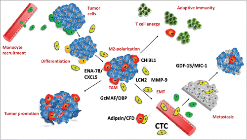
Figure 1. Induction of monocyte-macrophage differentiation in PBMNCs exposed to BHGc7-CM (A) shows the residual PBMNCs and debris in control cultures and (B) the CTC-primed recruitment of macrophages following 10 d of incubation.
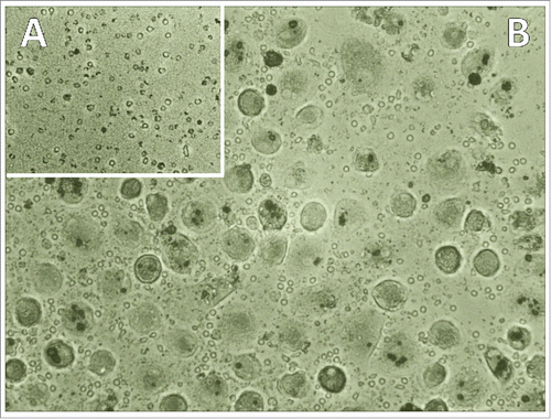
Figure 2. Flow cytometry of cell surface markers of CTC-primed macrophages. Typical experiments showing expression of CD14, CD163 and CD68 in indirect immunofluorescence staining experiments.
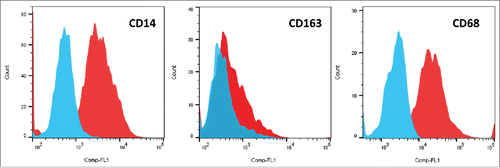
Figure 3. Expression of cytokines/chemokines of SCLC26A and BHGc7-primed macrophages. The figure shows the significantly expressed cytokines/chemokines expressed by macrophages induced by preincubation with CM of SCLC26A and BHBc7 CTC line, respectively (mean ± SD). All differences are statistically significant, except for PF4, RBP4 and IL-17A.
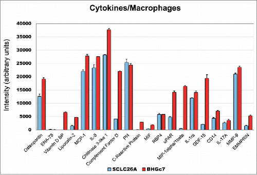
Figure 4. Expression of cytokines/chemokines of SCLC26A and BHGc7-primed macrophages. The figure shows the significantly expressed cytokines/chemokines expressed by macrophages induced by preincubation with CM of BHGc7 and BHBc10 CTC lines, respectively (mean ± SD). All differences are statistically non-significant, except for ENA-78, VDBP, LPC2, CRP, uPAR, MIP-1 and GDF-15.
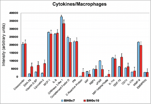
Figure 5. Exposure of PBMNCs to control HEK293-CM and recombinant CHI3L1. Incubation of normal PBMNCs with HEK-293 CM resulted in attachment of residual monocytes (A) in contrast to exposure with 2 ng/mL recombinant CHI3l1 which resulted in appearance of differentiated macrophages (B).
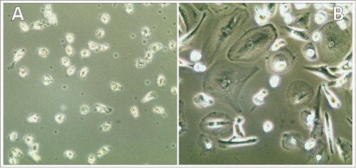
Figure 6. Tumor-associated macrophages and CTCs in tumor biology. This figure depicts the role and factors contributed by CTCs in tumor-macrophage interactions. Peripheral blood monocytes are recruited to the developing tumor (green; top, left) und undergo differentiation/polarization (yellow) under the influence of tumor-derived factors to TAMs of M2-like type (red) which results in suppression of immune responses (top, right) and enhanced neoangiogenesis/tumor progression (bottom, left). Inflammatory and tumor-derived cytokines/chemokines induce precursors of CTCs (light green) which secrete additional cytokines/chemokines, comprising ENA-78/CXCL5, LNC2, CHI3L1, CFD/adipsin, VDBP/GcMAF and MMP-9. Increased neoangiogenesis and degradation of ECM by MMP-9 and possibly other proteases induce intravasation of CTCs, most likely after EMT. At distant sites (bottom, right) CTCs extravasate capillaries and set up secondary lesions, possibly protected against immune system attack by CTC-educated macrophages. Highly effective recruitment of macrophages seems to be linked to CHI3L1 expression of CTCs as SCLC tumor cells lack expression of this pseudochitinase. CFD/adipsin may be associated with cachexia in advanced disease.