Figures & data
Table 1. C. elegans genes implicated in molting that are discussed in this review.
Figure 1. Structural organization of the cuticle in adult C. elegans. Indicated are the five layers of the cuticle together with the apical part of epidermis (right side), and composition of cuticle layers (left side). The upper enlarged region shows the surface coat (glycocalyx), whereas the lower enlarged region indicates the presence of hemidesmosome-like structures (HDLSs) at the interface between the cuticle and epidermis. © WormAtlas. Adapted by permission of WormAtlas. Permission to reuse must be obtained from the rightsholder.Citation15
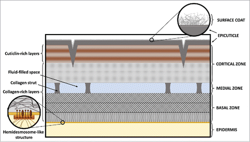
Figure 2. Attachments between the cuticle, epidermis, and muscles. Cellular and extracellular layers are indicated on the right side. Specific attachment structures are labeled in the figure, as well as important structural elements within the layers. © WormAtlas. Adapted by permission of WormAtlas. Permission to reuse must be obtained from the rightsholder.Citation15
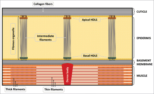
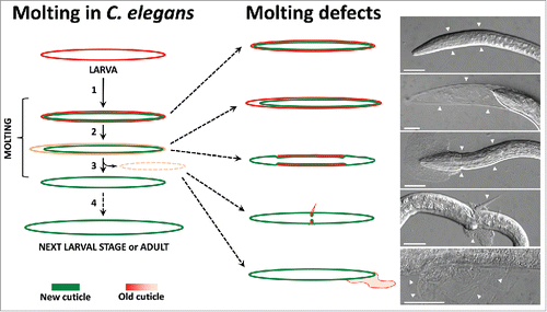
Figure 3. Schematic representation of the molting process and molting defects with representative micrograph examples. The normal physiological molting process is shown on the left. Schematic and micrograph examples of molting defects are shown on the right side, including (from top to bottom) the complete encasement phenotype of a mlt-3(fd72) mutant, the partially released cuticle in the head region of a qua-1(RNAi) larva, the corset phenotype of a nekl-3(sv3) mutant, a narrow constriction caused by the old cuticle in a nekl-3(sv3) mutant, and an old cuticle attached to the body surface after nekl-2(RNAi). White arrowheads indicate the presence of the old cuticle. Scale bars: 25µm.