Figures & data
Table 1. Primer sequences
Figure 1. Expression of VEGF and VEGFR2 was detected by immunofluorescence. (a): Magnification, × 200, (b): Magnification, × 400. Immunofluorescence staining showed that cells positive for VEGF and VEGFR2 were predominantly observed in the cytoplasm of all principle cells, some of the interstitial small vascular endothelial cells, and in some blood capillaries and sperm in the ductus epididymitis. VEGF, Vascular endothelial growth factor; VEGFR2, VEGF receptor 2
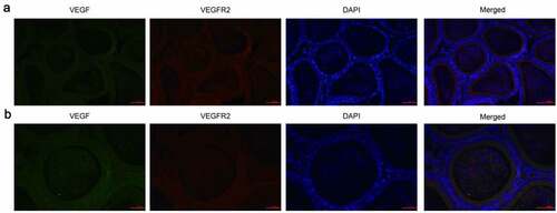
Table 2. Mean expression levels of VEGF and VEGFR2 in the epididymis of rats, as determined by immunohistochemical analysis (n = 10, Mean ± SD)
Figure 2. VEGF and VEGFR2 were downregulated in epididymal tissues of arsenic-exposed rats. Representative images of immunohistochemical staining using antibodies against VEGF (a) and VEGFR2 (b). VEGF, Vascular endothelial growth factor; VEGFR2, VEGF receptor 2
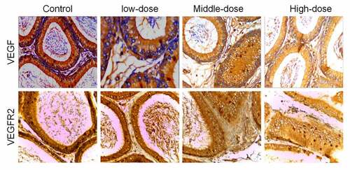
Figure 3. mRNA expression levels of VEGF and VEGFR2 in the epididymis of rats after transfection with VEGF-shRNA. Real-time PCR was used to measure the expression levels of VEGF and VEGFR2 in the epididymis of rats in different treatment groups following transfection with VEGF-shRNA. Data are represented as the mean ± SD. *P < 0.05 vs. control group. VEGF, Vascular endothelial growth factor; VEGFR2, VEGF receptor 2
Figure 4. Protein expression levels of VEGF and VEGFR2 in the epididymis of rats after transfection with VEGF-shRNA. (a) The protein expression of VEGF and VEGFR was measured by Western blot. (b) Quantification analysis of the protein expression based on immunoblotting results. Data are shown as the mean ± SD (n = 3) and were analyzed by one-way analysis of variance (ANOVA). *P < 0.05 vs. control group. VEGF, Vascular endothelial growth factor; VEGFR2, VEGF receptor 2
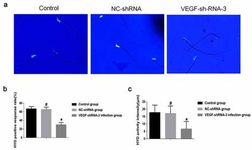
Figure 5. VEGF-shRNA decreased sperm hyaluronidase (HYD) activity in epididymis of rat. (a) The positive expression of HYD was detected by Fluorescent staining. Magnification, × 400., (b) The HYD positive response rate. (c) The HYD activity was measured by the mature substrate membrane method. *p < 0. 05 vs. control group. #P < 0.05 vs. VEGF-shRNA-3 group