
Figures & data
Figure 1. (A) En face scanning electron microscopy of brush border microvilli extending from the surface of mouse duodenal enterocytes. (B) Deep-etch electron microscopy reveals a dense network of adhesion complexes connecting the tips of adjacent microvilli. Scale bars are 1 μm and 1 nm in A and B, respectively.
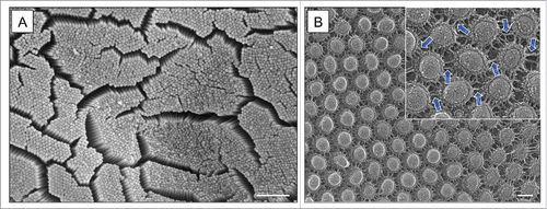
Figure 2. Localization and molecular architecture of the conserved apical adhesion complex.
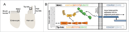
Table 1. Tip-link and IMAC components.