
Figures & data
Table 1. Therapeutic potential of EVs derived from various donor cells
Figure 1. Endogenous and exogenous loading strategies to obtain drug-loaded EVs.
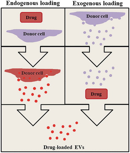
Figure 2. Generation of SPIONs containing EVs (Yuana Y, unpublished results). A. Exogenous loading were performed by growing cells in medium containing SPION and stimulating by sonoporation. B. EVs were harvested from conditioned medium containing SPIONs. C. Negative control EVs from cells grown without SPIONs and stimulated by sonoporation. Scale bars are 100 nm.
Figure 3. CFSE-containing cells stimulated with calcium ionophore (CaI) released EVs with CFSE (Yuana Y, unpublished results). A. 0.5 µM CFDA-SE were used to label cell using diffusion method. The CFDA-SE was cleaved by intracellular esterase enzymes to form an amine-reactive product, CFSE which can be detected by fluorescent microscopy. B. Negative control image (non-labelled cells). C. CFSE-labelled cells treated with 10 µM CaI have comparable CFSE signal to untreated CFSE-labelled cells as detected by flow cytometry. D. Detection of CD9-positive EVs using flow cytometry captured by CD9-antibody (AB) conjugated beads and stained with Alexa Fluor 647-tagged anti-CD9 AB. E. Detection of CD63-positive EVs using flow cytometry captured by CD63-antibody (AB) conjugated beads and stained with Alexa Fluor 647-tagged anti-CD63 AB. EVs were stained positive for CD9 or CD63 and CFSE as shown in the region Q2 (green rectangle) in D and E. E. Negative control (EVs captured with beads and unstained).
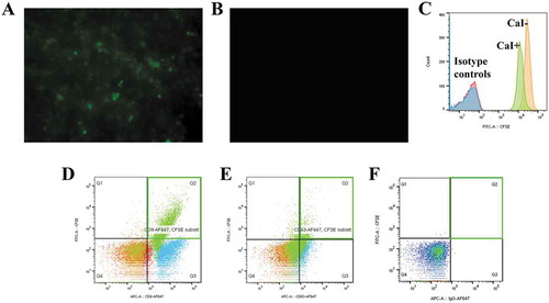
Table 2. Tissue distribution of EVs derived from different cell lines in mice model