Figures & data
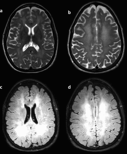
Figure 1. Magnetic resonance imaging of the brain showing diffuse white matter changes. (a) T2-weighted diffuse hyper-intensity involving the midbrain, insula, and basal ganglia bilaterally. (b) Bilateral symmetric reduced diffusion in periventricular white matter as seen on diffusion-weighted imaging (DWI). (c,d) FLAIR extent was graded severe.