
Figures & data
Figure 1. Tools for analysis of the dynamic conformation of IDPs. Atomic level descriptions of disordered protein ensembles are facilitated by quantitative observables from NMR spectroscopy and single molecule fluorescence techniques, which can be combined with computational methods (left panel). Overall molecular features, biophysical properties and morphologies of IDPs and IDP aggregates can be obtained from the experimental methods shown to the right.
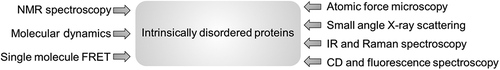
Figure 2. Targeting the Tau protein by small molecules. A. Schematic representation of the longest Tau isoform, 2N4R. 2N implies the number of inserts (I1 and I2) in the N-terminal projection region and 4R indicates the number of repeat regions in the C-terminal half of the protein. The microtubule assembly domain spans the proline-rich regions (P1 and P2), the pseudo-repeat sequences (R1–R4) and a region R’, which is partially similar to R1–R4. The hexapeptide stretches VQIINK and VQIVYK (marked in triangles) are essential for aggregation of Tau into paired helical filaments. B. Representative 2D-[1H-15N]-HSQC spectrum of 441-residue Tau protein. Small amide proton chemical shift dispersion is indicative of structural disorder. C. Superposition of a selected region of a 2D-[1H-15N]-HSQC spectrum of Tau in presence (grey) and absence (black) of methylene blue. Methylene blue specifically modifies the cysteine residues (C291 and C322, highlighted in panel A) in Tau monomers, resulting in broadening of adjacent glycine peaks (G292 and G323).[Citation130] D. Molecular structure of the Tau aggregation inhibitor methylene blue. Methylene blue stabilizes an aggregation incompetent monomer and prevents the formation of filaments and toxic oligomeric precursors.
![Figure 2. Targeting the Tau protein by small molecules. A. Schematic representation of the longest Tau isoform, 2N4R. 2N implies the number of inserts (I1 and I2) in the N-terminal projection region and 4R indicates the number of repeat regions in the C-terminal half of the protein. The microtubule assembly domain spans the proline-rich regions (P1 and P2), the pseudo-repeat sequences (R1–R4) and a region R’, which is partially similar to R1–R4. The hexapeptide stretches VQIINK and VQIVYK (marked in triangles) are essential for aggregation of Tau into paired helical filaments. B. Representative 2D-[1H-15N]-HSQC spectrum of 441-residue Tau protein. Small amide proton chemical shift dispersion is indicative of structural disorder. C. Superposition of a selected region of a 2D-[1H-15N]-HSQC spectrum of Tau in presence (grey) and absence (black) of methylene blue. Methylene blue specifically modifies the cysteine residues (C291 and C322, highlighted in panel A) in Tau monomers, resulting in broadening of adjacent glycine peaks (G292 and G323).[Citation130] D. Molecular structure of the Tau aggregation inhibitor methylene blue. Methylene blue stabilizes an aggregation incompetent monomer and prevents the formation of filaments and toxic oligomeric precursors.](/cms/asset/4c5ea898-a6cb-4d63-99e8-eea468cc67a6/iedc_a_1107041_f0002_b.gif)
Figure 3. Ensemble of conformations populated by the serine/arginine-rich region of SRSF1 in its phosphorylated state. SRSF1 is a classical serine/arginine (SR) protein, which plays a crucial role in RNA metabolism. Combination of NMR spectroscopy and MD simulations indicated a phosphorylation-induced conformational switch of the RS dipeptide repeat sequence from a highly disordered state to a partially rigid, arch-like structure [Citation113] (as shown in the figure). Phosphorylated serine residues are highlighted in the amino acid sequence.
![Figure 3. Ensemble of conformations populated by the serine/arginine-rich region of SRSF1 in its phosphorylated state. SRSF1 is a classical serine/arginine (SR) protein, which plays a crucial role in RNA metabolism. Combination of NMR spectroscopy and MD simulations indicated a phosphorylation-induced conformational switch of the RS dipeptide repeat sequence from a highly disordered state to a partially rigid, arch-like structure [Citation113] (as shown in the figure). Phosphorylated serine residues are highlighted in the amino acid sequence.](/cms/asset/b1de134b-e1da-4c50-8e2d-e11f8cef141a/iedc_a_1107041_f0003_b.gif)
Figure 4. Inhibition of the pathogenic aggregation of vesicle bound α-synuclein by PcTS. The N-terminal ~100 residues of α-synuclein adopt an α-helical conformation upon binding to vesicles. The small molecule aggregation inhibitor PcTS binds to the aromatic residues Tyr39 and Phe94 of α-synuclein and thereby stabilizes the α-helical conformation.[Citation126] This delays pathogenic aggregation of α-synuclein in a physiological vesicle environment.
![Figure 4. Inhibition of the pathogenic aggregation of vesicle bound α-synuclein by PcTS. The N-terminal ~100 residues of α-synuclein adopt an α-helical conformation upon binding to vesicles. The small molecule aggregation inhibitor PcTS binds to the aromatic residues Tyr39 and Phe94 of α-synuclein and thereby stabilizes the α-helical conformation.[Citation126] This delays pathogenic aggregation of α-synuclein in a physiological vesicle environment.](/cms/asset/a6085ed6-caa6-41b7-9dae-1c005d56d7dd/iedc_a_1107041_f0004_b.gif)